

Reinforced tissue shields
a tissue shield and reinforced technology, applied in the field of protective tissue shields, can solve the problems of increasing the incidence of ocular infection, i.e. endophthalmitis, and the potential toxic effects a recipient may be exposed to from residues and/or chemicals, and achieve the effect of inhibiting microbial contamination
- Summary
- Abstract
- Description
- Claims
- Application Information
AI Technical Summary
Benefits of technology
Problems solved by technology
Method used
Image

Examples
example 1
Fabrication of Self-Reinforced Ophthalmic Protective Shields of Single Curvature
[0066]1.5% gelatin solution is prepared by dissolving gelatin powder in distilled water. 1.5% chitosan solution is prepared by dissolving chitosan of 85% deacetylation degree in 1% acetic acid water solution. 1% insoluble collagen is swelled and dispersed in 1% acetic acid water. The solutions of gelatin and chitosan and collagen dispersion are combined together along with addition of polyethylene glycol 400 (PEG400) and therein mixed for 2 hours to yield a dispersion having a total solids concentration of 1.7%, and such that on drying the solution yields a gelatin concentration of 42%, chitosan 42%, PEG400 12%, and insoluble collagen 4%. The combined dispersion is then centrifuged for 2 minutes at 3000 rpm at room temperature.
[0067]The dispersion is placed into concave semi-spherically shaped molds having a radius of base curvature of 12.5 mm and a diameter of 20 mm The dispersion is allowed to dehydrat...
example 2
Fabrication of Self-Reinforced Therapeutic Protective Shields
[0069]A gelatin solution is prepared by dissolving 1.5 gram of gelatin powder in 100 ml distilled water. 1.5% chitosan solution is prepared by dissolving chitosan of 85% deacetylation degree in 1% acetic acid water solution. Insoluble collagen suspension is prepared by blending 1 gram of insoluble fibrous collagen in 100 ml of 1% acetic acid water at 4° C. for 4 minutes. The solutions of gelatin and chitosan, collagen dispersion and PEG 400 are combined together and then mixed for 2 hours, followed by addition of silver acetate and sodium chloride to yield 5 mM silver chloride in the final suspension that has a total solid concentration of 1.8%. The combined dispersion is then centrifuged for 2 minutes at 3000 rpm at room temperature. 12 ml of the dispersion is placed in a 2″×2″ flat polystyrene tray and allowed to dehydrate in a vibration free environment under controlled temperature (25±3° C.) and humidity (55-80%) condi...
example 3
Degradation of the Self-Reinforced Ophthalmic Shields (the Effect of Ratio of Gelatin To Chitosan On the Degradability)
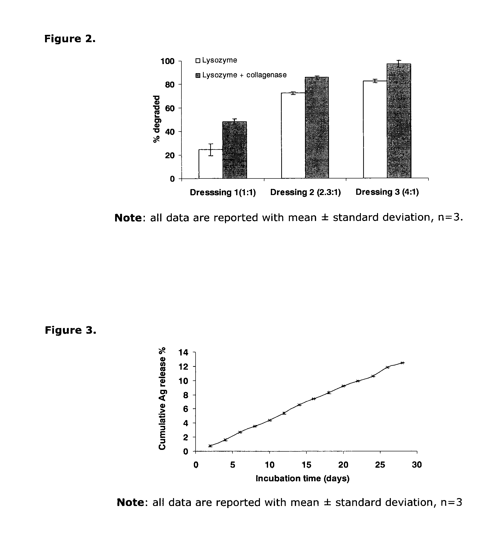
[0070]Three groups of concave shaped ophthalmic shields with ratios of gelatin to chitosan of 1:1, 2.3:1, and 4:1 are incubated in simulated tear fluid with 0.1% lysozyme with and without 12.5 unit / ml collagenase at 37° C. for 24 hours. The degradability of the shields is evaluated by comparing the dry weight of each shield before and after the incubation. Shields with the highest ratio of gelatin to chitosan (4:1) were almost completely dissolved in a simulated tear fluid (STF) containing both lysozyme and collagenase. The degradability of these shields in lysozyme-containing STF with or without collagenase is significantly influenced by the ratio of gelatin to chitosan (FIG. 2, p<0.05). Exemplary suitable polymer concentrations for formulating a 24 hr ophthalmic shield are 67% gelatin, 17% chitosan, 12% PEG400, and 4% insoluble collagen. For a 72 hr shield, a comp...
PUM
| Property | Measurement | Unit |
|---|---|---|
| Temperature | aaaaa | aaaaa |
| Temperature | aaaaa | aaaaa |
| Temperature | aaaaa | aaaaa |
Abstract
Description
Claims
Application Information