Preparation method of hydroxyapatite coating layer using aerosol deposition and hydrothermal treatment, and nanostructured hydroxyapatite coating layer prepared by the method
- Summary
- Abstract
- Description
- Claims
- Application Information
AI Technical Summary
Benefits of technology
Problems solved by technology
Method used
Image
Examples
experiment 1
Analysis of the Phase Evolution of Hydroxyapatite Coating Layer in Thermal Treatment
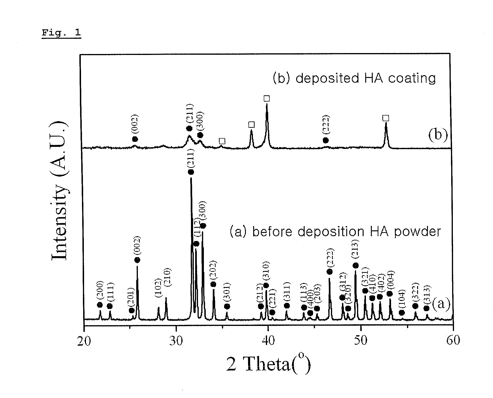
[0043]To analyze the phase evolution of hydroxyapatite coating layer in thermal treatment, X-ray diffractometer (XRD, X′pert MPD 3040, Philips Ltd., Eindhoven, Netherlands) was conducted with respect to the hydroxyapatite powder coating layer before deposition, and to the hydroxyapatite powder coating layer after deposition and before hydrothermal treatment respectively. The XRD data was acquired at 36 kV and 26 mA with single CuKa radiation and the result is illustrated in FIG. 1. In comparing waveforms (a) and (b) of FIG. 1, waveform (b) represents weaker and wider hydroxyapatite peak, indicating the low crystallinity of coating layer and small size of the crystallites.
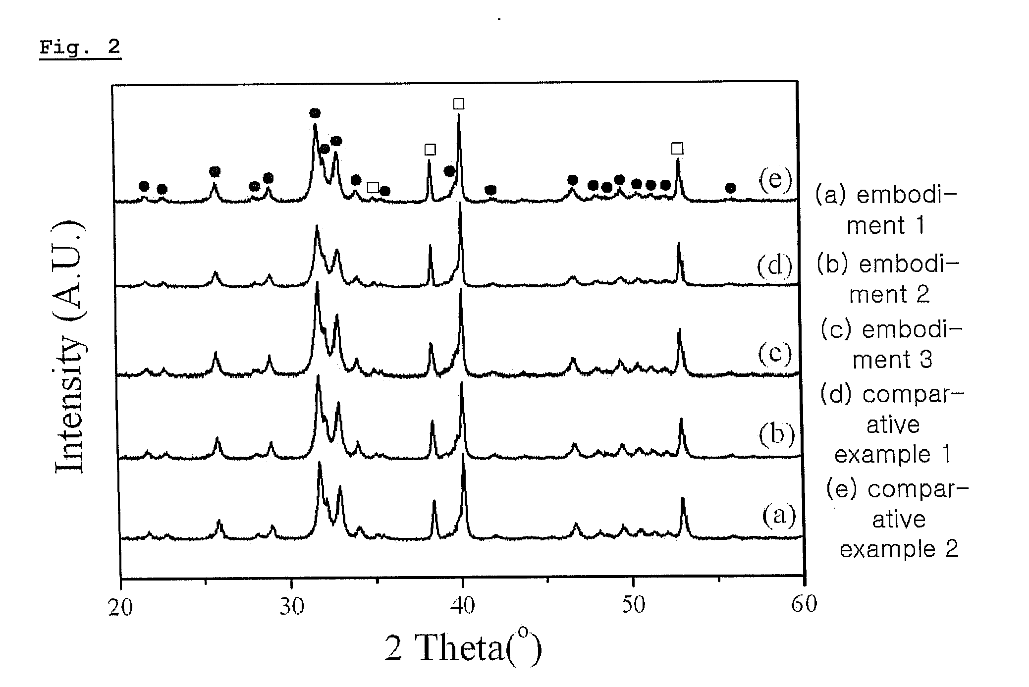
[0044]Also, to compare the effects by the hydrothermal treatment and the traditional heating of the coating layer by furnace, X-ray diffractometer was conducted with respect to Embodiments 1 and 3 and Comparative examples 1 and 2 in ...
experiment 2
Observation of Structural Changes of Hydroxyapatite Coating Layer After Thermal Treatment
[0045]Structural changes of hydroxyapatite coating layer after thermal treatment were observed with the infrared spectrophotometer (FT-IR, IFS 66, Bruker Optics, Ettlingen, Germany) in the range of between 400 and 4,000 cm−1. FIG. 3 illustrates the FT-IR spectra of hydroxyapatite coating deposited on Ti substrate. FIG. 3 shows that peaks of PO43− and OH− were distinct after hydrothermal treatments according to Embodiment 1 and 3 and Comparative example 1, 2. It shows that hydrothermal treatment has effect of improving crystallinity of coating layer, which corresponds with the result of X-ray diffractometer of Example 1.
experiment 3
Observation of Micro Structure of Hydroxyapatite Coating Layer
[0046]Micro structure of hydroxyapatite coating layer was observed with Scanning Electron Microscope (SEM, JSM-5800, Jeol Co., Tokyo, Japan) and Transmission Electron Microscope (TEM, JEM-2100F, Jeol Co., Japan).
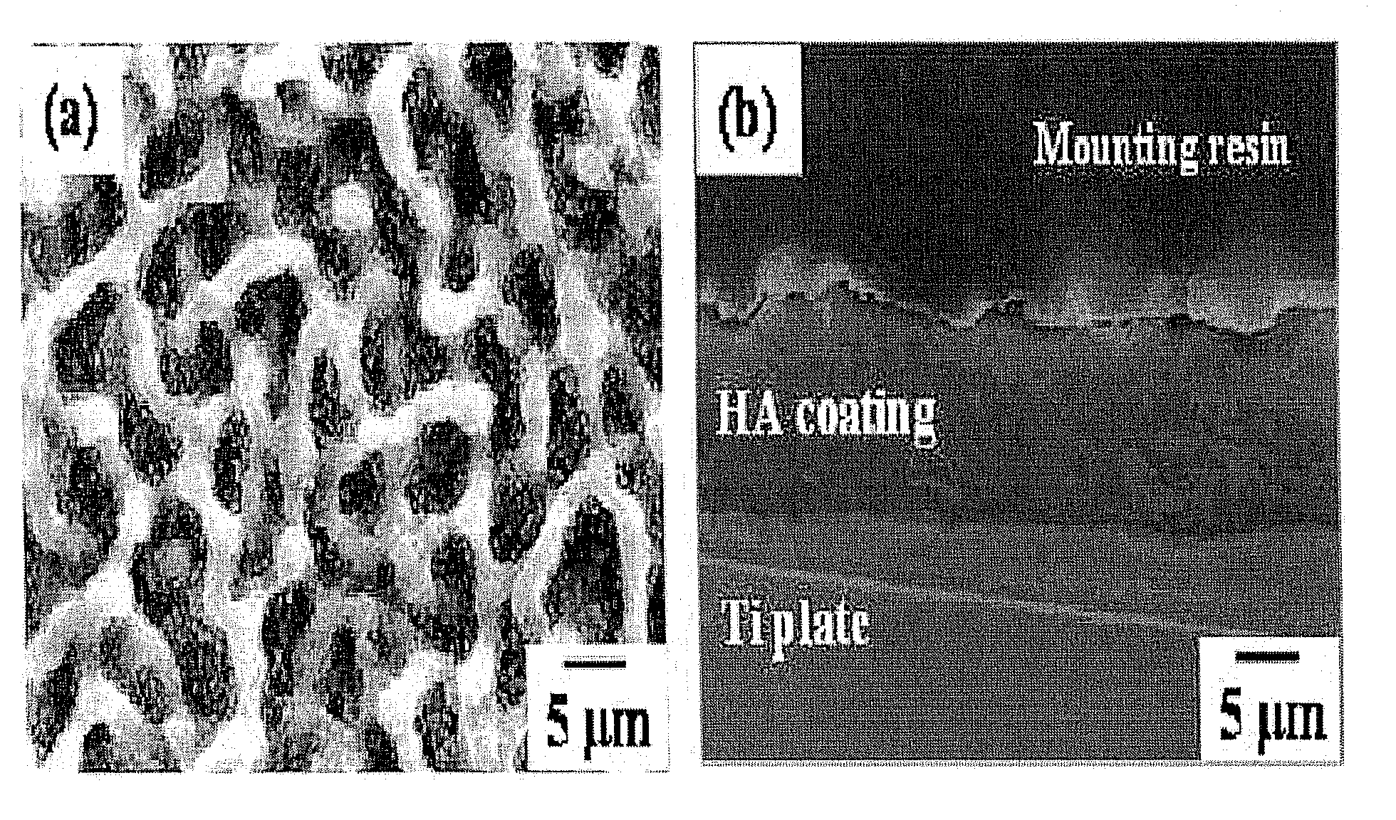
[0047]FIG. 4A illustrates the surface shape of SEM image of the hydroxyapatite coating layer prepared according to Embodiment 1. The hydroxyapatite coating layer shows the coarse surface with network-type micro-structure. The coarse surface of hydroxyapatite coating layer provides better environment for osteoblast in attachment, proliferation and segmentation. FIG. 4B illustrates the SEM cross sectional image of the hydroxyapatite coating layer prepared by the method of Embodiment 1. FIG. 4(b) shows that the interface between coating layer and Ti substrate is continuous without pore or defect. Therefore, hydroxyapatite coating layer according to the invention is expected to have superior biocompatibility and stron...
PUM
| Property | Measurement | Unit |
|---|---|---|
| Temperature | aaaaa | aaaaa |
| Temperature | aaaaa | aaaaa |
| Size | aaaaa | aaaaa |
Abstract
Description
Claims
Application Information
 - R&D
- Intellectual Property
- Life Sciences
- Materials
- Tech Scout
- Unparalleled Data Quality
- Higher Quality Content
- 60% Fewer Hallucinations
Browse by: Latest US Patents, China's latest patents, Technical Efficacy Thesaurus, Application Domain, Technology Topic, Popular Technical Reports.
© 2025 PatSnap. All rights reserved.Legal|Privacy policy|Modern Slavery Act Transparency Statement|Sitemap|About US| Contact US: help@patsnap.com


