Ratio based biomarkers and methods of use thereof

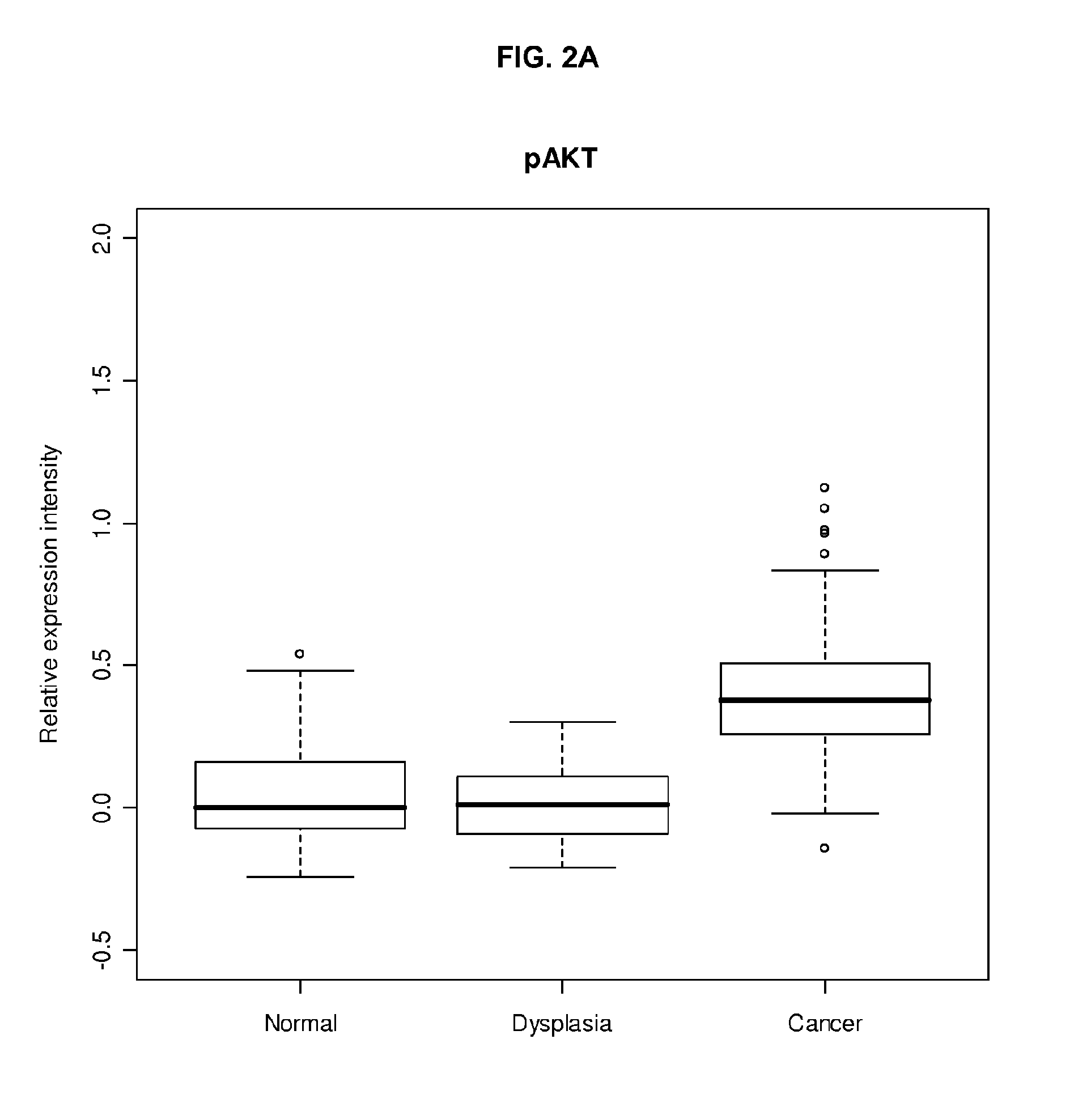
a biomarker and ratio technology, applied in the field of ratio-based biomarkers, can solve the problems of erratic tumor response, no simple answer to the question, and the success rate of tumor treatment or tumor treatment after administration of such non-cytotoxic agents is even more complex, so as to reduce the expression of tumor antigen, decrease the expression of pten, and increase the expression of p-akt.

Inactive Publication Date: 2011-12-15
UNITED STATES OF AMERICA
View PDF0 Cites 59 Cited by
  • Summary
  • Abstract
  • Description
  • Claims
  • Application Information

AI Technical Summary

Benefits of technology

[0015]In a further embodiment, the disclosure provides a method of determining relative cancer survival rates (or more generally prognosis) for a subject with a solid tumor by obtaining a biomarker indicator, the biomarker indicator being obtained by acquiring a solid tumor sample from the subject, extracting a first cancer associated protein from the solid tumor to produce a fraction comprising the first cancer associated protein, calculating the content of the first cancer associated protein in the fraction, normalizing the first cancer associated protein content against total cellular content in the fraction, extracting a second cancer associated protein from the solid tumor sample, calculating the content of the second cancer associated protein in the fraction, normalizing the second cancer associated protein content against total cellular protein content in the fraction, and correlating the normalized first cancer associated protein content against the normalized second cancer associated protein content to obtain a biomarker indicator, and comparing the biomarker indicator with relative survival rates, thereby determining the relative cancer survival rate for the subject with the solid tumor.
[0016]The present disclosure is further directed to methods for predicting relative cancer survival rates for a subject with a solid tumor by detecting the presence of an antibody to a tumor antigen in the solid tumor, wherein the tumor antigen involves increased expression of p-AKT and p-mTOR or decreased expression of PTEN as compared to a normal non-cancerous sample, thereby detecting the cancer in the subject, and correlating decreased expression of the tumor antigen in the subject as compared to a normal non-cancerous sample with a lower survival rate in the subject with the solid tumor. In another embodiment, the calculated tumor antigen involves increased HER2 relative to and / or along with decreased HER3 expression. Thus, the absolute value of the level of individual tumor (or other disease / condition) antigen is not necessarily determinative—rather, it is the relative amount compared to one or more other antigens that provides the predictive biomarker described herein.

Problems solved by technology

In addition to the variability of histopathologic subtypes, molecular study of tumors is even more complex.
However in practice, rates of success for the treatment of tumors or responsiveness of tumors after administration with such non-cytotoxic agents have been erratic.
Currently, there is no simple answer to the question of which cellular proteins or signaling pathways are responsible for making a cell cancerous.
While LCM does provide the capacity to perform a directed western blot on a tissue section, the methodology is time consuming and does not provide a global expression view of a targeted protein.
Immunohistochemistry while providing excellent localization, lacks quantification without sophisticated equipment such as high resolution tandem mass spectrometry, and lacks a normalization component.
Other “grind and bind” techniques for protein expression profiling provide quantification, but fail to provide a histo-morphological perspective of protein expression.
Although these techniques are generally superior in expression profiling and quantification of protein changes associated with disease states, each has significant limitations.

Method used


Image

  • Ratio based biomarkers and methods of use thereof
  • Ratio based biomarkers and methods of use thereof
  • Ratio based biomarkers and methods of use thereof

Examples


example 1

Patient Selection and Tumor Sample Collection

[0308]221 patients with EHCC who were surgically resected at Asian Medical Center, University of Ulsan College of Medicine in Seoul, South Korea were studied. Carcinomas with the epicenter in the extrahepatic bile duct were included, while carcinomas of the ampulla of Vater or pancreas, and those with obvious precancerous epithelial changes in the ampulla of Vater or pancreas were excluded. Carcinomas arising in the gallbladder, or intrahepatic bile duct with extension to the extrahepatic bile duct were also excluded in this study. Medical records were reviewed to obtain data including age and gender of patients, surgical procedure, survival time, and survival status. Data with tumor location, size, and growth pattern were obtained from reviewing pathology reports. Information on post-operative radiation and / or chemotherapy, and performance status of patients was not available for analysis. Material was obtained with appropriate human pro...

example 2

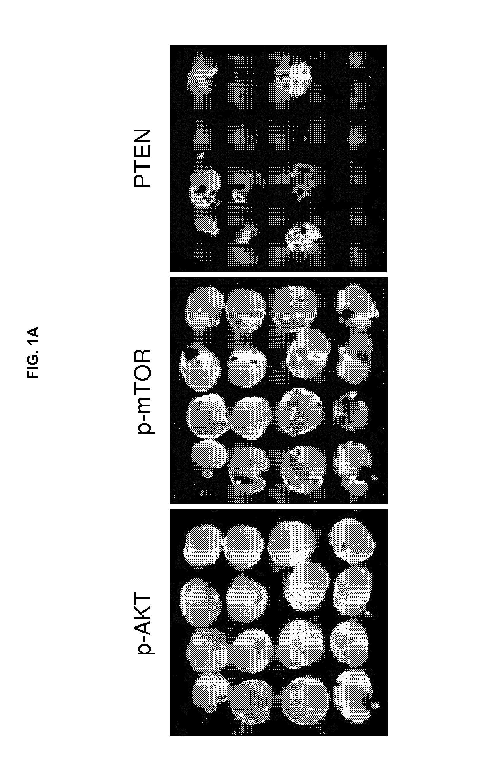
Proteomic Expression Profiling by Multiplex Tissue Immunoblotting

[0311]Multiplex tissue immunoblotting (MTI) was performed as known in the art (for example, Chung et al., Proteomics. 6:676-74, 2006 and Chung et al., Cancer Epidemiol. Biomarkers. Prev. 15:1403-08, 2006). In brief, TMA slides were deparaffinized and treated with an enzyme cocktail solution [0.001% trypsin plus 0.002% proteinase-K, 10% glycerol, 50 mM NH4HCO3 pH 8.2 (Fisher Scientific, Hampton, N.H.)] for 30 minutes at 37° C. Slides were subsequently incubated with Probuffer complete protease inhibitor solution [0.5 ml phosphatase inhibitor I (Sigma, St. Louis, Mo.), 0.5 ml phosphatase inhibitor II (Sigma), 1 protease inhibitor tablet (Roche Diagnostics, Indianapolis, Ind.) in 50 ml PBS (pH 7.2)] for 20 minutes at room temperature (RT). The proteins of treated slides were transferred to a 5-membrane stack (set) of P-FILM (20 / 20 GeneSystems, Rockville, Md.) using Tris-glycine transfer buffer (50 mM Tris, 380 mM Glycine)...

example 3

Immunohistochemistry

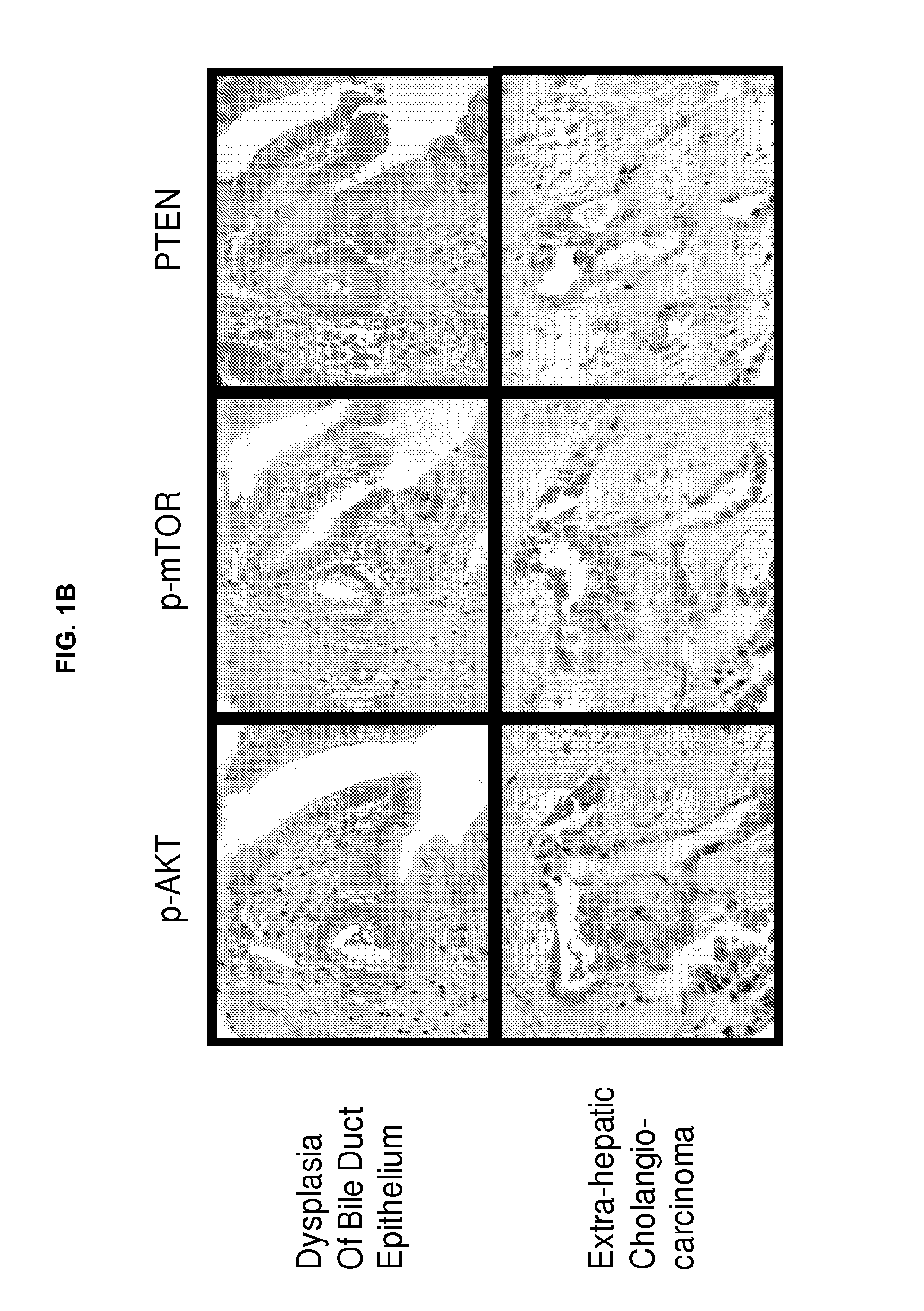
[0314]Tissue sections were deparaffinized and hydrated in xylene and serial alcohol solutions, respectively. Endogenous peroxidase was blocked by incubation in 3% H2O2 for 10 minutes. Antigen retrieval was performed in a steam pressure cooker with prewarmed antigen retrieval buffer pH 10 (Dako, Glostrup, Denmark) at 95° C., for 10 minutes. To minimize non-specific staining, sections were incubated with protein block (Dako) for 15 minutes. Primary antibodies were incubated overnight at 4° C. Antigen-antibody reactions were detected with DAKO LSAB+ peroxidase kit and DAB. Anti-p-AKT, anti-p-mTOR, and PTEN antibodies (Cell Signaling) were used at a dilution of 1:200. Immunostained sections were lightly counterstained with hematoxylin, dehydrated in ethanol, and cleared in xylene.


PUM

PropertyMeasurementUnit
depthaaaaaaaaaa
depthaaaaaaaaaa
freezing temperatureaaaaaaaaaa

Abstract

Compositions, methods and kits are described for identifying biomolecules (e.g., proteins and nucleic acids) expressed in a biological sample that are associated with the presence, development, or progression of a disease (such as cancer), or more generally determination of the etiology or risk factors associated with a disease. Sample types analyzed by the disclosed methods include but are not limited to archival tissue blocks that have been preserved in a fixative, tissue biopsy samples, tissue microarrays, and so forth. The methods disclosed herein correlate expression profiles of biomolecules with various disease types, and allow for the determination of relative survival rates; in some embodiments, the methods permit determination of survival rates for a subject with cancer. In other embodiments, the disclosure relates to methods for evaluating therapeutic regimes for the treatment, such as treatment of cancer.

Description

REFERENCE TO RELATED APPLICATION(S)[0001]This application claims the benefit of U.S. provisional application No. 61 / 144,501, filed Jan. 14, 2009, the entire content of which is incorporated herein in its entirety.FIELD OF THE DISCLOSURE[0002]This disclosure relates to identification of ratio-based biomarkers for the detection, progression and prognosis of disease, such as cancer, in a subject. Also provided are similar methods for determination of the etiology or risk associated with a disease or condition. This disclosure also relates to methods of predicting survival probabilities and prognosis for a subject, and to methods of stratifying patient therapeutic regimes.BACKGROUND[0003]Tumors are characterized by their extensive heterogeneity and histopathologic variability. Currently, more than 250 malignant tumors and thousands of subtypes and histologic variants have been described in humans. Nevertheless, the classic pathologic criteria, such as tumor size, grade of malignancy, an...

Claims


Application Information

Patent Timeline
Patent Type & Authority Applications(United States)
IPC IPC(8): C40B30/04C12Q1/34G01N21/64H01J49/26C12Q1/02C40B40/00G01N33/559G01N27/447C12Q1/48C40B30/00G16B25/10G16Z99/00
CPCG01N2800/60G01N33/57484G16H50/20G16Z99/00G16B99/00G16B25/10G01N33/582
Inventor HEWITT, STEPHEN M.CHUNG, JOON-YONG
Owner UNITED STATES OF AMERICA
Why Eureka
Try Eureka