
Method for Detecting Variations in Nucleic Acid Sequences

a nucleic acid sequence and variation technology, applied in the field of nucleic acid sequence variation detection, can solve the problems of limited combinations of fluorescence donor and acceptor, difficult selection of conserved regions for anchor probes, and limited number of fluorescence channels that can be used for labeling detection of this format of labeling

Active Publication Date: 2012-03-29
XIAMEN UNIV
View PDF20 Cites 12 Cited by
  • Summary
  • Abstract
  • Description
  • Claims
  • Application Information

AI Technical Summary

Benefits of technology

[0013]Said self-quenched probe generally refers to an oligonucleotide probe, with one end labeled by a fluorescent group and the other end labeled by a quenching group. Fluorescence intensity increases when said probe hybridizes with a target nucleic acid sequence. Said probe may be labeled at the 5′ end with a fluorescent group and at the 3′ end with a quenching group. Alternatively, said probe may be labeled at the 3′ end with a fluorescent group and at the 5′ end with a quenching group. When said probe is present alone, the fluorescent group and the quenching group is close to and thus interacts with each other, so that the fluorescence emitted by the fluorescent group is absorbed by the quenching group, leading to a decrease in the fluorescence of the probe. When said probe hybridizes to its target sequence, the fluorescent group is apart from the quenching group, so that the fluorescence emitted by the fluorescent group cannot be absorbed by the quenching group, thereby increasing the fluorescence of the probe.
[0067]3) Comparing to the existing probe techniques useful for melting curve analysis (such as FRET probe, single-labeled oligonucleotide probe, HyBeacon probe), this technique has the following advantages: easy to synthesize and purify (labeling a probe in the terminals is the most commonly employed approach), the fluorescence background is relatively low and the signal-to-noise ratio is relatively high (the probe is able to quench itself), easy to perform multiple detections (labeling with multiple different fluorescent groups, each fluorescent group corresponds to one probe, multiple probes may be added into one reaction tube), the cost for detection is low (one probe can be used to examine multiple variation sites that it covers).

Problems solved by technology

The fluorescent dye and fluorescent probe in combination method (regardless whether this combination involves fluorescence enhancing format or fluorescence quenching format) is limited to certain special fluorescent dyes, and the number of fluorescence channels that can be used for detection of this format of labeling is limited, so as to the number of sites that can be detected.
When sequence variation exists in a wide range, the selection of a conserved region for the anchor probe would become difficult.
However, only limited combinations of fluorescence donor and acceptor are currently available for carrying out effective FRET.
Meanwhile, the optical channel for the detection of FRET is different from that for detection of conventional, single fluorescent dyes: with the exception of the dedicated instruments, most mainstream real-time PCR machines can not be used for the detection of FRET.
Moreover, the number of channels useful for detecting FRET is limited as well, making the FRET technology greatly restrained in the application of detecting multiple genes in a single tube.
However, in this fluorophore only single-labeling manner, no quencher group exists and quenching efficiency of the probes depends on specific nucleic acid sequence or the guanine residue.
Moreover, in the case of the HyBeacon probe, the fluorescent group is labeled internally, making it difficult to synthesize and label the probes, thereby restricting wide application of such probes in the detection of nucleic acid sequence variation by melting curve analysis.
In addition, the synthesis of this kind of probes is more difficult and more expensive than the synthesis of probes without MGB.

Method used


Image

  • Method for Detecting Variations in Nucleic Acid Sequences
  • Method for Detecting Variations in Nucleic Acid Sequences
  • Method for Detecting Variations in Nucleic Acid Sequences

Examples


example 1

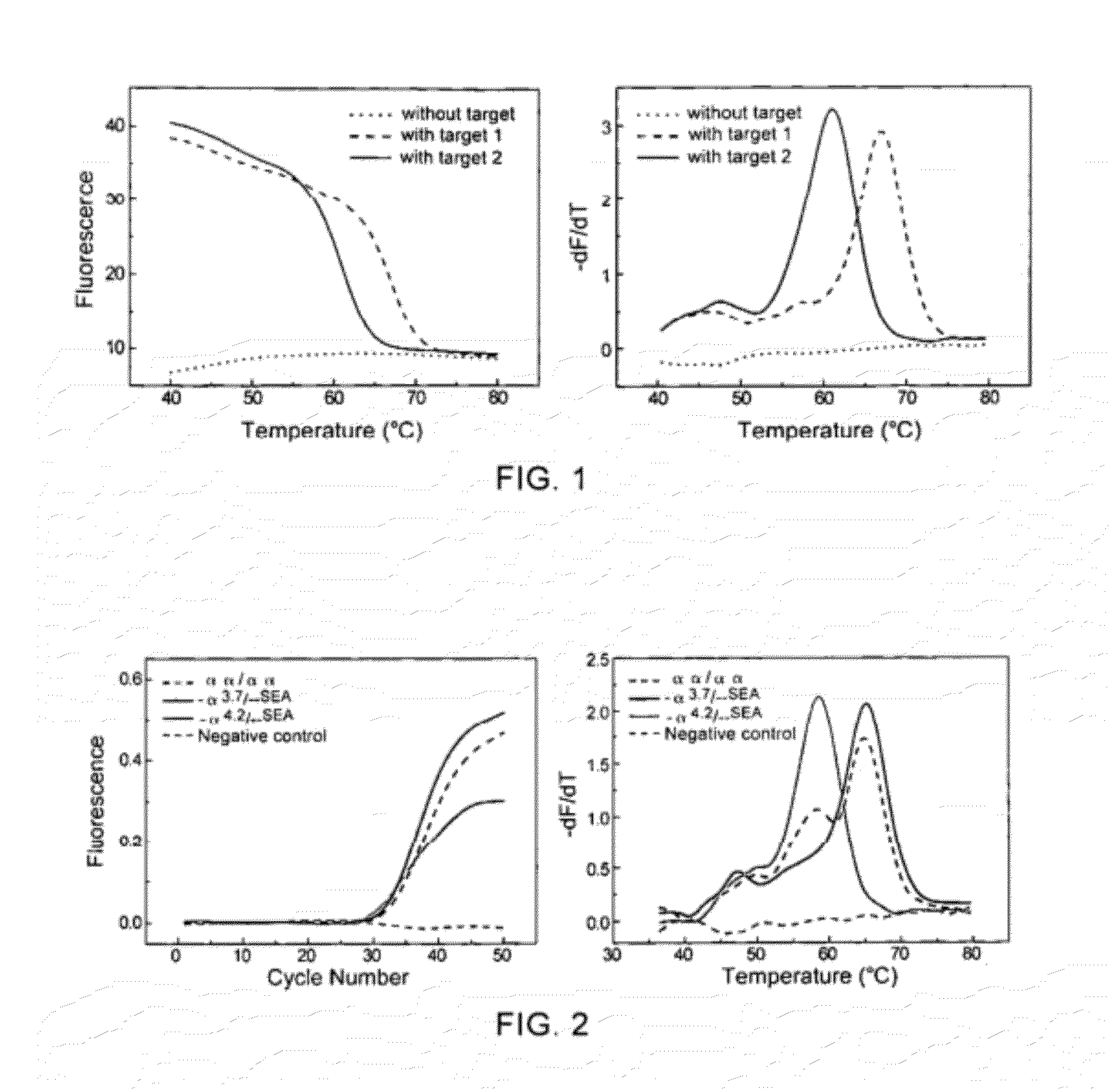
[0079]Artificially designing different complementary target nucleic acid sequences to examine the ability of using the method of linear self-quenched probe melting curve to detect nucleic acid sequence variations.

[0080]In this example, a self-quenched probe directed to the 5′ untranslated region of the α-globin gene was designed. By artificially synthesizing target nucleic acid sequences completely complementary to the probe or with a point mutation, the ability of the method of self-quenched probe melting curve in distinguishing different target nucleic acid sequences was examined. The self-quenched probe and the target nucleic acid sequences used are:

(SEQ ID NO: 1)Probe 1: 5′-FAM-CCTGGTGTTTGTTCCTTCCC-BHQ-3′,the linkage between the first and the secondbases in the 5′ end is a phosphorothioatedlinkage.(SEQ ID NO: 2)(SEQ ID NO: 3)

wherein, the underlined part of the target nucleic acid sequence is complementary to the probe, the boxed part of Target 2 indicates the mutated base, the t...

example 2

[0083]Detecting specimens of different genotypes using the method of linear self-quenched probe PCR-melting curve analysis.

[0084]In this example, a self-quenched probe directed to the 5′ untranslated region of the α-globin gene was designed (probe 1, see example 1). The α1-globin gene was distinguished from the α2-globin gene based on the differences of probe melting temperatures; human genomic DNA was used as the template, after real-time PCR amplification, melting curve analysis was performed for Probe 1, to illustrate that the method of self-quenched probe melting curve may be used for genotyping. The primers used were:

P1:(SEQ ID No: 4)5′-GCAAGCCCTCACGTAGCGAAGTAGAGGAGTCTGAATCTGGA-3′andP2:(SEQ ID No: 5)5′-GCAAGCCCTCACGTAGCGAATCCCTCTGGCGATAGTCA-3′.

[0085]The PCR reaction system was: in 25 μL reaction solution, there were 2.5 μL 10× PCR buffer (without Mg2+), 4.0 mM MgCl2, 5 pmol probe 1, 0.2 mM dNTP, 1 U Hotstart Taq DNA polymerase, 0.1 μM upstream primer P1, 1 μM downstream primer ...

example 3

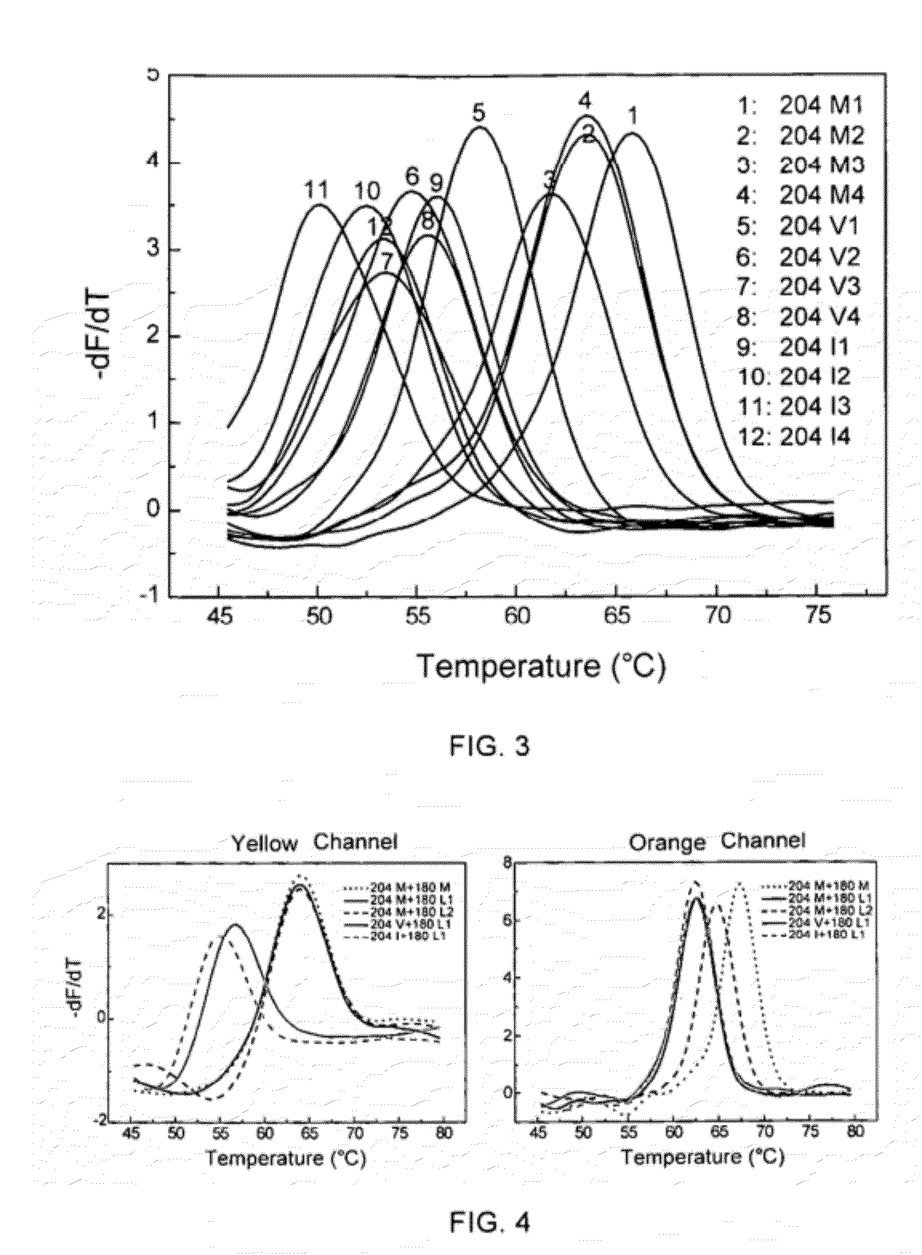
[0087]The ability of the method of self-quenched probe melting curve with different artificially synthesized complementary sequences to detect nucleic acid sequence variations,

[0088]The codon at position 204 in the C region of the coding region of the DNA polymerase from hepatitis B virus was mutated from methionine (M) into valine (V) (ATG→ATT) or isoleucine (I) (ATG→GTG) will result in resistance to the first-line drug lamivudine, and may be accompanied by a mutation of the codon at position 180 from leucine (L) into methionine (M) (CTG / TTG→ATG).

[0089]In this example, a self-quenched probe covering the C region of the coding region of the DNA polymerase from hepatitis B virus was designed, and artificially synthesized target nucleic acid sequences were used to examine the ability of the method using LNA modified self-quenched probe melting curve to detect nucleic acid sequence variations. The self-quenched probes and the target nucleic acid sequences used were:

(SEQ ID No. 6)(SEQ I...


PUM

PropertyMeasurementUnit
melting temperatureaaaaaaaaaa
temperatureaaaaaaaaaa

Abstract

The present invention relates to a method and a kit for detecting nucleic acid sequence variation using melting curve analysis, especially relates to a method and a kit for detecting nucleic acid sequence variation by melting curve analysis using self-quenched probe. Said method provides the characteristics of the self-quenched probe employed, as well as the corresponding nucleic acid amplification conditions, so that the probe can bind to the amplified target sequence, and variations of the target sequence can be detected by melting curve analysis. The present invention also encompasses a kit assembled according to the method described.

Description

[0001]The present invention claims priority of the Chinese patent applications No. 200910143480.6 filed on May 26, 2009 in the name of Xiamen University, this priority application is incorporated herein in its entirety by reference, as if the entire contents thereof are explicitly described in the present application.FIELD OF THE INVENTION[0002]The present invention relates to a method for detecting nucleic acid sequence variation, as well as to probes and kits for said method. Particularly, it relates to a method for detecting nucleic acid sequence variation by self-quenched probe-based melting curve analysis, and to self-quenched probes and kits.TECHNICAL BACKGROUND[0003]Melting curve analysis for detecting nucleic acid sequence variation is to include an additional temperature increasing step (sometimes may also be a temperature decreasing step) in the real-time PCR program, then, information about amplification products or sequence variations can be detected by recording the cha...

Claims


Application Information

Patent Timeline
Patent Type & Authority Applications(United States)
IPC IPC(8): C12Q1/68C07H21/04
CPCC12Q1/6827C12Q1/6858C12Q2527/107C12Q2531/107C12Q2535/131
Inventor LI, QINGGEHUANG, QIUYING
Owner XIAMEN UNIV
Learn More