
Vesicular system and uses thereof

a technology of vesicle and membrane, applied in the field of vesicle system, can solve the problems of difficult to maintain the robustness of a conventional membrane layer on the surface, the approach does not allow the incorporation of complex membrane proteins, and the task is extremely difficul

Active Publication Date: 2012-05-24
AGENCY FOR SCI TECH & RES
View PDF1 Cites 21 Cited by
  • Summary
  • Abstract
  • Description
  • Claims
  • Application Information

AI Technical Summary

Benefits of technology

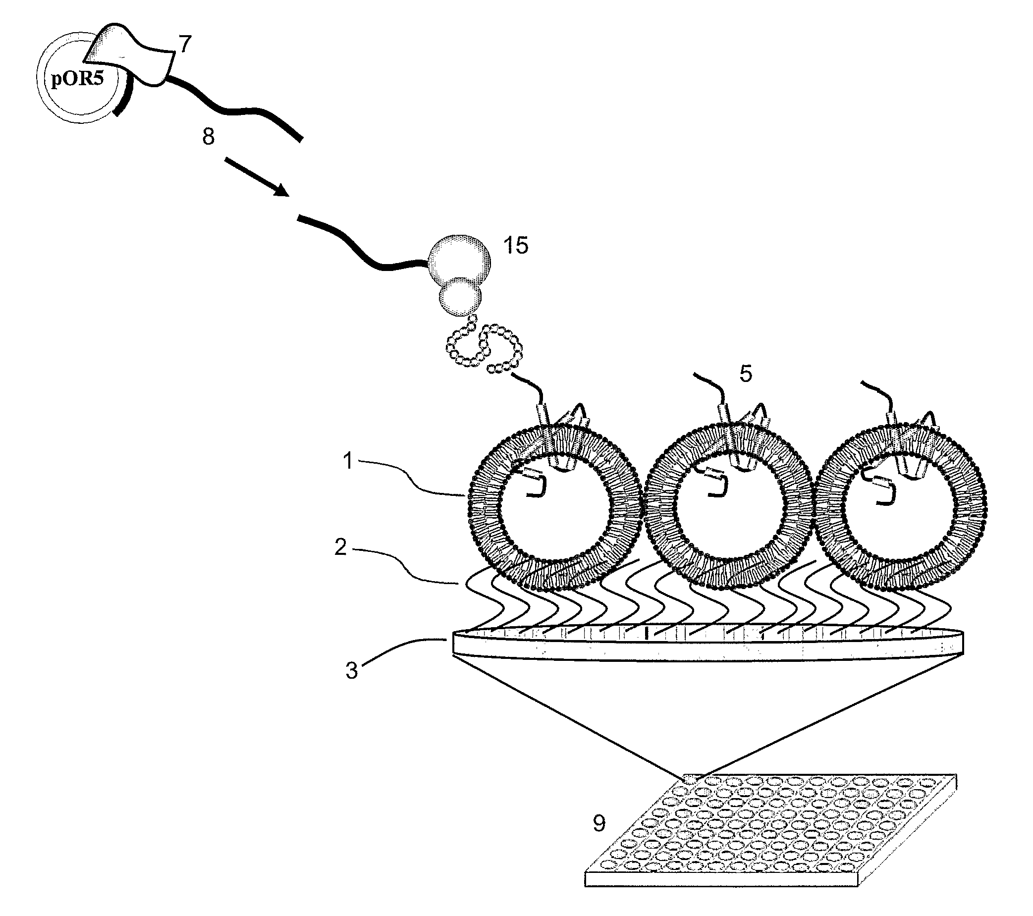
[0015]According to a fifth aspect, the invention provides an in-vitro method of identifying a compound that is capable of modulating the function of an ion channel protein. The ion channel protein is capable of allowing a known ion to pass. The method includes providing a vesicular system according to the first aspect. The immobilized vesicle of the vesicular system includes a membrane protein. This membrane protein is associated to the circumferential membrane of the amphiphilic polymer of the immobilized vesicle. The membrane protein is an ion channel protein, which is the ion channel protein that is capable of allowing a known ion to pass. The method includes contacting the immobilized vesicle with a candidate compound. The candidate compound is suspected to modulate the function of the ion channel protein. The method also includes detecting the passage of ions into or out of the vesicle. The ion channel protein may be an ion channel protein of a cell membrane, i.e. a cell surface ion channel, or of an organelle membrane.

Problems solved by technology

It is an extremely challenging task to provide a biomimetic structure that allows preserving the functional structure of a membrane protein, which has a portion that is included in a cellular membrane in vivo.
The conventional approach does not allow a complex membrane protein to be incorporated in a form which maintains the availability and functional presentation of the protein.
Furthermore, it is difficult to maintain the robustness of a conventional membrane layer on a surface when it is formed from traditional phospholipid materials.
Despite their importance, detailed functional studies of membrane proteins are still scarce because of their sensitivity and notorious difficult procedures of isolation and functional re-constitution.
While a variety of assay techniques is meanwhile available for screening of membrane receptors (for an overview see e.g. Cooper, M A, J. Mol. Recognit. (2004) 17, 286-315), a reconstitution system for easily handling the same is still unavailable.
However, mechanical strength is compromised as a result of the fragile nature of the membrane formed by phospholipids.
In addition, it is difficult to incorporate this technology into a multi-well format.

Method used


Image

  • Vesicular system and uses thereof
  • Vesicular system and uses thereof
  • Vesicular system and uses thereof

Examples


example 1

Preparation of Polymer Vesicles

[0083]Polybutadiene-Polyethyleneoxide (PB21-PEO12) diblock copolymer was kindly provided by Prof. Jan van Hest, Radboud University Nijmegen, Netherlands.

[0084]10 mg of PB21-PEO12 di-block copolymer is dissolved in 300 μl of THF and added into 700 μl of MilliQ H2O. The solution is dialyzed using 50,000 MW ready to use membrane (Spectrum Labs) against MilliQ H2O for 24 hours.

example 2

Preparation of Anchored Polymer Vesicles on a Multiwell Plate

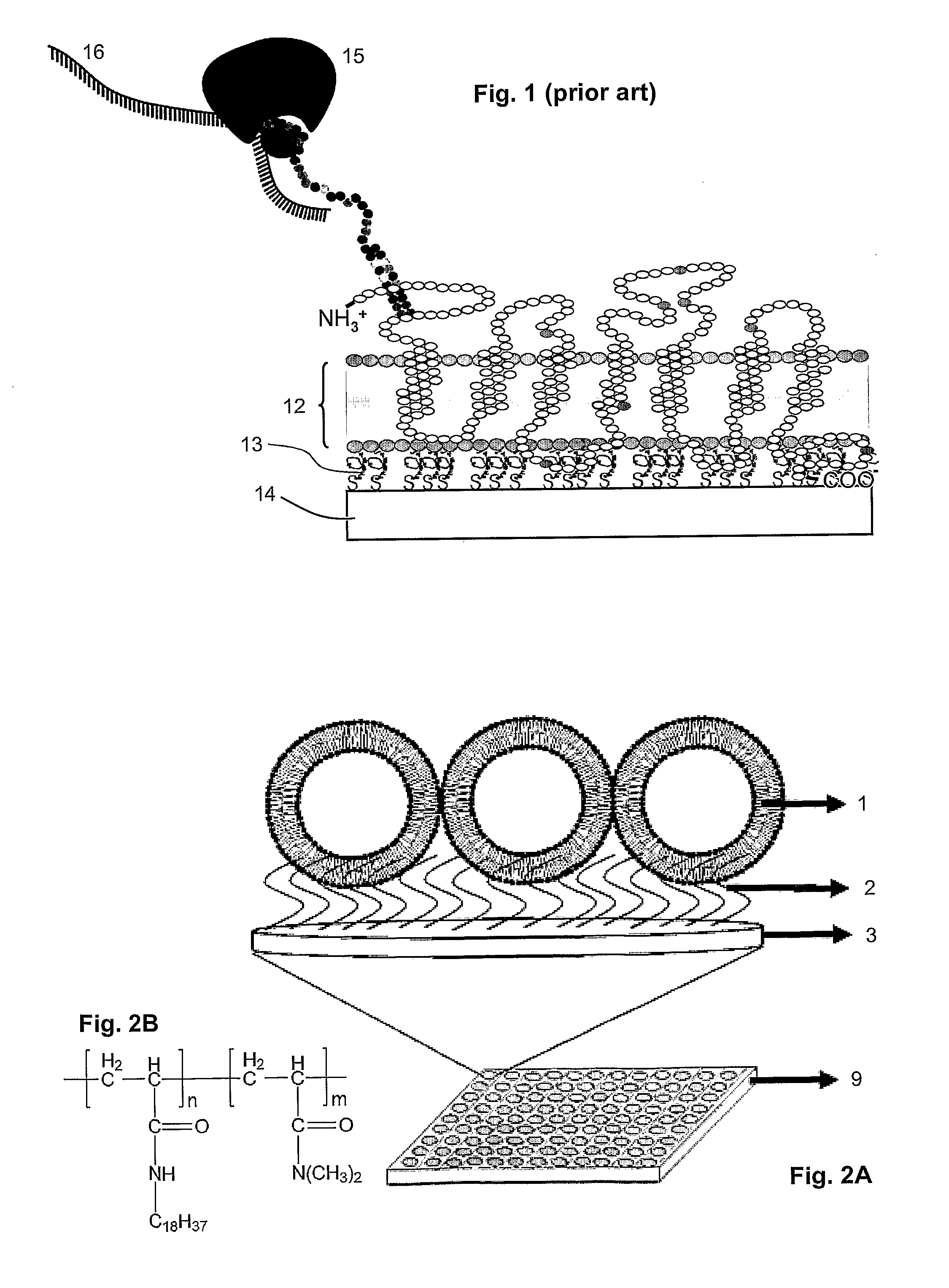
[0085]Octadecylacrylamide and N,N-dimethyl acrylamide with a molar ratio of 99:1 (polymer A) were synthesized according to the procedures of Hara et al. (Supramolecular Science (1998) 5, 777-781). A Microtiter® plate from BD Biosciences was used.

[0086]Acrylamide solution (3.5 ml of 50 mM Tri-Cl (pH 7.2), 0.5 ml of 40% acrylamide, 5 μl of N,N,N′,N′Tetramethylethylenediamine (TEMED), 35 μl of ammonium persulphate (APS) with and without polymer A is prepared. 100 μl of above solution is added to each well of the Microtiter® plate. 75 μl of polymer vesicles prepared in Example 1 are added into these wells before the acrylamide polymerizes. The Microtiter® plate is shaken gently at room temperature for two hours for even spreading of polymer vesicles on to the Polyacrylamide gel. Microtiter® plate wells were rinsed with MilliQ several times to remove the non-anchored polymer vesicles.

example 3

Calcein Leakage Assay

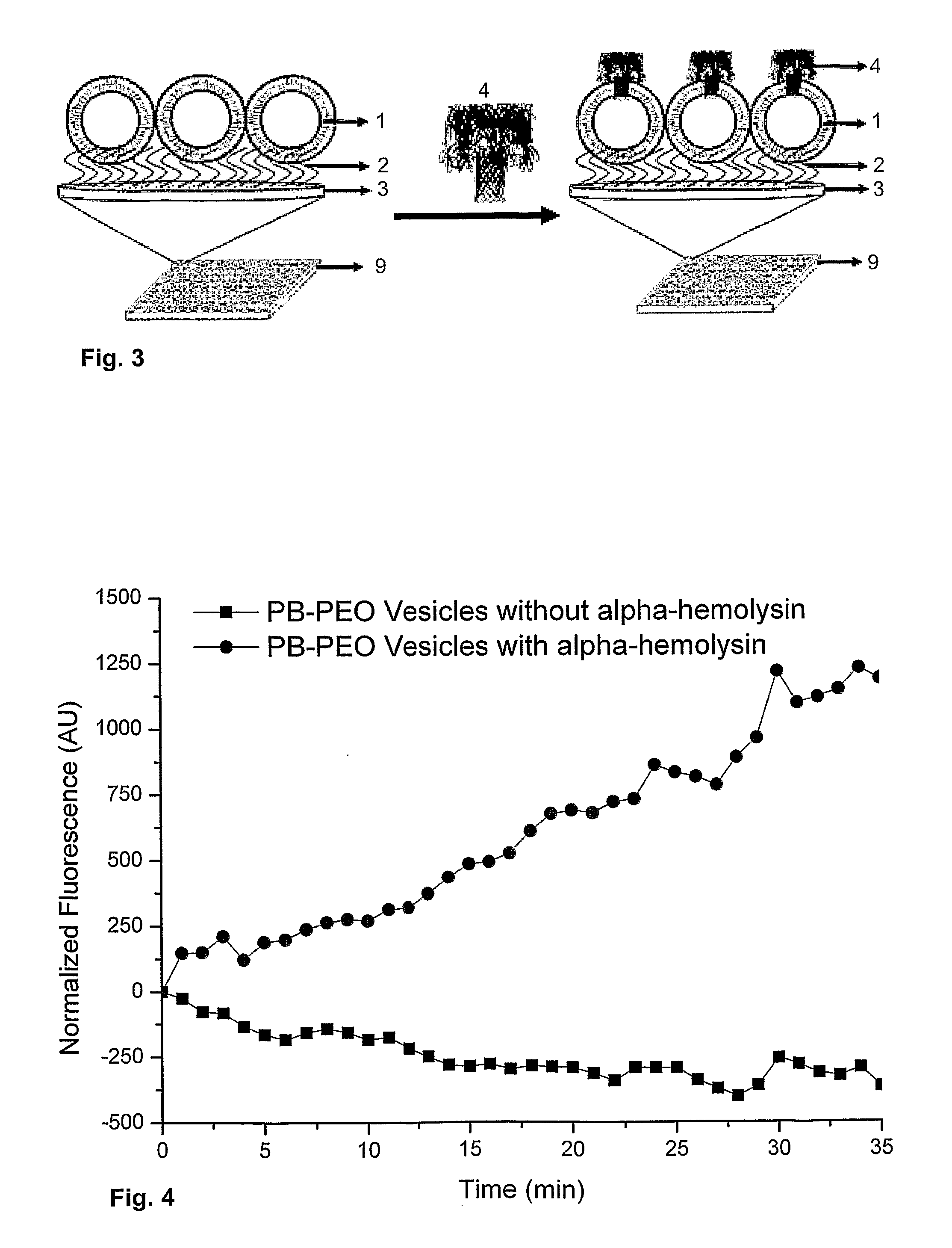
[0087]In order to investigate the insertion of membrane proteins into the polymer vesicles embedded on the Microtiter® plate wells, calcein was encapsulated inside the polymer vesicles at self-quenching concentrations. The schematic of this protocol is illustrated in FIG. 3. This calcein based assay provides a quick assay to test the integrity of the spherical structures. A standard calcein based assay can be used to test the integrity of the polymersomes.

[0088]30 mM of calcein was encapsulated inside the polymersomes. The solution is dialyzed using 50,000 MW ready to use membrane (Spectrum Labs) against MilliQ H2O for 24 h to remove the non-encapsulated calcein and to attach the polymersomes to the poly-acrylamide matrix. These polymer vesicles encapsulated with calcein were added to the microtiter plate and shaken gently at room temperature to anchor to the polyacrylamide matrix. After incubation, the microtiter plate was rinsed with MilliQ to remove the non a...


PUM

PropertyMeasurementUnit
Temperatureaaaaaaaaaa
Amphiphilicaaaaaaaaaa
Glass transition temperatureaaaaaaaaaa

Abstract

Disclosed is a vesicular system comprising a surface with a vesicle immobilized thereon. The immobilized vesicle has a circumferential membrane of an amphiphilic polymer. The vesicle is coupled to a surface by means of a molecule with a non-polar moiety. The non-polar moiety comprises a main chain of 3 to about 30 carbon atoms and 0 to about 12 heteroatoms selected from Si, O, S, and Se. The molecule with the non-polar moiety is coupled to the surface via a covalent or non-covalent bond. A portion of the non-polar moiety is integrated in the circumferential membrane.

Description

CROSS REFERENCE TO RELATED APPLICATIONS[0001]This application makes reference to and claims the benefit of priority of a provisional application for a “Vesicular Platform For High Throughput Membrane Protein Expression” filed on Apr. 20, 2009 with the United States Patent and Trademark Office, and there duly assigned Ser. No. 61 / 202,920. The content of said application filed on Apr. 20, 2009 is incorporated herein by reference for all purposes, including an incorporation of any element or part of the description, claims or drawings not contained herein and referred to in Rule 20.5(a) of the PCT, pursuant to Rule 4.18 of the PCT.FIELD OF THE INVENTION[0002]The present invention relates to a vesicular system and uses thereof. The system is defined by a surface with a vesicle immobilized thereon. Disclosed is also a method of associating a membrane protein with the membrane of the vesicle, including integrating the membrane protein into the same. In some embodiments the system is defin...

Claims


Application Information

Patent Timeline
IPC IPC(8): G01N33/566C08G81/02
CPCC08G65/329C08L2203/02C08L71/02C08L15/00C08L9/00C08F220/52
Inventor NALLANI, MADHAVANSINNERKUNNAVAKKAM, MADANAGOPAL
Owner AGENCY FOR SCI TECH & RES
Learn More