Surface Passivation Methods for Single Molecule Imaging of Biochemical Reactions

a biochemical reaction and surface passivation technology, applied in the field of surface passivation methods for biochemical reactions, can solve the problems of reducing the availability and activity of fluorescent molecules on the surface, increasing background noise, and interference with the signal from the specific field

Inactive Publication Date: 2012-09-13
HOWARD HUGHES MEDICAL INST
View PDF4 Cites 13 Cited by
  • Summary
  • Abstract
  • Description
  • Claims
  • Application Information

AI Technical Summary

Benefits of technology

[0009]In one aspect, a method for passivating a surface for single molecule imaging is described. Such a method typically includes contacting the surface with an effective amount of a hydrophobic organosilane / siloxane for a time sufficient to passivate the surface for single molecule imaging. Representative surfaces include a slide, coverslip, plate, chip, sphere, microparticle, bead, microwell, and microfluidic device.
[0010]Representative hydrophobic organosilane / siloxane include, without limitation, 1,7-dichlorooctamethyltetrasiloxane, tert-butyltrichlorosilane, 1,3-dichlorotetramethyldisiloxane, butyltrichlorosilane, dichlorodimethylsilane, trimethylchlorosilane, butyltrichlorosilane, tert-butyltrichlorosilane, octadecyltrichlosilane, dichlorodimethylsilane, and 1,5-dichlorohexamethyltrisiloxane. In some embodiments, the hydrophobic organosilane / siloxane is 1,7-dichlorooctamethyltetrasiloxane. In some embodiments, the effective amount of the hydrophobic organosilane / siloxane is about 1% to about 50% in solution. In some embodiments, the time sufficient to passivate the surface for single molecule imaging is between 6 hours and 24 hours.

Problems solved by technology

Although convenient for imaging, glass surfaces are well known for their ability to absorb organic molecules and bio-molecules non-specifically, thus reducing the availability and activity of these molecules.
Thus, non-specific adsorption of fluorescent molecules to the surface increases the background noise and interferes with the signal from the specifically immobilized molecules (see, for example, FIG. 1).
Furthermore, performing single-molecule reactions with surface-immobilized components (e.g., monitoring interactions of a DNA-binding protein with a surface-immobilized DNA through fluorescence detection or optical / magnetic tweezers) dramatically increases the probability of inactivation of the reagents by the surface due to repetitive interactions between the tethered molecule and the surface.
Finally, with the expansion of the single-molecule methods into complex, poorly controlled biological systems (for example, detection of antigens in cell extracts or blood samples), the robustness of existing surface passivation methods, optimized for simple systems involving only one or a few components, is likely to be challenged.
Current imaging surface passivation methods (e.g., polyethylene glycol (PEG)-based protocols) are not suitable for complex biological systems due, at least in part, to surface fouling and the resulting effects.

Method used


Image

  • Surface Passivation Methods for Single Molecule Imaging of Biochemical Reactions
  • Surface Passivation Methods for Single Molecule Imaging of Biochemical Reactions
  • Surface Passivation Methods for Single Molecule Imaging of Biochemical Reactions

Examples


example 1

Passivation Methods

Passivation Procedure

[0056]Borosilicate glass cover slips (VWR 48393 230) were placed in ceramic racks and treated with Piranha solution (3:1 H2SO4:H2O2) twice for 30 min, and rinsed with deionized water until pH had stabilized (according to an indicator paper test). H2SO4 was concentrated, and H2O2 was a 30% solution. The slides were then treated with 0.5 M potassium hydroxide for 1 hour in a ultrasound water bath and rinsed with deionized water until pH had stabilized (according to an indicator paper test).

[0057]The cleaned substrates were briefly rinsed with acetone and soaked in a 3% solution of aminopropyltriethoxysilane (APTES, CAS #919-30-2) in acetone for 45 min with shaking The substrates were then rinsed briefly with acetone, and rinsed copiously with deionized water. The substrates were then immediately blown dry with clean nitrogen. Functionalization of the APTES-modified slides was achieved by incubation of coverglasses with freshly prepared 10% solut...

example 2

Characterization of Surface Passivation Using a “Stickiness Assay”

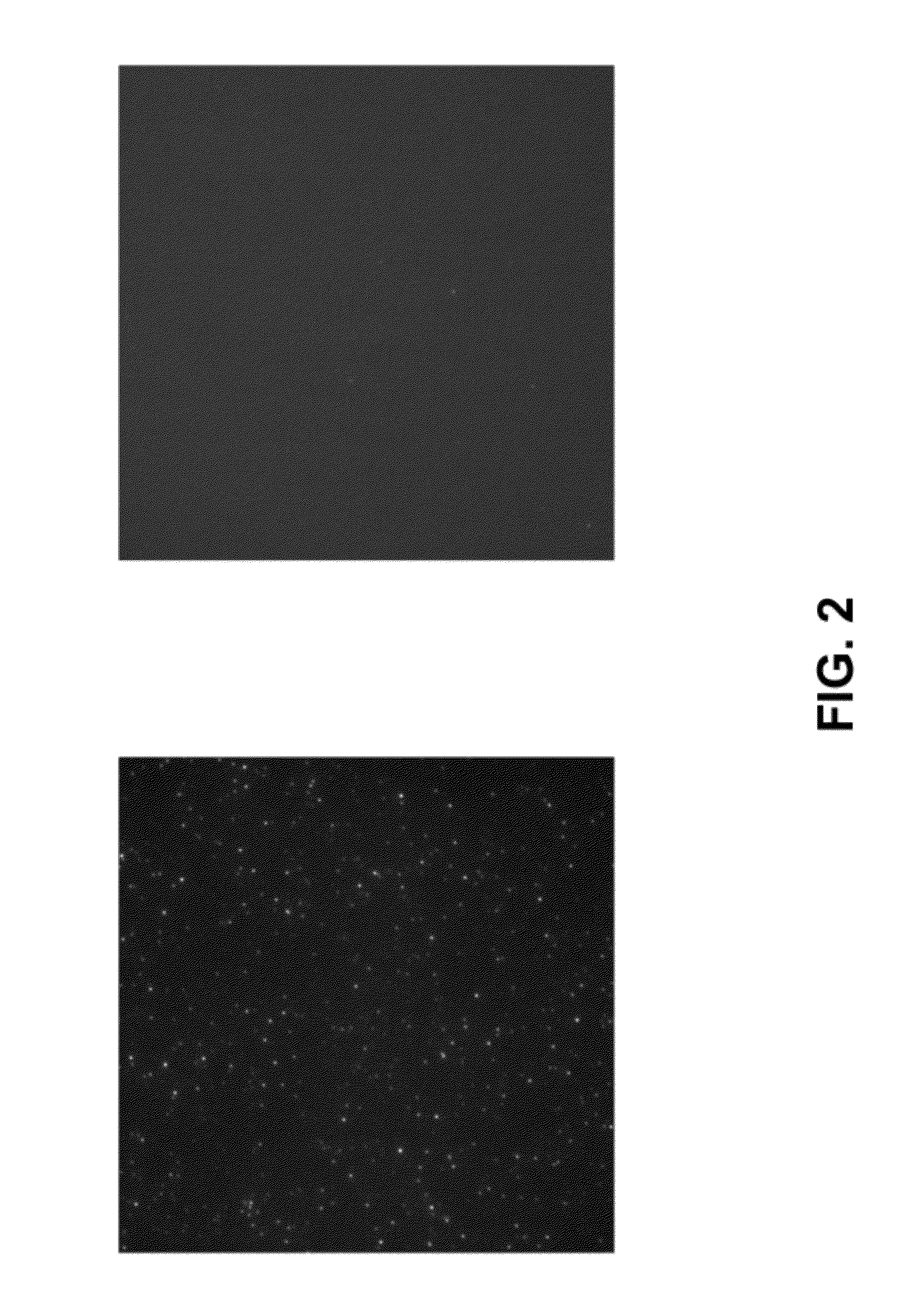
[0061]Glass coverslips (either modified with PEG alone, or with PEG followed by passivation with the hydrophobic siloxane) were prepared as described herein. An imaging “sandwich” flow cell was assembled using two coverslips and double-sided tape. Several “channels” were created with the double-sided tape to allow imaging of different test proteins on the same surface. Phosphate-buffered saline (pH 7.5) containing 0.1% Tween 20 (“PBST”) was injected into the channel and incubated for at least 5 min. As a first test protein, a 10 nM solution in PBST of E coli RNA polymerase holoenzyme labeled with Alexa555 at the sigma subunit was injected into the “channel”. As another test protein, a 10 nM solution in PBST of the highly basic U1A RNA-binding protein was injected into another channel. Interactions of the labeled protein with the surface were detected with real-time single-molecule TIRF microscopy (objective type). Bri...

example 3

Characterization of Surface Passivation Using an Activity Decay Assay

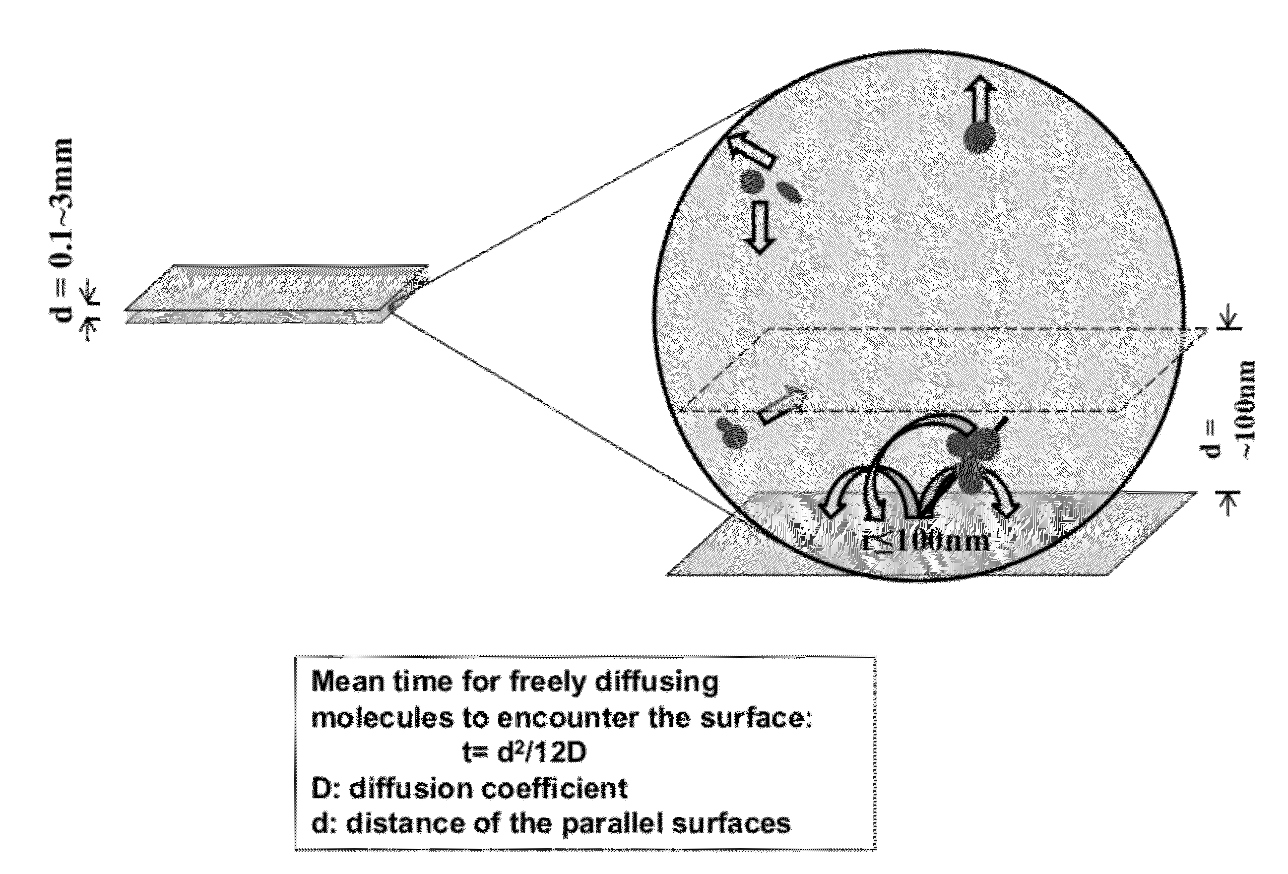
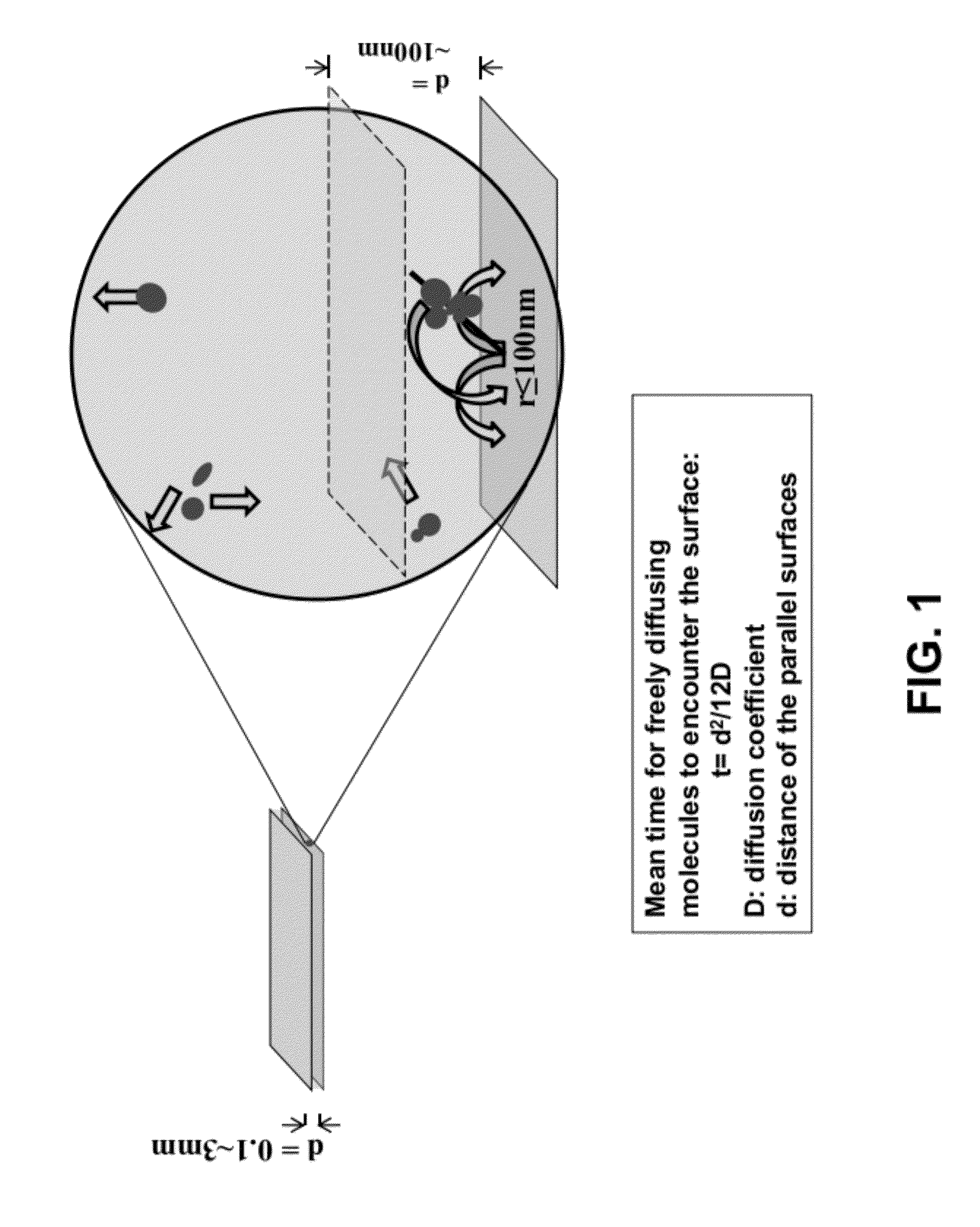
[0064]The following “transcription activity decay” experiment was used to monitor the loss of activity of factors that make up the RNA polymerase II transcription system. Due to the high complexity of the Pol II system, this assay is very sensitive to the quality of surface passivation and can be similarly applied to other complex systems.

[0065]A “surface decay machine” shown in FIG. 4A was set up to measure transcription activity decay between two modified glass cover slips as follows: (1) the bottom glass coverslip was glued to a stationary base with an adhesive, with the passivated side facing up; (2) the top glass coverslip, with the passivated side facing down, was placed above the bottom coverslip, and glued to a support that can be moved up and down with a micrometer. The resulting even narrow space between the two coverslips holds the “transcription reaction mixture” for the decay assay (see below for the d...


PUM

PropertyMeasurementUnit
timeaaaaaaaaaa
timeaaaaaaaaaa
diameteraaaaaaaaaa

Abstract

The present disclosure provides methods for treating a surface for single-molecule imaging.

Description

CROSS-REFERENCE TO RELATED APPLICATIONS[0001]This application claims benefit under 35 U.S.C. §119(e) to U.S. Application No. 61 / 443,016 filed Feb. 15, 2011. The prior application is incorporated by reference herein in its entirety.TECHNICAL FIELD[0002]The present disclosure relates to the use of reagents for the treatment of surfaces for single molecule imaging.BACKGROUND[0003]Imaging biochemical reactions at the single molecule resolution has a huge impact on basic research and has great potential in new diagnostic approaches. Single-molecule reactions are usually carried out in optically transparent chambers, frequently made of glass. Although convenient for imaging, glass surfaces are well known for their ability to absorb organic molecules and bio-molecules non-specifically, thus reducing the availability and activity of these molecules.[0004]In addition, single-molecule imaging frequently involves immobilization of biological molecules on a surface (to restrict their Brownian m...

Claims


Application Information

Patent Timeline
Patent Type & Authority Applications(United States)
IPC IPC(8): B05D1/36C07F7/12B05C11/00G01N21/64
CPCG01N21/6428C07F7/188G01N21/6458
Inventor ZHANG, ZHENGJIANREVYAKIN, ANDREYCHU, STEVETIJAN, ROBERT
Owner HOWARD HUGHES MEDICAL INST
Why Eureka
Try Eureka