
Targeted nanoclusters and methods of their use

a nanocluster and nanotechnology, applied in the field of compositions and methods can solve the problems of reducing the sensitivity of detection, and reducing so as to improve the efficiency of imaging and detection, improve the sensitivity of detection, and improve the effect of sensitivity

Inactive Publication Date: 2012-10-25
RGT UNIV OF CALIFORNIA
View PDF7 Cites 69 Cited by
  • Summary
  • Abstract
  • Description
  • Claims
  • Application Information

AI Technical Summary

Benefits of technology

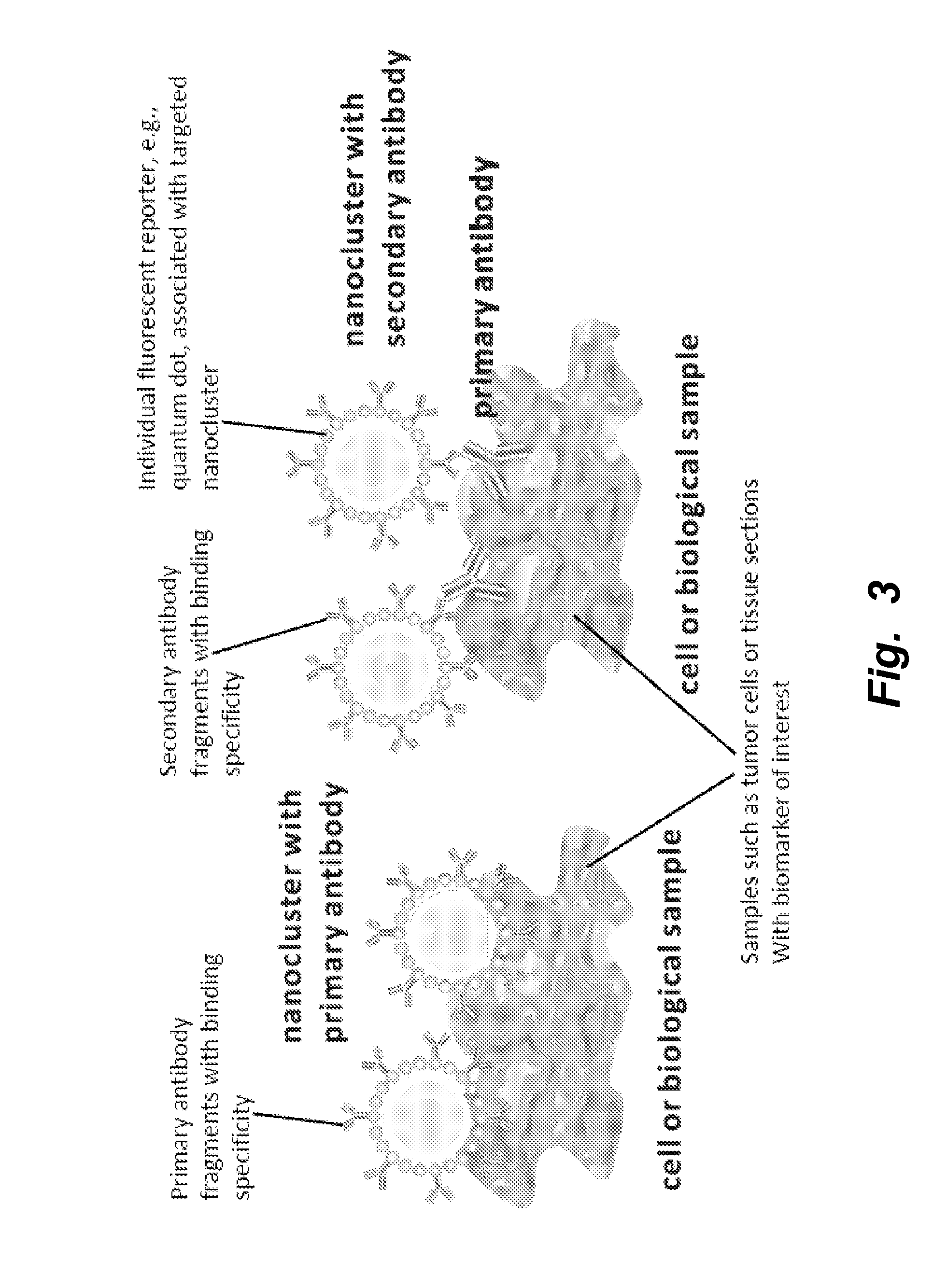
[0008]The present invention provides a targeted nano-molecular complex, i.e., a nanocluster, comprised of stably associating a multiplicity of targeting moieties (e.g., antibodies and fragments thereof) and a multiplicity of detectable labels in an aggregation of a plurality of crosslinked nanoscaffold core structures. The targeted nanoclusters or nanoaggregates improve and simplify known methods for imaging and detection. The targeted nanoclusters or nanoaggregates described herein provide a higher sensitivity for detection due to an enhanced avidity effect by multiple anchoring points to a target and due to the amplification of detectable signal by multiple attachment to a plurality of detectable labels, within a single nanoparticle and multiplied by the aggregation of a plurality of crosslinked nanoparticle core units. Accordingly, enhanced signal amplification due to multiple reporting agents, e.g., for use in flow cytometry, immunocytochemistry / immunohistochemistry and in vivo imaging methods, are provided.

Problems solved by technology

A conventional approach is to attach probes with strong signals to antibody without affecting the affinity and specificity of the antibody, which is technically challenging due to the linking chemistry and intrinsic steric limitation of the molecules.
While antibody-antigen coupling occurs in the first binding reaction, it involves multiple time-consuming steps and each additional step embeds the risk of deviating from accuracy and consistency.
More importantly, due to the biophysical properties of the reagents used, such as the most commonly adopted diaminobenzidine (DAB), quantitation and standardization of the assays often present significant challenges.1 Assessments of various antigens and biomarkers in tumor tissues are important in determining tumor subtypes and response to therapy.
As the body of knowledge in molecular signatures associated with tumor subtypes expanded vastly in recent years,2-4 the intrinsic disadvantages with the current method become serious obstacles for more efficient, quantitative, and consistent biomarker profiling.
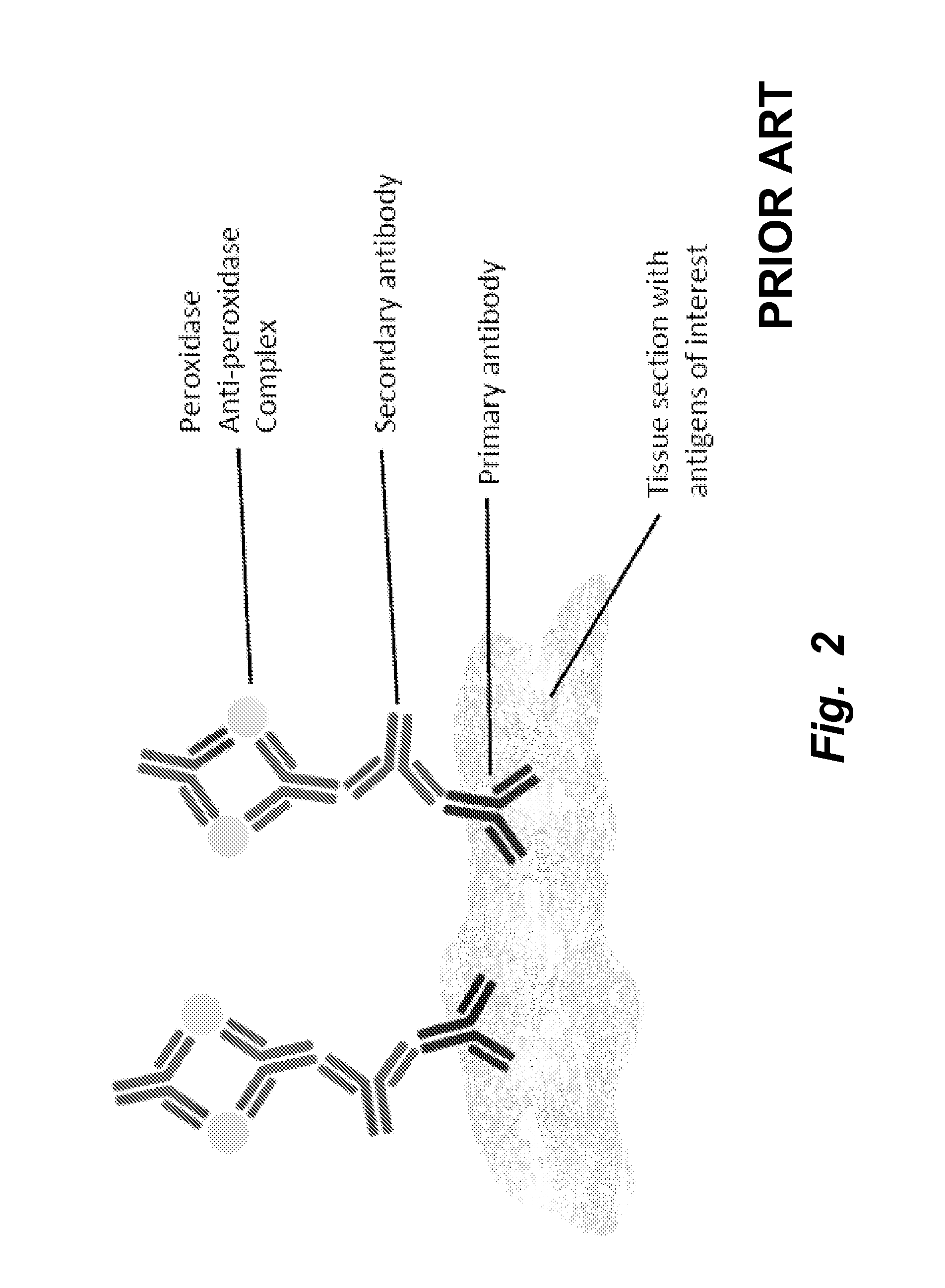
Several detection systems such as avidin-biotin complex (ABC), peroxidase anti-peroxidase (PAP), or polymer-based reagents have been used in traditional chromogenic techniques.5 They provide enhanced sensitivity through amplification; however, these systems also involve three or more steps, are not easy to quantify, and lack dynamic range.
Fluorescence-based immunodetection could potentially overcome the limitations and simplify the multi-step chromogenic methods by labeled primary or secondary antibodies; however, the trade-offs include need for optimizing conjugation for each primary antibody and loss of amplification due to non-crosslinked fluorophores on the secondary antibodies.
Furthermore, the unstable and photobleachable nature of conventional fluorophores make them unpractical for long term storage and observation, especially in tissue banking for clinical studies.
In fact, QDs have been demonstrated for use in immunohistochemistry by Nie et al.14 However, conjugation was reported to be inconsistent and required optimization for individual antibodies.

Method used


Image

  • Targeted nanoclusters and methods of their use
  • Targeted nanoclusters and methods of their use
  • Targeted nanoclusters and methods of their use

Examples


example 1

Sample Prior Art or Conventional Immunostaining

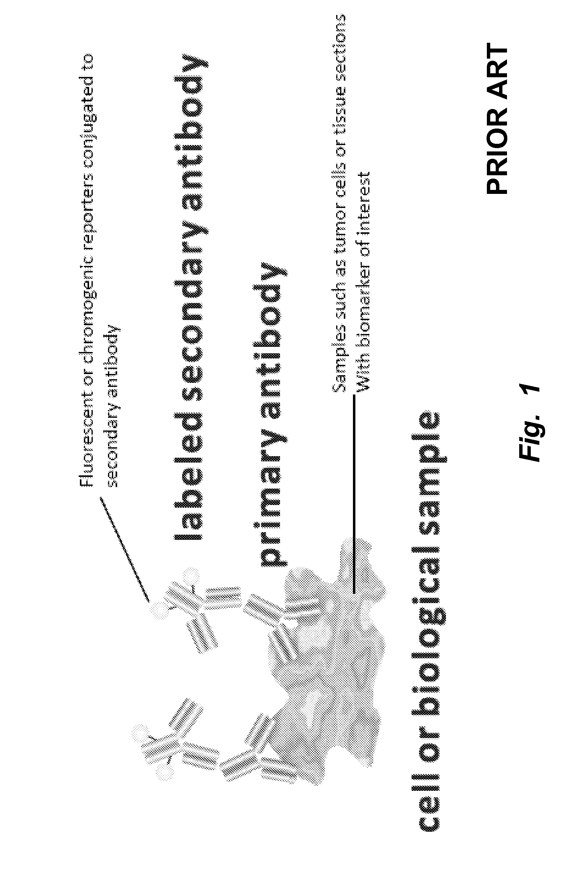
[0263]Conventional methods of immunostaining are illustrated in FIGS. 1 and 2. In the method illustrated, tissues, cells, or cell material including an antigen of interest are immobilized on a solid support, such as a glass slide, well of a multi-well plate, or the like and incubated in the presence of a primary antibody specific for the antigen of interest. After removing unbound primary antibody by washing, secondary antibody is allowed to bind to the primary antibody, followed by addition of antibody-color development reagent complexes capable of acting on a substrate to produce a detectable signal that corresponds, indirectly, to the present of the antigen of interest.

[0264]The conventional method is time-consuming, with typical processing times being about two days. Several discrete binding steps are required, e.g., for binding of primary antibody, secondary antibody, colorimetric development agents, and counterstaining. Moreover, ...

example 2

Flow Cytometry of Live Breast Tumor Cells Using Commercially Available Qdot IgG Conjugate and Targeted Nanoclusters or Nanoaggregates

[0265]Synthesis of targeted nanoclusters or nanoaggregates was achieved by incorporating reduced antibody binding fragments and luminescent quantum dots into a surface-bifunctionalized and cross-linked lipid vesicle scaffold. An illustrative protocol using the present composition and method for labeling live breast tumor cells is described.[0266]1. Take ˜70% confluent flasks of cancer cells.[0267]2. Aspirate the old medium from the flask, wash with PBS and trypsinize the cells with 0.25% trypsin-EDTA. Finally, neutralize trypsin by adding back media.[0268]3. Collect the cell suspension in a 50 ml centrifuge tube. Count the cells with hemocytometer.[0269]4. Aliquote appropriate volume of cell suspension into labeled Eppendorf tubes, typically 150,000 cells / tube.[0270]5. Spin down the cells at 400×g for 5 min, remove the supernatant and add 100 μl of 1% ...

example 3

Method and Protocol for Immunostaining FFPE Sections Using Targeted Nanoclusters or Nanoaggregates

[0281]An exemplary protocol using the present composition and method for labeling cells in formalin-fixed, paraffin-embedded sections is described. FFPE slides coated with HER2 human breast carcinoma cells were stained to visualize the erbB2 receptor as follows:

Part 1.

[0282]1. Bake slides in oven at 60° C. for 30 minutes prior to staining[0283]2. Deparaffinize and Rehydrate the tissues on slides[0284]3. Incubate with Ficin for 10 minutes at 37° C.[0285]4. Wash in PBS for 3.5 minutes three times[0286]5. Block in 3% H2O2 for 15 minutes[0287]6. Wash in PBS for 3.5 minutes three times[0288]7. Incubate with normal goat serum for 30 minutes at room temperature[0289]8. Incubate with erbB2 antibody (primary antibody)

Part 2.

[0290]9. Wash off coverslips with PBS for 8 minutes[0291]10. Wash in PBS for 3.5 minutes twice[0292]11. Incubate with goat anti-mouse targeted nanoclusters or nanoaggregates ...


PUM

PropertyMeasurementUnit
Lengthaaaaaaaaaa
Lengthaaaaaaaaaa
Lengthaaaaaaaaaa

Abstract

This invention provides targeted nanoclusters comprising multiple polyvalent nanoparticle core units or nanoscaffolds, each nanoparticle core unit attached to multiple targeting moieties and multiple detectable moieties. The nanoclusters find use in a broad range of analytical assays, diagnostic assays and as targeted therapeutics.

Description

CROSS-REFERENCE TO RELATED APPLICATIONS[0001]This application claims priority to U.S. Provisional Patent Application No. 61 / 250,793, filed on Oct. 12, 2009, hereby incorporated by reference in its entirety.STATEMENT OF GOVERNMENTAL SUPPORT[0002]This invention was made with government support under Contract No. DE-AC02-05CH11231 awarded by the Department of Energy and LG013 Agilent Foundation Gift. The government has certain rights to this invention.FIELD OF THE INVENTION[0003]The present invention relates to the field of compositions and methods of imaging and detection, using targeted nanoclusters or nanoaggregates comprising a plurality of nanoparticles attached to targeting moieties and detectable labels.BACKGROUND OF THE INVENTION[0004]Imaging and detection techniques are widely used in molecular biology and clinical diagnosis. One particularly important application is characterization of cells or microscopic particles by flow cytometry or fluorescence-activated cell sorting (FA...

Claims


Application Information

Patent Timeline
IPC IPC(8): G01N33/574A61K51/00A61K49/00G01N21/64B82Y15/00
CPCA61K47/488A61K49/0002A61K49/0058A61K49/0067G01N33/588G01N33/54346G01N33/574G01N33/57415G01N33/587B82Y15/00A61K47/6907
Inventor WENG, KEVIN C.CHEN, FANQING FRANKGRAY, JOE W.
Owner RGT UNIV OF CALIFORNIA
Learn More