Single-shot full-field reflection phase microscopy

a reflection phase and single-shot technology, applied in the field of quantitative study of mechanical properties of live cells, can solve the problems of limited usefulness, insufficient acquisition rate of cellular dynamics, and limitations of time domain optical coherence tomography (oct) based implementation of reflection phase microscopy, and achieve the effect of improving measurement sensitivity

Active Publication Date: 2012-12-06
HAMAMATSU PHOTONICS KK +1
View PDF7 Cites 35 Cited by
  • Summary
  • Abstract
  • Description
  • Claims
  • Application Information

AI Technical Summary

Benefits of technology

[0005]The present invention relates to full-field reflection-based phase microscopy. Preferred embodiments of systems and methods of the present invention involve the measurement of structures having small features, such as the plasma and / or nuclear membrane dynamics, in general cell types. Due to the 3-D cytoskeleton, these cells are much stiffer than red blood cells, for example, indicating that corresponding membrane fluctuations are much smaller than can be detected with transmission phase microscopy. In this respect, reflection-based optical methods can provide a 2n / Δn advantage in measurement sensitivity over the transmission-based optical techniques. Preferred systems and methods utilize a portion of light from a light source to interfere with light that is also used to illuminate the material to be measured. A selected diffraction order of the light from the light source is coupled to a two dimensional detector array along with an image of a selected field of view, or image field, of the material. This provides a full field interferogram of the material.
[0009]A preferred embodiment of the invention provides a quantitative reflection phase microscope based on en-face optical coherence tomography and off-axis digital holography. The system can utilize a diffraction grating in the reference arm to provide the desired angular tilt to the reference beam for off-axis interferometry. The full-field illumination allows single-shot phase measurement of multiple points on the surface of interest and enables the use of a self phase-referencing method to reject common-mode noise occurring in interferometric systems using a separate reference arm. In this full-field reflection phase microscope, the self-phase referencing suppressed phase noise down to as low as 21(pm / √{square root over (HZ)}). With such high phase sensitivity, the system can resolve thermal motion of the cell surface in the field of view, which can be on the order of 100 picometers to 150 nanometers, for example. An application of the full-field reflection phase microscope is to use plasma or membrane fluctuations to estimate the mechanical properties of cell membranes or the bulk visco-elastic properties of the cell cytoskeleton or nucleoskeleton. These variations in cell's mechanical properties can serve as non-invasive biomarker to measure the pathophysiology of general cell types. The system can also provide full-field and multi-cell imaging of cellular electromotility, including cell membrane motion driven by the action potential in single mammalian cells.

Problems solved by technology

In the past, both spectral domain as well as time domain optical coherence tomography (OCT) based implementations of reflection phase microscopy have had limitations that limit their usefulness.
However, the acquisition rate (25 ms integration time per wavelength) was not sufficient to observe cellular dynamics.
Prior attempts using a time-domain reflection phase microscope based on phase shifting interferometry limited time resolution (1.25 sec) due to the need for taking multiple images.
There was an attempt to use off-axis digital holography with a low-coherence source to take a full-field phase image in a single shot, but the tilting of the reference mirror caused uneven interference contrast and thereby impeded full-field imaging.

Method used


Image

  • Single-shot full-field reflection phase microscopy
  • Single-shot full-field reflection phase microscopy
  • Single-shot full-field reflection phase microscopy

Examples


Embodiment Construction

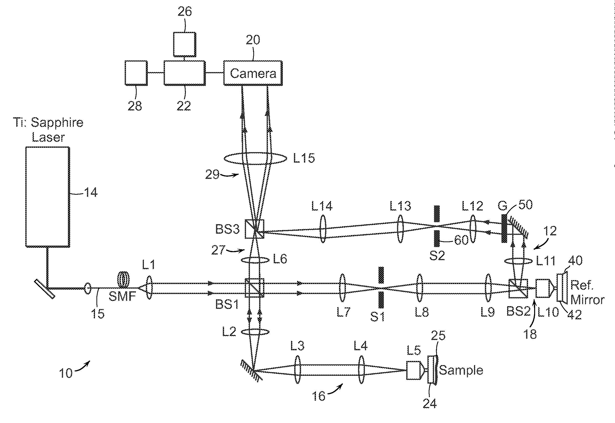
[0026]FIG. 1A shows the schematic of a preferred embodiment of the invention providing single-shot full-field reflection phase microscope (FF-RPM). Light from a mode-locked Ti:Sapphire laser (center wavelength, λc=800 nm) is coupled into a single-mode fiber 15 for delivery as well as for spectrum broadening. The full-width-half-maximum spectral width, AA at the fiber output measures 50 nm, which yields a round trip coherence length of 4 μm in a typical culture medium with refractive index, n, equal to 1.33. The sample beam that travels along the first beam path 16 through lenses L2, L3, L4, and a water immersion 60× objective lens L5 (NA=1.2), reflects off the sample surface 24 and makes an image of the sample on a high-speed complementary metal oxide semiconductor (CMOS) camera via lenses L6 and L15. The camera can be a pixelated imaging detector 20 that is connected to a data processor or computer 22 which can process images, provide the images to a display 26 or to a memory 28 fo...


PUM


Abstract

The present invention relates to a full-field reflection phase microscope. In a preferred embodiment, the invention can combine low-coherence interferometry and off-axis digital holographic microscopy (DHM). The reflection-based DHM provides highly sensitive and a single-shot imaging of cellular dynamics while the use of low coherence source provides a depth-selective measurement. A preferred embodiment of the system uses a diffraction grating in the reference arm to generate an interference image of uniform contrast over the entire field-of-view albeit low-coherence light source. With improved path-length sensitivity, the present invention is suitable for full-field measurement of membrane dynamics in live cells with sub-nanometer-scale sensitivity.

Description

CROSS REFERENCE TO RELATED APPLICATION[0001]This application claims the priority to U.S. Application No. 61 / 436,026, filed Jan. 25, 2011. The entire contents of the above application being incorporated herein by reference.STATEMENT REGARDING FEDERALLY SPONSORED RESEARCH OR DEVELOPMENT[0002]This work was funded by the National Center for Research Resources of the National Institutes of Health (P41-RR02594-18), the National Science Foundation (DBI-0754339).BACKGROUND OF THE INVENTION[0003]Bio-microrheology is the quantitative study of mechanical properties of live cells. Variations in mechanical properties are intrinsic indicators of ongoing cellular processes such as increase in elasticity of certain cancer cells, change of membrane stiffness in malaria-infected red blood cells, and changes in cellular adhesion, for example. The measurement of rheological properties of cell membranes is advantageous since it may also indirectly provide information on the internal structures of cell. ...

Claims


Application Information

Patent Timeline
Patent Type & Authority Applications(United States)
IPC IPC(8): H04N7/18
CPCG01B9/04G01N21/45G01B9/02064G01B9/0207G01B9/02091G02B27/1086G03H1/0443G01B9/02032G01B9/02047G01B9/02084G03H2001/005G03H2001/0456G03H2222/24G02B27/50
Inventor YAQOOB, ZAHIDCHOI, WONSHIKYAMAUCHI, TOYOHIKOFELD, MICHAELFELD, DAVIDHEARN, ALISONFELD, JONATHAN
Owner HAMAMATSU PHOTONICS KK
Why Eureka
Try Eureka