Engineered lumenized vascular networks and support matrix

a technology of lumenized capillaries and support matrix, which is applied in the field of biomedical tissue engineering, can solve the problems of inability to rapidly create lumenized capillary networks with properties, current methods of tissue engineering are limited to relatively thin and/or avascular, and vascularization remains a critical obstacle for engineering thicker, metabolically demanding organs, etc., to achieve the effect of reducing the chance of rejection, and increasing the proliferation of seeded end

Active Publication Date: 2013-01-03
THE TRUSTEES OF INDIANA UNIV
View PDF13 Cites 21 Cited by
  • Summary
  • Abstract
  • Description
  • Claims
  • Application Information

AI Technical Summary

Benefits of technology

[0007]Engineering thicker, metabolically demanding organs, such as heart and liver, requires techniques for manufacturing microvessels with highly controlled geometries. Existing microcirculation engineering techniques are not capable of constructing controlled-geometry microvessels capable of connection to the host vessels, expansion of vascular volume accompanying growing tissue, and prevention of excessive vascular regression.
[0015]The capillary fabrication devices disclosed herein can produce highly controlled functional lumenized capillary networks that enhance blood perfusion in the engineered tissue.
[0020]In the capillary fabrication devices described herein, only endothelial lineage cells incorporated in capillary cords are not motile and show reduced proliferation potency because of strong junctional complexes; the rest of the cells proliferate and are motile. Thus, the capillary fabrication devices described herein are less sensitive to the seeded cell density than the standard quasi-two-dimensional Matrigel™ capillary fabrication device. This allows formation of capillary networks from limited numbers of stem cells or from an autograft. An autograft capillary network has a lower chance of rejection in tissue repair and engineering applications.
[0021]In the capillary fabrication devices described herein, similar to long-term in vitro devices, the lumenized capillary networks remain viable for at least one week after tubulogenesis, and often remain viable for four weeks or more. In contrast, in most in vitro capillary fabrication devices, the capillary-like networks degrade and disappear after tubulogenesis. The longer viability of the engineered capillary networks described herein allows for assays to study the effects of pro / anti-angiogenic factors on both established lumenized capillary networks and the initial stages of tubulogenesis. The steps of endothelial-cell tubulogenesis can be recapitulated in quasi-in vivo conditions.
[0022]Use of cell-culture-treated dishes, rather than non-treated dishes, does not produce a capillary-like pattern, but rather increases proliferation of seeded endothelial resulting in a confluent layer of undifferentiated endothelial cells. Typically, normally adherent cells cultured in non-treated polystyrene dishes without coating undergo anoikis / apoptosis after a few hours and die. However, in the capillary fabrication device disclosed herein, the use of a support-generating medium to form the support medium on a non-treated polystyrene surface as a cell-culture surface allows the cells to survive and form capillary networks. Without being bound by theory, it is believed that the binding of specific integrin receptors in the endothelial cells contribute to the initiation of transcription of anti-apoptotic genes, differentiation and tubulogenesis.
[0024]In medically-oriented tissue engineering applications autologous resources of cells are limited. Thus engineering tissues from autologous components is often not practical. Extracellular components are highly conserved and known to enhance and regulate growth / regrowth and differentiation of cells in engineered tissues. Use of organically-fabricated ECM and basement membrane (formed using the devices disclosed herein) of non-autologous or non-human (like mouse) origin can be useful for medical tissue-repair and tissue engineering applications with minimal transfer of external factors and thus significant reduction of the likelihood of rejection after implant.

Problems solved by technology

Existing in vitro angiogenesis techniques are not capable of rapidly creating lumenized capillary networks with properties substantially similar to in vivo networks.
Current methods of tissue engineering are limited to relatively thin and / or avascular tissues like skin, cartilage, and bladder, where post-implantation vascularization from the host is sufficient to meet the implant's demand for oxygen and nutrients.
Vascularization remains a critical obstacle for engineering thicker, metabolically demanding organs, such as heart and liver.
Existing microcirculation engineering techniques are not capable of constructing controlled-geometry microvessels capable of connection to the host vessels, expansion of vascular volume accompanying growing tissue, and prevention of excessive vascular regression.
A high concentration of growth factors in Matrigel™ (or even Growth Factor Reduced (GFR) Matrigel™) results in artifacts and over-stimulation of cells.
Further, cell motility is restricted by adhesion of cells to the solid Matrigel™ and resultant capillaries have endothelial cells that are abnormally elongated compared to the in vivo morphology of endothelial cells.
81(4):439-452); thus these devices are not suitable for applications requiring viable capillary networks.
Long-term devices require 2 to 8 weeks of cell culture, making them unsuitable for high-throughput experiments.
Cell distribution in three-dimensional capillary fabrication devices is not uniform which may cause over-crowded regions and increased cell death.
The transport of oxygen and other nutrients to three-dimensional matrices via diffusion limits the thickness of the three-dimensional matrices in those capillary fabrication devices.
The cell resources in capillary fabrication devices are prohibitively limited and expensive.
Since the cells are embedded in a solidified gel which limits cell motility, the formation of capillary-like patterns is slow, taking 1 to 8 weeks.
Thus three-dimensional capillary fabrication devices are inefficient and slow which make them not suitable for industrial, drug-development and high throughput applications.
This ingrowth process is often limited to tenths of microns per days, meaning that the time needed for complete vascularization of an implant of several millimeters is in the order of weeks.
However, anastomosis of the vessels in the in vitro-pre-vascularized tissues is not as fast as in vivo pre-vascularization.
Capillary networks produced using the standard quasi-two-dimensional Matrigel™ capillary fabrication device are not viable for more than 48 hours.
Tubes less than 4 microns in diameter do not support natural blood flow.
Thus they are non-functional.
Use of cell-culture-treated dishes, rather than non-treated dishes, does not produce a capillary-like pattern, but rather increases proliferation of seeded endothelial resulting in a confluent layer of undifferentiated endothelial cells.
Current methods of tissue engineering are limited by the difficulties of forming functional vascular networks in the engineered tissues.
Engineered blood vessels using prosthetic conduits are not suitable for vessel diameters of less than 6 mm due to formation of thromboses.
Engineered blood vessels which have a lining of endothelial cells, using cell-sheet engineering or bioprinting, are limited to large blood vessels.
Thus existing tissue engineering methods are unable to produce functional vascular networks for tissue engineering.
Existing microcirculation engineering techniques are not capable of constructing controlled-geometry microvessels capable of connecting to the host vessels, expansion of vascular volume accompanying growing tissue, and prevention of excessive vascular regression.
In medically-oriented tissue engineering applications autologous resources of cells are limited.
Thus engineering tissues from autologous components is often not practical.

Method used


Image

  • Engineered lumenized vascular networks and support matrix
  • Engineered lumenized vascular networks and support matrix
  • Engineered lumenized vascular networks and support matrix

Examples


example 1

Sample Capillary Fabrication Devices Using Matrigel™ in the Support-Generating Medium to Culture HUVECs or ECFCs

[0124]Materials.

[0125]Matrigel™ is purchased from BD Biosciences. Single-donor human umbilical cord endothelial cells (HUVECs) are cultured according to protocols provided by PromoCell™. It is recommended to use HUVECs passaged less than 4 times. Alternatively, endothelial colony forming cells (ECFCs) are used. ECFCs may be extracted from human peripheral and umbilical cord blood.

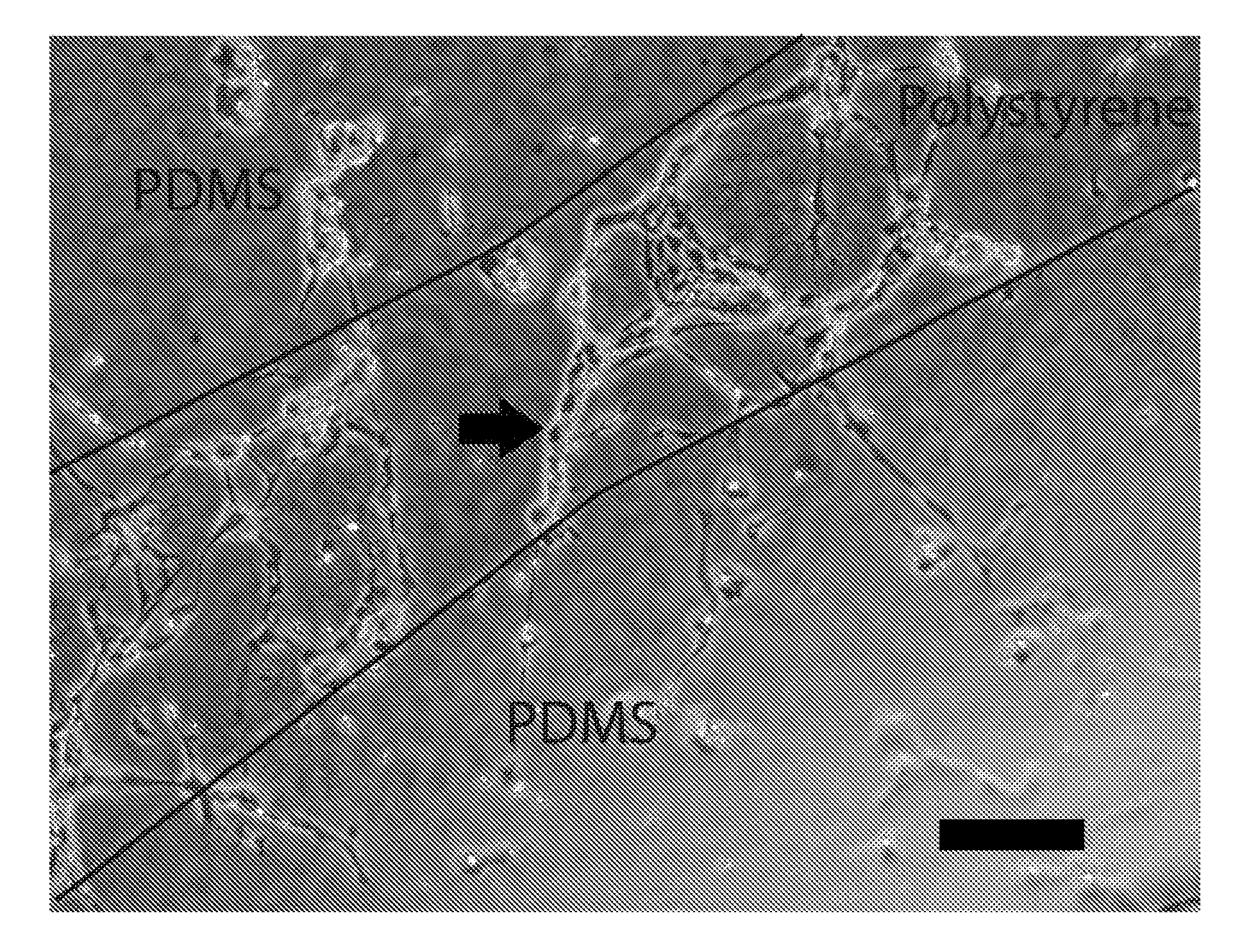
[0126]A stock of 1× Matrigel™ is diluted about 30 to about 60 times with about 4° C. cell-culture medium (from PromoCell) to produce a homogeneous support-generating medium. The support-generating medium is plated in non-treated polystyrene petri dishes as the cell-culture surface which has a hydrophobic surface. Components of the mixture settle / stick to the bottom of the dish and form a loosely connected low density environment. About 3 ml of the support-generating medium is enough for 35 mm dish...

example 2

Sample Custom-Patterned / Controlled-Geometry Capillary Fabrication Devices

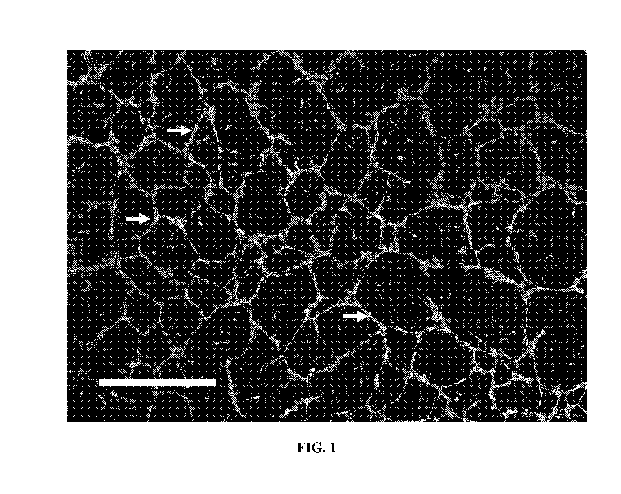
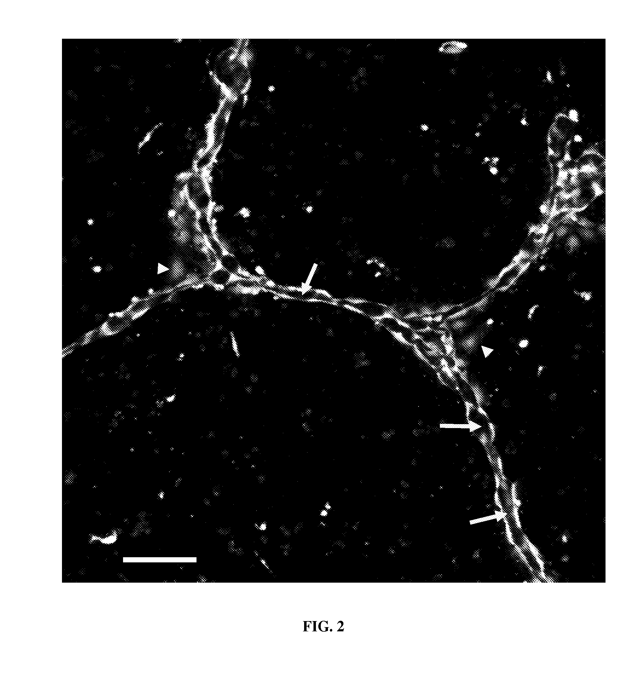
[0134]In this example, the cell-culture surface in the capillary fabrication device has hydrophobicity gradients. For example, the non-treated polystyrene petri dish in Example 1 can be replaced with a patterned ultra low adhesive polystyrene dish before plating support-generating medium. The ultra low adhesive polystyrene dish provides a cell-culture surface which is more hydrophilic than regular non-treated polystyrene petri dishes. The pattern on the ultra low adhesive polystyrene dish comprises a hexagonal network of narrow bands of 5 to 15 microns in width and 200 to 250 microns in length of less hydrophilic cell-culture surface. Cells of endothelial lineage can be used. The custom-patterned / controlled-geometry capillaries form on top of the predefined hexagonal network pattern of less hydrophilic cell-culture surface.

[0135]Optionally, the non-treated polystyrene cell-culture surface in Example 1 can be pr...

example 3

Sample Capillary Assay Devices

[0138]Capillary networks are grown as described in Example 1 using the capillary fabrication device or Example 2 using the custom-patterned capillary fabrication device combined with one or modifications with the aim of determining the effect of those modifications on the cultured cells or on the resulting capillary networks.

[0139]The capillary assay device can be used for determining the proangiogenic effects of different isoforms of vascular endothelial growth factor (VEGF) including soluble and ECM-bound isoforms. The cells in the capillary assay device are cultured inside the support medium, thus the device provides quasi-in vivo conditions for the interaction of VEGF isoforms and the cells. Cells in the 2-dimensional standard Matrigel™ capillary fabrication device are cultured on top of a thick solidified gel which differs significantly from in vivo conditions.

[0140]The capillary assay device also can be used for determining the effects of various ...


PUM

PropertyMeasurementUnit
thicknessaaaaaaaaaa
diameteraaaaaaaaaa
diameteraaaaaaaaaa

Abstract

Disclosed herein are capillary fabrication devices comprising living cells within a support medium. Culture of the cells produces viable lumenized capillary networks with natural or pre-determined geometries and ECM and basement membrane associated with the capillary networks. The capillary networks and the ECM and basement membrane detachable from the capillary networks are useful for tissue engineering applications.

Description

FIELD[0001]This disclosure generally relates to the field of biomedical tissue engineering. More specifically, the disclosure relates to devices for the synthesis of capillary networks with natural or controlled geometries and their associated extracellular matrix and basement membrane.BACKGROUND AND SUMMARY[0002]Under the proper conditions, endothelial cells or their precursors / progenitor cells, spontaneously assemble into networks and differentiate to form lumenized connected capillary networks.[0003]Both the forming capillary network and the final capillary network and its associated extracellular matrix (ECM) have numerous scientific, industrial and medical applications. In vitro angiogenesis assays employing the forming capillary network (or capillary fabrication devices) play a crucial role in identifying factors involved in vascular development. Such assays are used in drug development as moderate-throughput screens for angiogenesis promoters and inhibitors related to wound h...

Claims


Application Information

Patent Timeline
Patent Type & Authority Applications(United States)
IPC IPC(8): C12N5/071A61P9/00A61K35/44C07K14/78C12M3/00C12Q1/02
CPCC12M21/08C12M23/20C12M23/10A61P9/00A61K35/44C12N5/0068C12N2533/90
Inventor GLAZIER, JAMES A.ALILECHE, ABDELKRIMSHRINIFARD, ABBASAMARIE, DRAGOS
Owner THE TRUSTEES OF INDIANA UNIV
Why Eureka
Try Eureka