Microfluidics Sorter For Cell Detection And Isolation

a cell detection and cell technology, applied in biochemistry apparatus and processes, laboratory glassware, instruments, etc., can solve the problems of unsuitable clinical blood sample processing and low processing throughput, and achieve the effect of reducing processing time and cost, high flow rate, and faster processing of clinical samples

Active Publication Date: 2013-05-23
MASSACHUSETTS INST OF TECH +1
View PDF1 Cites 72 Cited by
  • Summary
  • Abstract
  • Description
  • Claims
  • Application Information

AI Technical Summary

Benefits of technology

[0009]This invention has many advantages, including continuous operation at a relatively high flow rate, enabling faster processing of clinical samples, with no chemical modification of the sample, which reduces processing time and cost, and the collection of viable cells for subsequent biological assays.

Problems solved by technology

However, a major limitation in these microfluidics systems is the low processing throughput, either due to sample dilution or to slow flow rates, making them unsuitable for processing clinical blood samples which are usually milliliters in volume.

Method used


Image

  • Microfluidics Sorter For Cell Detection And Isolation
  • Microfluidics Sorter For Cell Detection And Isolation
  • Microfluidics Sorter For Cell Detection And Isolation

Examples


example 1

Deformability Based Sorting for Cell Separation and Isolation

Materials and Methods

Malaria Culture

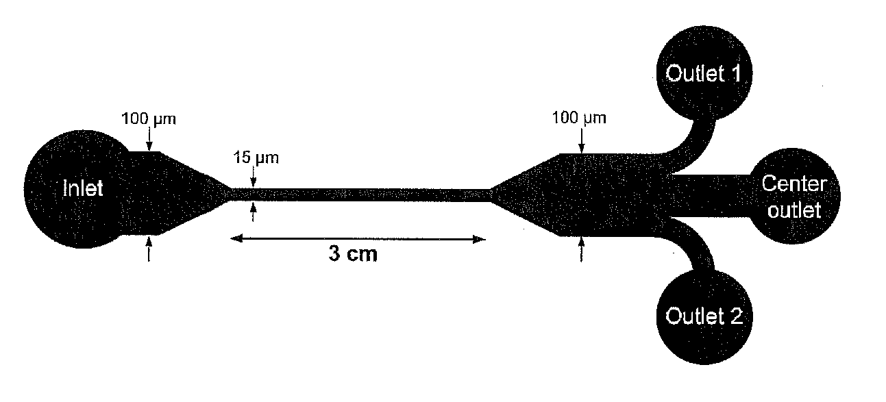
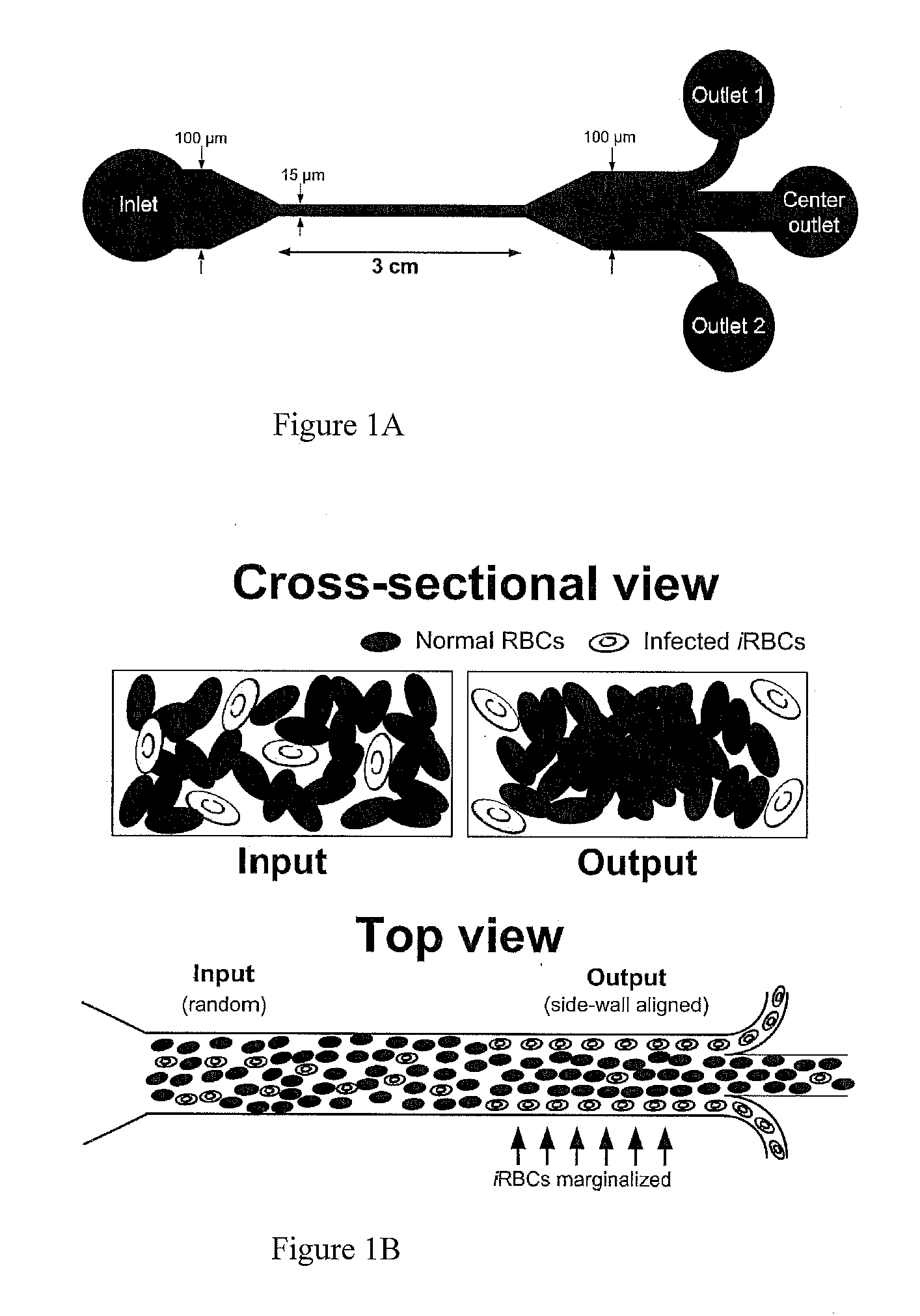
[0076]P. falciparum 3D7 strain was used in this study. Parasites were cultured in RPMI medium 1640 (Invitrogen, USA) supplemented with 0.3 g of L-glutamine, 5 g of AlbuMAX II (Invitrogen, USA), 2 g NaHCO3, and 0.05 g of hypoxanthine (Sigma-Aldrich, USA) dissolved in 1 ml of 1 M NaOH, together with 1 ml of 10 mg / ml of Gentamicin (Invitrogen, USA). Parasites were synchronized at ring stage using 2.5% sorbitol to maintain a synchronous culture. Cultures were stored at 37° C. after gassing with a 5% CO2, 3% O2 and 92% N2 gas mixture and their hematocrit maintained at 2.5%. Cells were harvested at the ring stage, late trophozoite and schizont stage. Whole blood for parasite culture was obtained from healthy donors and was spun down to separate the RBCs. The RBC pellet was treated with CPDA for 3 days before being washed three times with RPMI 1640 and stored for use.

Sample Preparation

[0077]The...

example 2

Cell Cycle Synchronization in Spiral Microfluidics

Materials and Methods

Cell Culture

[0091]Mesenchymal stem cells (Lonza, Switzerland) were cultured in low-glucose Dulbecco's modified Eagle's medium (DMEM) (Invitrogen, USA) supplemented with 10% fetal bovine serum (FBS) (Invitrogen, USA) together with 1% penicillin-streptomycin (Invitrogen, USA). The Chinese hamster ovary cells transfected with human CD36, CHO-CD36 (ATCC, USA), were cultured in RPMI 1640 medium (Invitrogen, USA) supplemented with 10% FBS together with 1% penicillin-streptomycin. The cervical cancer cells HeLa (CCL-2™, ATCC, USA) were cultured in low-glucose DMEM supplemented with 10% FBS and 1% penicillin-streptomycin. The cholangiocarcinoma cell line, KKU-100 (received as a gift), were cultured in Ham's F-12 medium containing 10% FBS, 3% HEPES buffer and 1% penicillin-streptomycin. All cultures were maintained at 37° C. in a humidified atmosphere containing 5% (v / v) CO2. The MSCs were seeded at 500 cells / cm2 and cult...

example 3

Shear Modulated Abstraction of Rare-Cells Technology Biochip for High Throughput Isolation of Circulating Tumor Cells

Materials and Methods

Cell Culture and Sample Preparation

[0112]Two human breast adenocarcinoma cell lines, MCF-7 and MDA-MB-231, were tested in this work. The MCF-7 cells (HTB-22™, ATCC, USA) and MDA-MB-231 cells (HTB-26™, ATCC, USA) were cultured in low-glucose Dulbecco's modified Eagle's medium (DMEM) (Invitrogen, USA) supplemented with 10% fetal bovine serum (FBS) (Invitrogen, USA) together with 1% penicillin-streptomycin (Invitrogen, USA). The culture was maintained at 37° C. in a humidified atmosphere containing 5% (v / v) CO2. The cells were cultured in sterile 25 cm2 flasks (Corning) and subcultivated (1:4) three times a week with media replaced every 48 h. Sub-confluent monolayers were dissociated using 0.01% trypsin and 5.3 mM EDTA solution (Lonza, Switzerland). For the control and recovery experiments, the cancer cells were diluted in buffer containing 1× phosp...


PUM

PropertyMeasurementUnit
heightaaaaaaaaaa
heightaaaaaaaaaa
heightaaaaaaaaaa

Abstract

A method of detecting one or more diseased blood cells in a blood sample includes introducing a blood sample into at least one inlet of a microfluidic device comprising one or more linear channels wherein each channel has a length and a cross-section of a height and a width defining an aspect ratio adapted to isolate diseased blood cells along at least one portion of the cross-section of the channel based on reduced deformability of diseased blood cells as compared to non-diseased blood cells, wherein diseased blood cells flow along a first portion of the channel to a first outlet and non-diseased blood cells flow along a second portion of the channel to a second outlet. The one or more channels can be adapted to isolate cells along portions of the cross-section of the channel based on cell size. In some embodiments, the one or more channels can be spiral channels.

Description

RELATED APPLICATION(S)[0001]This application claims the benefit of U.S. Provisional Application No. 61 / 310,387, filed on Mar. 4, 2010 and U.S. Provisional Application No. 61 / 383,881 filed on Sep. 17, 2010. The entire teachings of the above application(s) are incorporated herein by reference.BACKGROUND OF THE INVENTION[0002]Convectional macroscale methods for separation of cells include physical filtration using membrane-based filter and density gradient centrifugation which exploit differences in cell size, deformability, and density to filter out target cells. These techniques are labor-intensive and require multi-step sample preparations which may introduce artifacts or lead to loss of desired cells. Membrane filtration methods are also easily susceptible to clogging and require frequent cleaning. Further, evidence of mechanical stress-induced changes in original phenotype of target cells subjected to filtration and centrifugation techniques has also been reported.[0003]Hence, the...

Claims


Application Information

Patent Timeline
Patent Type & Authority Applications(United States)
IPC IPC(8): C12Q1/04C12Q1/02
CPCB01L3/50273B01L3/502761B01L2200/0636B01L2300/0864C12Q1/04B01L2400/086G01N2015/1415G01N2015/149C12Q1/025B01L2400/0457C12M1/34C12M3/00
Inventor LIM, CHWEE TECKHAN, JONGYOONHOU, HAN WEIBHAGAT, ALI ASGARVLIET, KRYSTYN J. VANLEE, WONG CHENG
Owner MASSACHUSETTS INST OF TECH
Why Eureka
Try Eureka