Use of bacterial beta-lactamase for in vitro diagnostics and in vivo imaging, diagnostics and therapeutics

a technology of beta-lactamase and in vitro diagnostics, applied in the field of pathogenic microorganisms and imaging technologies, can solve the problems of difficult diagnosis of many of these infections, inability to detect penicillin resistance, and inability to quantify and assess the viability the current methods of tuberculosis in the laboratory, can only be used to determine the viability of colony forming units (cfu) and/or

Active Publication Date: 2013-06-27
THE BOARD OF TRUSTEES OF THE LELAND STANFORD JUNIOR UNIV +1
View PDF1 Cites 5 Cited by
  • Summary
  • Abstract
  • Description
  • Claims
  • Application Information

AI Technical Summary

Benefits of technology

[0017]The present invention is directed further still to a method for detecting a pathogenic bacteria in real time in a subject. The method comprises introducing into the subject a substrate radiolabeled with an isotope associated with gamma emission where the substrate is for a beta-lactamase or other enzyme or protein specific to the pathogenic bacteria. The subject is imaged for gamma emissions from the radiolabeled substrate during activity thereon and signals generated by the emitted gamma rays are acquired. A 3D reconstruction of the concentration in the subject of the radiolabel based on intensity of the gamma ray generated signals is produced thereby detecting the pathogenic bacteria. The present invention is directed to a related method further comprising diagnosing in real time a pathophysiological condition associated with the pathogenic bacteria based on detection thereof. The present invention is directed to another related method further comprising administering one or more therapeutic compounds effective to treat the pathophysiological condition. The present invention is directed to yet another related method further comprising readministering the radiolabeled substrate to the subject and reimaging the subject to monitor the efficacy of the therapeutic compound; wherein a decrease in gamma emission compared to gamma emission at diagnosis indicates a therapeutic effect on the pathophysiological condition.

Problems solved by technology

Diagnosis of many of these infections and the presence of penicillin resistance is often difficult and requires extensive diagnostic laboratory culturing prior to susceptibility determination.
For example, tuberculosis currently affects nearly one-third of the world's population and remains a critical public health threat.
Current methods to quantify and assess the viability of tuberculosis in the laboratory, tissue culture cells and during infection in animal models and humans are limited to determination of colony forming units (CFU) and / or microscopy of tissues and sputum.
These methods are time-consuming, often difficult to interpret and relatively insensitive.
These inadequacies make it difficult to follow disease progression, vaccine efficacy and therapeutic outcome, both in animal models and patients.
More specifically, the prior art is deficient in sensitive and specific real-time optical imaging methods for beta-lactamase positive bacteria that can be used in vitro and in live subjects to diagnose and locate the bacterial infection, to rapidly screen for new therapeutics and to identify new drug targets.

Method used


Image

  • Use of bacterial beta-lactamase for in vitro diagnostics and in vivo imaging, diagnostics and therapeutics
  • Use of bacterial beta-lactamase for in vitro diagnostics and in vivo imaging, diagnostics and therapeutics
  • Use of bacterial beta-lactamase for in vitro diagnostics and in vivo imaging, diagnostics and therapeutics

Examples


example 1

Detection of Bla in M. tuberculosis in Culture

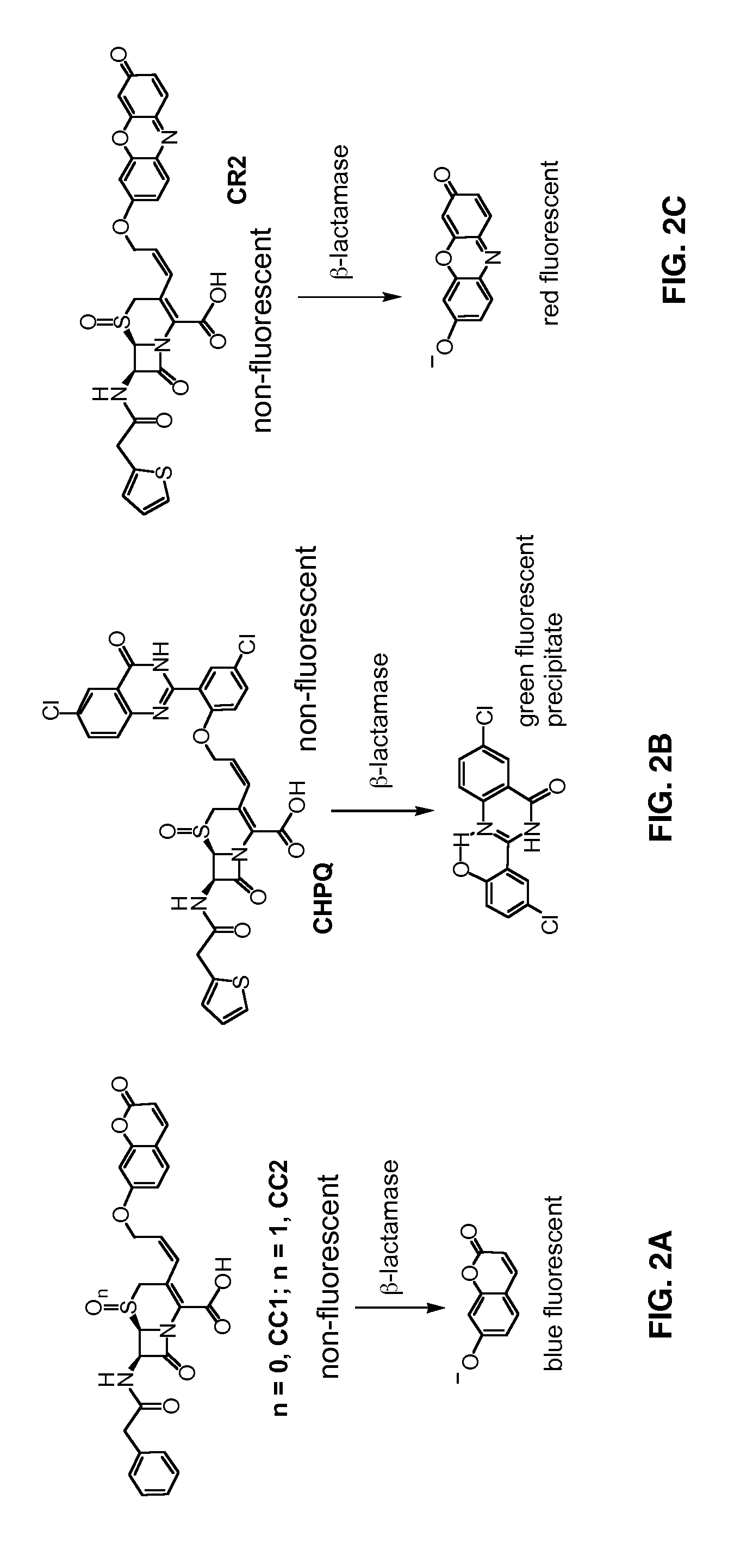
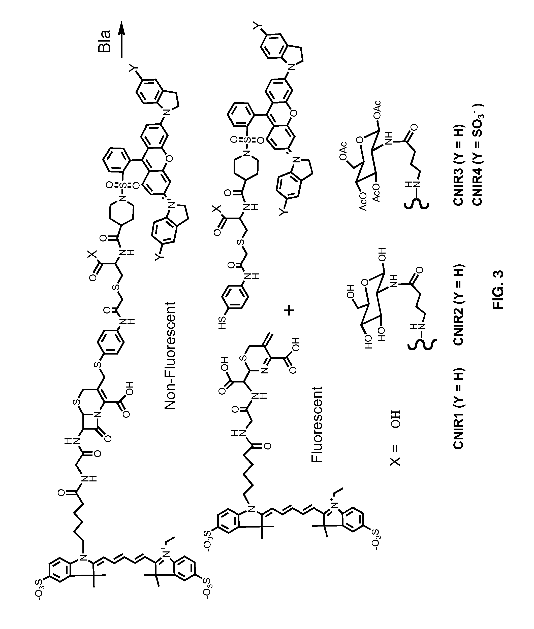
[0104]Potential fluoregenic substrate compounds and known compounds, including Nitrocefin (Calbiochem), CENTA Bla substrate (Calbiochem), Fluorocillin Green (Molecular Probes), CCF2-AM (Invitrogen) and CCF4-AM (Invitrogen), are compared for detection of Bla in Mtb using whole cells and whole cell lysates grown to early log-phase. Dilutions are assayed for all of these samples to determine the minimal number of bacteria or amount of lysate that results in significant signal. Titers are carried out to determine the number of actual CFU used, before and after assays with intact cells and before lysis for lysates. Both the sensitivity and reproducibility are evaluated in quadruplicate spectrophotometrically using 96-well plates incubated at 37° C. in bacterial culture medium from 15-120 min. Initially, compounds are used at concentrations recommended by the manufacturer and for CNIR5, 2 nM, i.e., that used for in vivo imaging. Different conc...

example 2

Intra-Vital Microscopy Imaging Using the Cell Transplantation Model

[0123]Universal donor Tr, CD8+ T cells, monocytes, macrophages and dendritic cells are transplanted into syngeneic mice infected with BCG, and the distribution of these cells over time are imaged with in vivo bioluminescence imaging (BLI) and image-guided intravital microscopy (IVM). A line of transgenic mice in which luciferase is produced by the beta-actin promoter, provide a source of tissues and cells that will emit light in non-transgenic animals (11-12). This mouse line (L2G85), shows bright bioluminescence from the firefly luciferase (Fluc), but weak GFP fluorescence, so it was mated with a separate line exhibiting strong GFP expression and fluorescence in lymphocytes. The spatial distribution of universal donor stem cells and other cells can thus be followed by BLI in the recipient as they expand, re-distribute or are cleared, and the cells detected can be subsequently visualized by IVM utilizing GFP.

[0124]Th...

example 3

Crystallization of M. Tuberculosis BlaC and BlaC Mutant Enzymes

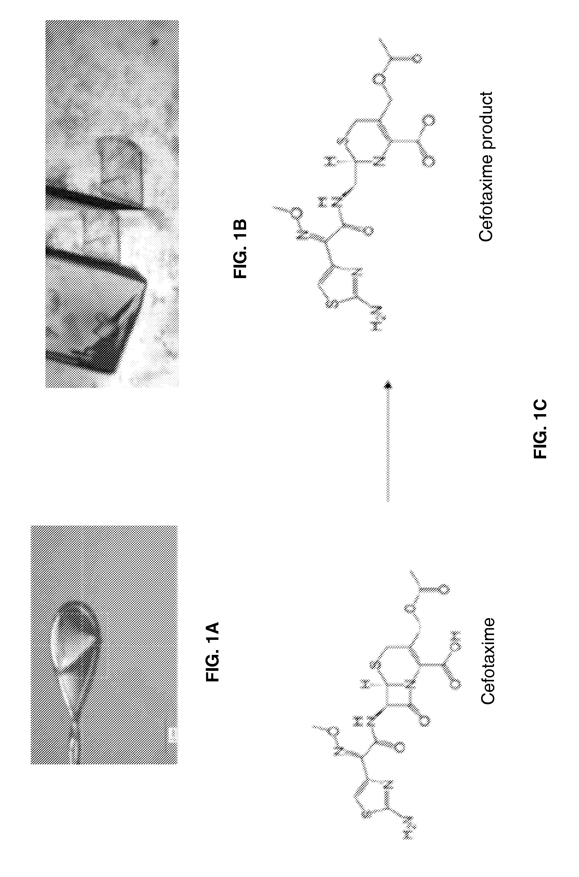
[0130]Very good crystals of BlaC were obtained after a few months of crystallization. Co-crystals with penicillin were produced using crystallization conditions of 0.1M Tris-HCl, pH 8.0, 20.M NH4H2PO4. These crystals allowed visualization of the intact protein active site and intermediate, but the initial bound substrate was not visible due to turnover in the crystal itself. To overcome this barrier, a Mtb BlaC mutant enzyme was constructed with the mutation in the Glu residue involved in hydrolysis (E166A) that allows trapping of the acy-intermediate on the enzyme and visualization of the specific interactions required for catalysis. This mutant has now been crystallized with a rapid, i.e., about two weeks, crystallization process yielding high quality crystals of Mtb BlaC mutant that are ready to be soaked with substrate (FIG. 1A). It is demonstrated that substrate can be incorporated into the Mtb BlaC mutant crystals ...


PUM

PropertyMeasurementUnit
emission wavelengthaaaaaaaaaa
imaging wavelengthaaaaaaaaaa

Abstract

Provided herein are methods for detecting, quantifying, differentiating, diagnosing and imaging pathogenic bacteria or condition associated therewith using substrates for bacterial enzymes. Fluorescent, luminescent or colorimetric signals emitted by substrates or enzyme products in the presence of the bacteria are compared to controls to detect and locate the pathogenic bacteria. Provided is a method for screening therapeutic agents to treat the pathophysiological conditions by measuring a signal emitted from the substrates or products in the presence and absence of the potential therapeutic agent and a diagnostic method for detecting a mycobacterial infection in a subject by contacting biological samples with a substrate and imaging for signals emitted from a mycobacterial beta-lactamase product. Also provided are fluorogenic substrates or substrates comprising a colored dye or a chemical reagent effective to induce a color or pH change.

Description

CROSS-REFERENCE TO RELATED APPLICATIONS[0001]This continuation application claims benefit of priority under 35 U.S.C. §120 of pending international application PCT / US2011 / 001018, filed Jun. 3, 2011, which claims benefit of priority under 35 U.S.C. §120 of pending continuation-in-part patent application U.S. Ser. No. 12 / 802,340, filed Jun. 4, 2010, which claims benefit of priority under 35 U.S.C. §120 of pending non-provisional U.S. Ser. No. 12 / 462,644, filed Aug. 6, 2009, which claims benefit of priority under 35 U.S.C. §119(e) of provisional U.S. Ser. No. 61 / 203,605, filed Dec. 24, 2008, now abandoned, and of provisional U.S. Ser. No. 61 / 188,112, filed Aug. 6, 2008, now abandoned, the entirety of all of which are herein incorporated by reference.BACKGROUND OF THE INVENTION[0002]1. Field of the Invention[0003]The present invention relates generally to the fields of medicine, pathogenic microbiology and imaging technologies. More specifically, the present invention relates to compoun...

Claims


Application Information

Patent Timeline
Patent Type & Authority Applications(United States)
IPC IPC(8): A61K49/00G01N21/64
CPCA61K49/0021G01N21/6486A61K49/0028A61K49/0032G01N2333/986A61K49/0052C12Q1/04C12Q1/34A61K49/0041A61K49/0013C09B57/02
Inventor CIRILLO, JEFFREY D.SACCHETTINI, JAMES C.RAO, JIANGHONGXIE, HEXIN
Owner THE BOARD OF TRUSTEES OF THE LELAND STANFORD JUNIOR UNIV
Why Eureka
Try Eureka