

Device For High Throughput Investigations Of Multi-Cellular Interactions
a multi-cellular interaction and device technology, applied in the field of cellular interaction research, can solve problems such as the inability to image blood vessels from the sid
- Summary
- Abstract
- Description
- Claims
- Application Information
AI Technical Summary
Benefits of technology
Problems solved by technology
Method used
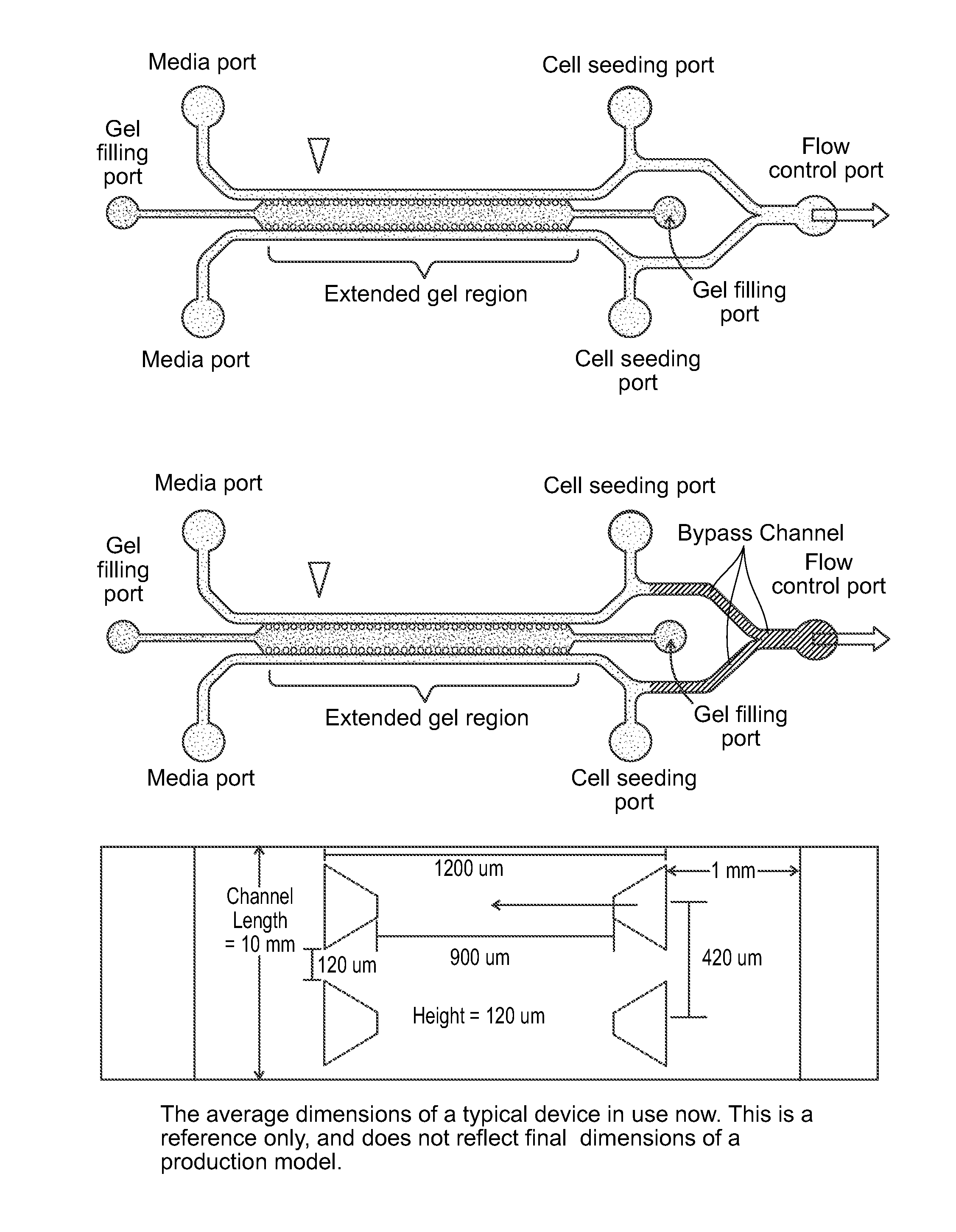
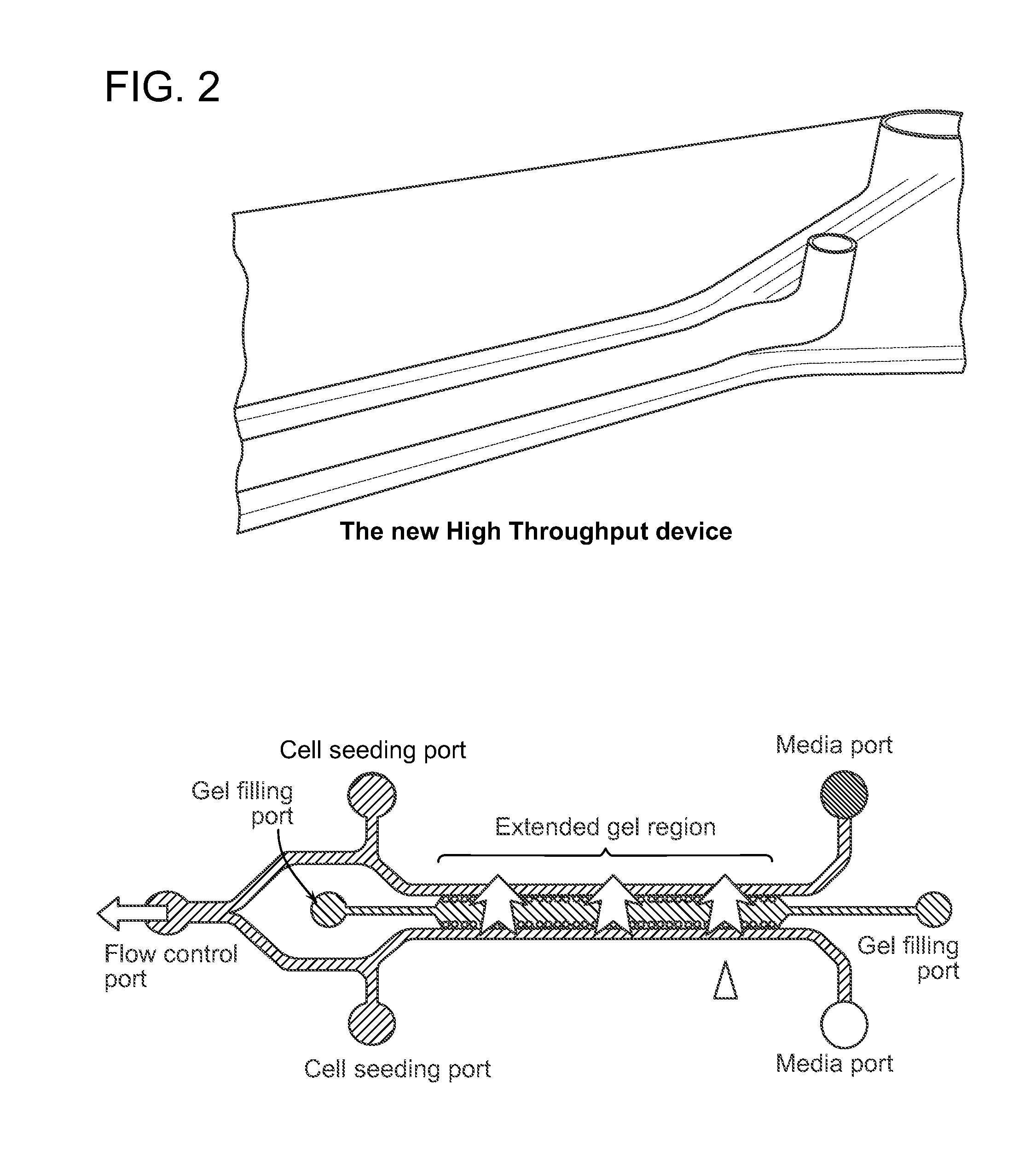
Image

Examples
example 1
Testing Concentrations of Anti-Metastatic Drugs to Keep Cancer Clusters from Migrating
Methods
[0144]Red colure cells are from Human Lung Cancer Cell Line A549 were transfected with H2B protein (m-Cherry), and partially disassociated, yielding cell aggregates of random sizes, between 40 um and 70 um. This was aspirated through a 100 micrometer strainer, yielding a media with cell aggregates with radii under 100 micrometers. This was poured through a second 40-micrometer filter, yielding a media with A549 aggregates between 40 and 100 micrometers. The cells were then centrifuged at 800 rpm for 2 minutes, and the media aspirated.
[0145]Collagen gel was prepared by adding 9 microliters of 1 M NaOH to 20 microliters of Phosphate Buffered Solution (BD Bioscience), and then adding 46 microliters of ultrapure H2O. Finally, 125 microliters of Type I Rat Tail Collagen at 2.5 mg / mL (BD Biosciences) were added, and mixed thoroughly.
[0146]The A549 aggregates were then mixed with the collagen gel, ...
example 2
Spider Device—a High-Throughput Microfluidic Assay to Study Axonal Response to Growth Factor Gradients
[0156]Studying axon guidance by diffusible or substrate bound gradients is challenging with current techniques. Described in this example is the design, fabrication and utility of one embodiment of the microfluidic devices provided herein used to study axon guidance under chemogradients. Experimental and computational studies demonstrated the establishment of a linear gradient of guidance cue within 30 min which was stable for up to 48 h. The gradient was found to be insensitive to external perturbations such as media change and movement of device. The effects of netrin-1 (0.1-10 μg / mL) and brain pulp (0.1 μL / mL) were evaluated for their chemoattractive potential on axonal turning, while slit-2 (62.5 or 250 ng / mL) was studied for its chemorepellant properties. Hippocampal or dorsal root ganglion (DRG) neurons were seeded into a micro-channel and packed onto the surface of a 3D colla...
example 3
Interstitial Flow Influences Direction of Tumor Cell Migration Through Competing Mechanisms
[0189]Interstitial flow is the convective transport of fluid through tissue extracellular matrix. This creeping fluid flow has been shown to affect the morphology and migration of cells such as fibroblasts, cancer cells, endothelial cells, and mesenchymal stem cells. However, due to limitations in experimental procedures and apparatuses, the mechanism by which cells detect flow and the details and dynamics of the cellular response remain largely unknown. A microfluidic cell culture system was designed to apply stable pressure gradients and fluid flow, and allow direct visualization of transient responses of cells seeded in a 3D collagen type I scaffold. This system was employed to examine the effects of interstitial flow on cancer cell morphology and migration and to extend previous studies showing that interstitial flow increases the metastatic potential of MDA-MB 231 breast cancer cells (Shi...
PUM
| Property | Measurement | Unit |
|---|---|---|
| Length | aaaaa | aaaaa |
| Thickness | aaaaa | aaaaa |
| Permeability | aaaaa | aaaaa |
Abstract
Description
Claims
Application Information