System and method for image guided medical procedures

a medical and surgical technology, applied in the field of system and method for image guided medical and surgical procedures, can solve the problems of incontinence and erectile dysfunction, low tissue discrimination ability of ultrasound imaging, and serious side effects, so as to achieve the effect of minimizing corrections and effectively using them

Inactive Publication Date: 2014-03-13
CONVERGENT LIFE SCI
View PDF0 Cites 93 Cited by
  • Summary
  • Abstract
  • Description
  • Claims
  • Application Information

AI Technical Summary

Benefits of technology

[0056]It is noted that as an alternate, the MRI or CAT scan data may be used to provide a coordinate frame of reference for the procedure, and the TRUS image modified in real-time to reflect an inverse of the ultrasound distortion. That is, the MRI or CAT data typically has a precise and undistorted geometry. On the other hand the ultrasound image may be geometrically distorted by phase velocity variations in the propagation of the ultrasound waves through the tissues, and to a lesser extent, by reflections and resonances. Since the biopsy instrument itself is rigid, it will correspond more closely to the MRI or CAT model than the TRUS model, and therefore a urologist seeking to acquire a biopsy sample may have to make corrections in course if guided by the TRUS image. If the TRUS image, on the other hand, is normalized to the MRI coordinate system, then such corrections may be minimized. This requires that the TRUS data be modified according to the fused image volume model in real time. However, modern graphics processors (GPU or APU, multicore CPU, FPGA) and other computing technologies make this feasible.
[0057]According to another aspect, the urologist is presented with a 3D display of the patient's anatomy, supplemented by and registered to the real-time TRUS data. Such 3D displays may be effectively used with haptic feedback.

Problems solved by technology

Such treatments are costly and can cause serious side effects, including incontinence and erectile dysfunction.
However, ultrasound imaging has relatively low tissue discrimination ability.
Accordingly, ultrasound imaging provides adequate imaging of the prostate organ, but it does not provide adequate imaging of tumors within the organ due to the similarity of cancer tissue and benign tissues, as well as the lack of tissue uniformity.
Due to the inability to visualize the cancerous portions within the organ with ultrasound, the entire prostate must be considered during the biopsy.
As with ultrasound imaging, MRI also has limitations.
For instance, it has a relatively long imaging time, requires specialized and costly facilities, and is not well-suited for performance by a urologist at a urology center.
Furthermore, performing direct prostate biopsy within MRI machines is not practical for a urologist at a urology center.
Until now, however, such systems have not been adequate for enabling MRI-ultrasound fusion to be performed by a urologist at a urology center.
Such MRI data, however, is not readily available to urologists and it would be commercially impractical for such MRI data to be generated at a urology center.
This is due to many reasons, including urologists' lack of training or expertise, as well as the lack of time, to do so.
Also, it is uncertain whether a urologist can profitably implement an image-guided biopsy system in his or her practice while contemporaneously attempting to learn to perform MRI scans.
Furthermore, even if a urologist invested the time and money in purchasing MRI equipment and learning to perform MRI scans, the urologist would still be unable to perform the MRI-ultrasound fusion because a radiologist is needed for the performance of advanced MRI assessment and manipulation techniques which are outside the scope of a urologist's expertise.
The use of imaging modalities other than trans-rectal ultrasound (TRUS) for biopsy and / or therapy typically provides a number of logistic problems.
For instance, directly using MRI to navigate during biopsy or therapy can be complicated (e.g. requiring use of nonmagnetic materials) and expensive (e.g., MRI operating costs).
This need for specially designed tracking equipment, access to an MRI machine, and limited availability of machine time has resulted in very limited use of direct MRI-guided biopsy or therapy.
CT imaging is likewise expensive and has limited access, and poses a radiation risk for operators and patient.
However, the available real-time medical imaging modalities for guiding the localized treatment visualize the organ, but do not clearly delineate the portion of the organ to be treated.
A further complication is that the non-real time image may have a different intrinsic coordinate system from the real time imaging, leading to artifacts.
The availability of “soft-correspondence” permits or facilitates automated or semi-automated labeling of objects, since the real-time imaging is typically not used by a fully automated system to perform a procedure, and the skilled medical professional can employ judgment, especially if the labeling indicates a possible degree of unreliability, in relying on the automated labeling.
Likewise, in some cases the pre-operative imaging labeling boundaries are imprecise, and therefore that the medical professional might wish to treat such boundaries as being advisory and not absolute.
The registration / fusion of images obtained from different modalities creates a number of complications.
Typically, well-defined and invariant anatomical landmarks may be used to register the images, though since the margins of landmarks themselves vary with imaging modality, the registration may be imperfect or require discretion in interpretation.
A further difficulty with these different modalities is that the intensity of objects in the images do not necessarily correspond.
As a result, rigid registration is not sufficient to account for difference between MRI and TRUS images.
Finally, the resolution of the images may also impact registration quality.

Method used


Image

  • System and method for image guided medical procedures
  • System and method for image guided medical procedures
  • System and method for image guided medical procedures

Examples


Embodiment Construction

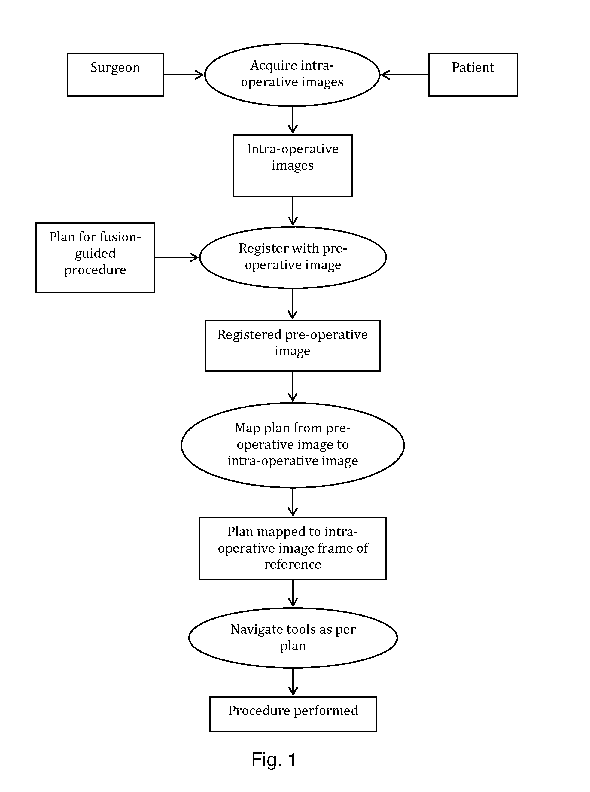
[0082]The present invention will be described with respect to a process, which may be carried out through interaction with a user or automatically. One skilled in the art will appreciate that various types of imaging systems, including but not limited to MRI, ultrasound, PET, CT, SPECT, X-ray, and the like may be used for either pre-operative or intra-operative imaging, but that a preferred scheme employs a fusion of MRI and / or CT and / or PET and ultrasound imaging for the pre-operative imaging, and trans-urethral ultrasound for intra-operative real time imaging in a prostate diagnosis or therapeutic procedure.

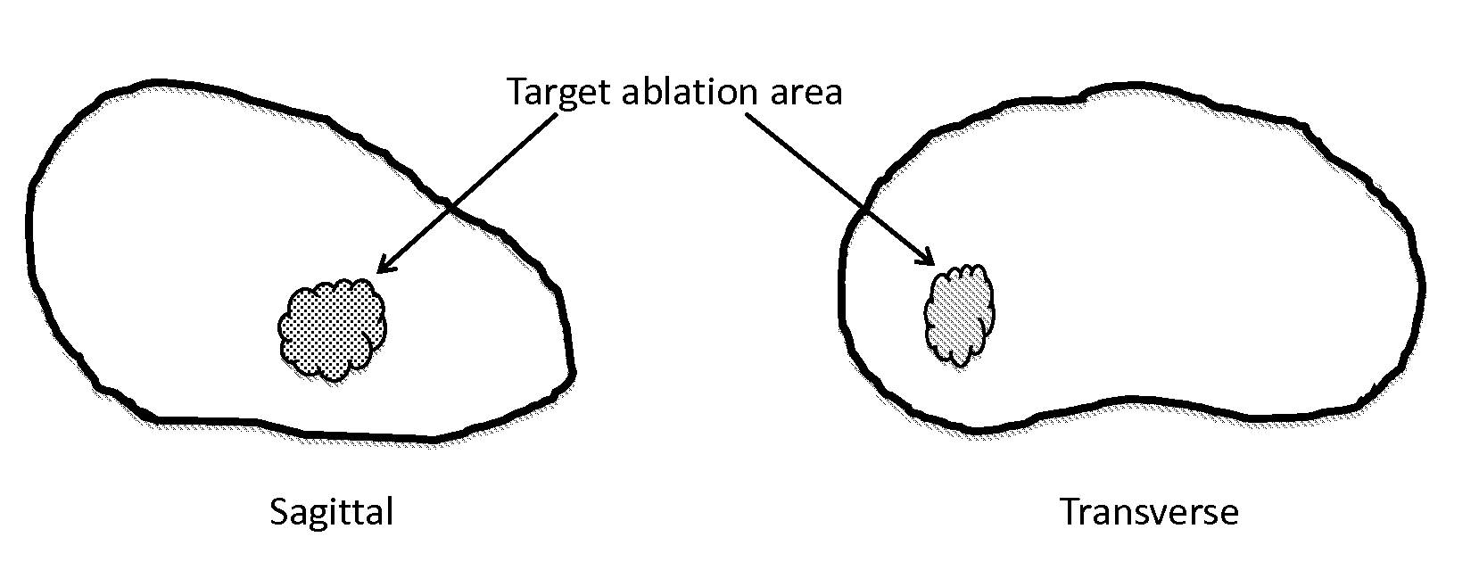
[0083]According to an embodiment of the present technology, one or more pre-procedure “planning” images are used to plan a procedure and one or more intra-procedure “live” images used to guide the procedure. For example, prostate biopsy and ablation is typically done under ultrasound guidance. While speed of imaging and cost make ultrasound an ideal imaging modality for guiding...


PUM


Abstract

A system and method combines information from a plurality of medical imaging modalities, such as PET, CT, MRI, MRSI, Ultrasound, Echo Cardiograms, Photoacoustic Imaging and Elastography for a medical image guided procedure, such that a pre-procedure image using one of these imaging modalities, is fused with an intra-procedure imaging modality used for real time image guidance for a medical procedure for any soft tissue organ or gland such as prostate, skin, heart, lung, kidney, liver, bladder, ovaries, and thyroid, wherein the soft tissue deformation and changes between the two imaging instances are modeled and accounted for automatically.

Description

CROSS REFERENCE TO RELATED APPLICATIONS[0001]The present application is a non-provisional of U.S. Provisional Patent Application 61 / 691,758, filed Aug. 12, 2012, the entirety of which is expressly incorporated herein by reference.BACKGROUND OF THE INVENTION[0002]1. Field of the Invention[0003]The present disclosure relates to systems and methods for image guided medical and surgical procedures.[0004]2. Description of the Art[0005]U.S. Pat Pub. 2009 / 0054772 (EP20050781862), expressly incorporated herein by reference, entitled “Focused ultrasound therapy system”, provides a method for performing a High Intensity Focused Ultrasound (HIFU) procedure for specific clinical application. Basic image registration is performed for fusion from a diagnostic modality such as Computed Tomography (CT) or Magnetic Resonance Imaging (MRI) to ultrasound, only through body positioning, referred to as “immobilization”, resulting in only image registration via horizontal movement and zoom factor. See al...

Claims


Application Information

Patent Timeline
Patent Type & Authority Applications(United States)
IPC IPC(8): A61B19/00
CPCA61B19/5212A61B10/00A61B2017/00274A61B10/0241A61B2090/364A61B90/361A61B34/10A61B2034/107A61B2090/378A61B2090/365A61B10/02A61B2018/2005A61B18/20A61B18/22A61N5/0601A61N2005/0612A61N2005/063A61N5/0625A61N2005/1012A61B34/20A61B90/39
Inventor KUMAR, DINESHVOHRA, AMITSPERLING, DANIEL S.
Owner CONVERGENT LIFE SCI
Why Eureka
Try Eureka