
PCR diagnostics of dermatophytes and other pathogenic fungi

a technology of dermatophytes and pathogenic fungi, applied in the field of pcr diagnostics of dermatophytes and other pathogenic fungi, can solve the problems of low sensitivity of techniques, false negatives, and long-term systemic treatment, and achieves the effects of reducing the number of cases, and improving the diagnostic accuracy

Active Publication Date: 2014-06-05
STATENS SERUM INST
View PDF1 Cites 0 Cited by
  • Summary
  • Abstract
  • Description
  • Claims
  • Application Information

AI Technical Summary

Benefits of technology

The patent describes a method called pan-dermatophytes PCR that can detect all species in three genera of dermatophytes using a common gene sequence. This suggests that the method can be used as a reliable and efficient way to identify these organisms.

Problems solved by technology

Topical therapy is sufficient in most of cases of dermatophyte skin infection, but long term and often expensive systemic treatment is necessary in cases of tinea capitis, tinea barbae and onychomycosis.
Furthermore, genus and in some cases even the species-specific diagnosis is necessary due to different susceptibility patterns and contagious potential of the various dermatophytes, thus e.g. Microsporum species are less susceptible than Trichophyton spp. to terbinafine and should be treated with griseofulvin, T. rubrum strains are more susceptible to some antifungal drugs than T. mentagrophytes and some dermatophytes may cause epidemic outbreaks in schools and institutions due to transfer from man to man while others may not as their primary host is an animal (1
Direct microscopic examination of skin and nail material is often sufficient for the preemptive diagnosis of a fungal infection, but it does not give specific species diagnosis.
Furthermore, although rapid and cheap, this technique has a relatively low sensitivity and shows false negative results in up to 15% cases (2).
Such cases are especially cost- and time-consuming and require specialist skills (2).
Such methodologies are inconvenient for routine diagnostic purposes for several reasons.
First, the necessary initial cultivation of the patient samples is time consuming (up to 10 days) (13).
Next, the phenol / chlorophorm DNA extraction method applied in the methods of the DNA extraction directly from skin and hair samples (14) is toxic and thus not applicable for routine diagnostic use in laboratories receiving a large number of samples per day.
Finally, the previously published methods all involve a number of handlings (e.g. grinding or bead beating, and washing of the DNA pellets or columns) that increase the risk of the contamination of the samples and false PCR results.

Method used


Image

  • PCR diagnostics of dermatophytes and other pathogenic fungi
  • PCR diagnostics of dermatophytes and other pathogenic fungi
  • PCR diagnostics of dermatophytes and other pathogenic fungi

Examples


example 1

Primer Designing

[0108]The dermatophyte, Trichophyton rubrum, Microsporum canis, Trichophyton mentagrophytes-Trichophyton tonsurans complex or Epidermophyton floccosum specific primers were selected from available DNA sequences listed in Table 1. The alignment of respective sequences allowed the design of primer-pairs detecting all of the dermatophyte species (in case of panDerm1 and panDerm2 oligonucleotides), species belonging to Microsporum genus (in case of Micr773-for and Micr885-rev oligonucleotides), species belonging to Trichophyton genus (in case of Trichopyros-for and Trichopyros-rev oligonucleotides) and for specific detection of Trichophyton rubrum, Microsporum canis, Trichophyton mentagrophytes-Trichophyton tonsurans complex or Epidermophyton floccosum.

TABLE 1The sequenced used in primers designing.Gene Bank ™ accession number(www.ncbi.nih.gov)Arthoderma benhamiaeAB044155Arthoderma benhamiaeAB003558Arthoderma gypseumAB003568Arthoderma simiiAB003564Arthoderma vanbreuseghe...

example 2

Evaluation of the Specificity of Dermatophyte, Genus Trichophyton, Genus Microsporum, Trichophyton rubrum, Epidermophyton floccosum, Microsporum canis and Trichophyton mentagrophytes-Trichophyton tonsurans Complex Specific Primers

Materials and Methods

[0109]Strains. A list of the type, references and clinical fungal strains used in the study is presented in Table 2 Twelve strains were obtained from the National Collection of Pathogenic Fungi (United Kingdom). Clinical strains were obtained from Mycology Laboratory of Statens Serum Institute (SSI, Denmark). All clinical isolates were identified by colony characteristics and micro-morphology.

TABLE 2Microorganism used in the study.Number of clinicalMicroorganismNCPF numberisolatesMicrosporum gypseumNCPF-402Microsporum canisNCPF-17710Microsporum nanum—1Microsporum audouiniiNCPF-4365TrichophytonNCPF-22410mentagrophytes var.TrichophytonNCPF-780mentagrophytes var.Trichophyton schoenleniniiNCPF-124—Trichophyton terrestreNCPF-6028Trichophyton...

example 3

DNA Extraction Method—the Protocol for Invented DNA Extraction Method from Dermatophyte Infected Nails

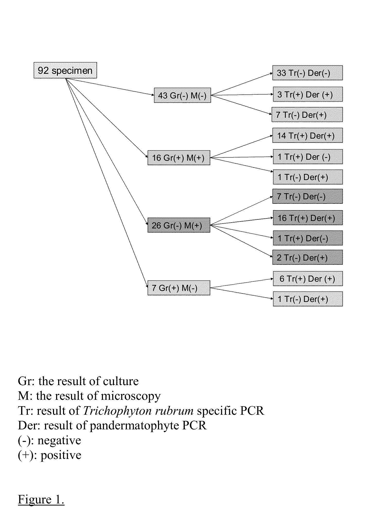
[0125]Sixty-four nail samples were randomly chosen from samples received at the Laboratory of Mycology at SSI for microscopy and culture of dermatophytes and Candida (Table 3). Fourteen were diagnosed as T. rubrum, two as T. mentagrophytes, one as a T. tonsurans, two as Aspergillus sp., two as Candida sp., one as Alternaria sp., three as Acremonium sp. and 39 were found negative.

TABLE 3Microorganisms used in the studySpecimen diagnosed byroutine Mycology Lab asNumber of clinical isolatesTrichophyton rubrum14Trichophyton mentagrophytes2Aspergillus sp.2Candida sp.2Acremonium sp.3Alternaria1Trichophyton tonsurans1Negatives (no growth of any fungi)39

[0126]DNA from 64 clinical specimens listed in table 3 were extracted according to the following protocol:[0127]1. The nails were placed in 2 ml Eppendorf tubes[0128]2. 100 μl (can be increased up to 500) lysis buffer L1 (250 mM KCl, 60 mM N...


PUM


Abstract

Dermatophytes which belong to one of the three genera Epidermophyton, Trichophyton and Microsporum are the main cause of fungal infections of skin, hair and nails. Traditional diagnostic procedures consist of microscopy and culture, but due to the slow growth rate of dermatophytes typically two to four weeks are needed before a final diagnosis is obtained. The present invention is a rapid DNA extraction method extracting nucleic acids from fungi (e.g. dermatophytes and other pathogenic fungi) which can be performed from directly on hair, nail or skin specimens from humans, from naturally or experimentally infected animals or from cultured fungal colonies for the use in PCR amplification and detection assays. The present invention also includes specific primer sets for detection of any dermatophyte and for species specific detection of Trichophyton rubrum and Epidermophyton floccosum by PCR and a kit for diagnosing fungal infections.

Description

CROSS-REFERENCE TO RELATED APPLICATIONS[0001]This application is a continuation of U.S. patent application Ser. No. 11 / 917,437, filed Dec. 13, 2007, which is a national stage of International Patent Application No. PCT / DK06 / 000332, filed Jun. 13, 2006, which applications are incorporated by reference herein in their entireties.FIELD OF INVENTION[0002]A method for extracting nucleic acids from fungi, a PCR method for detecting fungi in patient samples and a PCR kit for detecting dermatophytes and diagnosing infections by the three genera Trichophyton, Microsporum and Epidermophyton. GENERAL BACKGROUND[0003]Human pathogenic dermatophytes, which belong to the three genera Trichophyton, Microsporum and Epidermophyton, are fungi that infect human skin, nails, and hair. While the genus Epidermophyton is represented only by a single species (E. floccosum), the genera Microsporum and Trichophyton include several different species (1). Depending on the site of the infection the dermatophytos...

Claims


Application Information

Patent Timeline
Patent Type & Authority Applications(United States)
IPC IPC(8): C12Q1/68C12N15/10
CPCC12N15/1003C12Q2600/16C12Q1/6895
Inventor BRILLOWSKA-DABROWSKA, ANNA H.
Owner STATENS SERUM INST
Learn More