Nanostructured Biomimetic Device with Contour Map of Multiple Variable Correlation Method to Visually Display the Cancer Progresses

a biomimetic device and contour map technology, applied in the field of cancer progress monitoring devices, can solve the problems of lack of selectivity and selectivity of conventional biopotential methods used for diagnosing cancer, time-consuming large computer algorithm for modeling, lack of selectivity and sensitivity, etc., to selectively induce a bio communication and small impact on potentials

Active Publication Date: 2014-06-26
CHEN ELLEN T
View PDF2 Cites 2 Cited by
  • Summary
  • Abstract
  • Description
  • Claims
  • Application Information

AI Technical Summary

Benefits of technology

[0034]FIGS. 5A and 6A illustrate current change effect on the DSCPO profiles under 5 breast cancer cell/mL concentration with or without 1 day incubation, respectively, against the controls that did not have cancer cell as shown in FIG. 7A. Both figures were without inhibitors. It is obvious that with 1 day incubation, the DSCPO's action and resting potential profiles moved up to all positive potential fields, especially for resting potential, indicating the cancer cells are not in a normal “resting potential” stage, i.e, about −70 mV, have critically impacted the ratio of action/resting potential (RAPRP), hence the results of RAPRP are larger than that of without incubation. The action potential signals were increased as current increased drastically than that of without incubation. For without incubation, there were superimposed curves for resting potentials regardless the current changes. Current changes had smaller impact on the potentials for without incubation than that of with 24 hrs incubation.
[0035]Current change effect on a living normal breast cell was illustrated in FIG. 7A. The amplitude of curves at the action and resting potential fields are symmetric alone the zero line, indicating the RAPRP values are close to 0.75-0.9 range, which is at a normal electrophysiological situation [6]. The sensor is attachable on to the live human breast skin noninvasively. This sensor demonstrated its capability to selectively induce a bio communication more favorably to cancer cell rather than to a normal cell at very sensitive concentration level, because cancer cells with high negative charge density tend to direct hydrogen bonding to the positive imidazolium receptor in the sensor membrane.
[0036]The discharge potential was defined as “action potential”, and the charge potential defined as “resting potential. The duration time is 2 s for action or resting potential for the model cancer sensor study. The absolute value of action potential divided by the...

Problems solved by technology

The conventional biopotential method used for diagnosing cancer is lacking in sensitivity and selectivity [3].
Biologists measure cell membrane action and resting potentials with burdensome instrumentation with time consuming procedures.
However, the method requires a time consuming larg...

Method used


Image

  • Nanostructured Biomimetic Device with Contour Map of Multiple Variable Correlation Method to Visually Display the Cancer Progresses
  • Nanostructured Biomimetic Device with Contour Map of Multiple Variable Correlation Method to Visually Display the Cancer Progresses
  • Nanostructured Biomimetic Device with Contour Map of Multiple Variable Correlation Method to Visually Display the Cancer Progresses

Examples


example 1

Fabrication of the Nanostructured Biomimetic Self-Assembling Membranes (SAM)

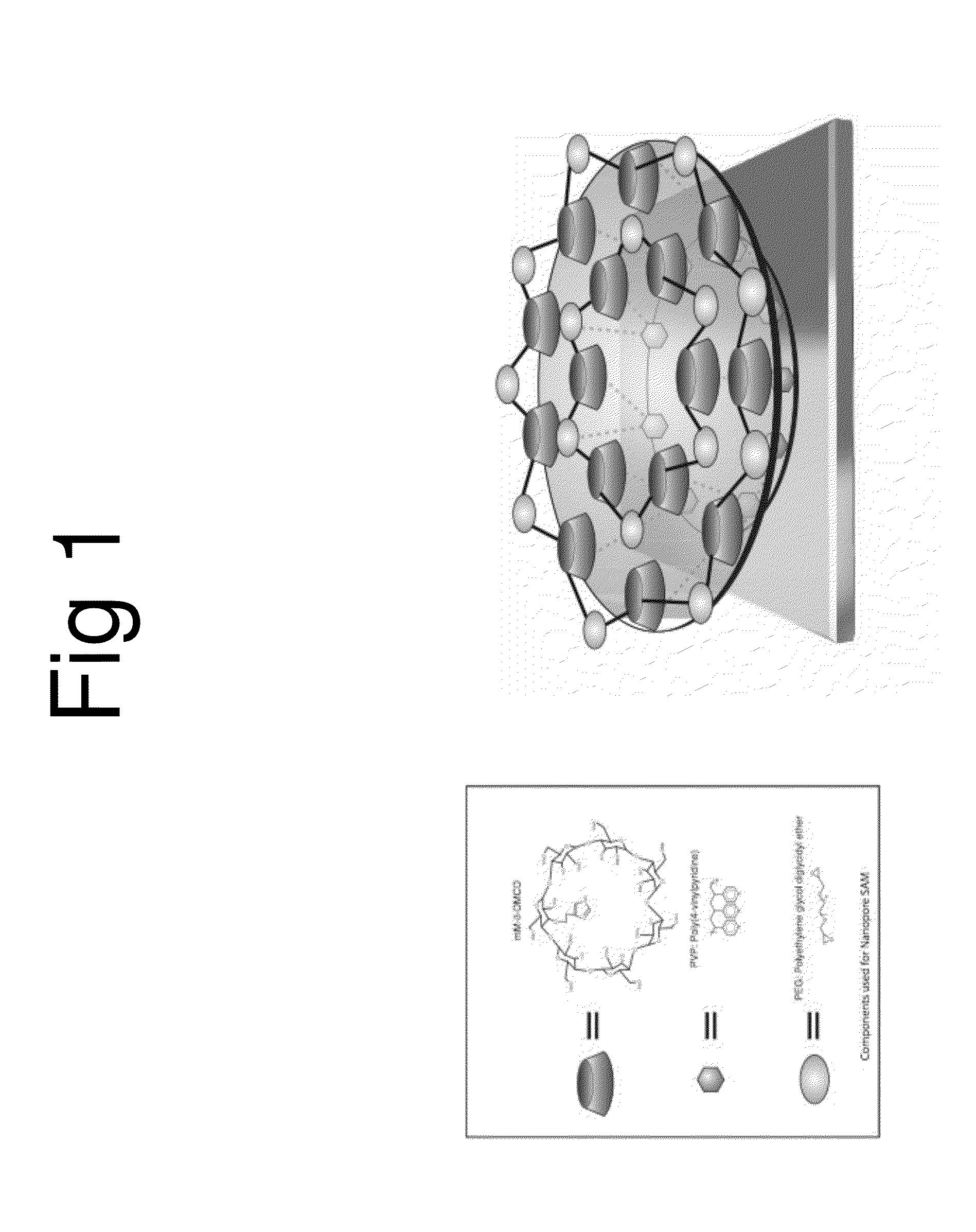
[0026]Reagent grade poly (4-vinylpyridine) (PVP), polyethylene glycol diglycidyl ether (PEG), were purchased from Aldrich-Sigma. The PVP was recrystallized in methanol. The mono imidazol derivative dimethyl β-cyclodextrin (mM-β-DMCD) was generally synthesized according to the published procedures [8]. The gold chips were purchased (Fisher Scientific) and the mixture solutions with proper compositions and procedures were followed by published literature in [9].

example 2

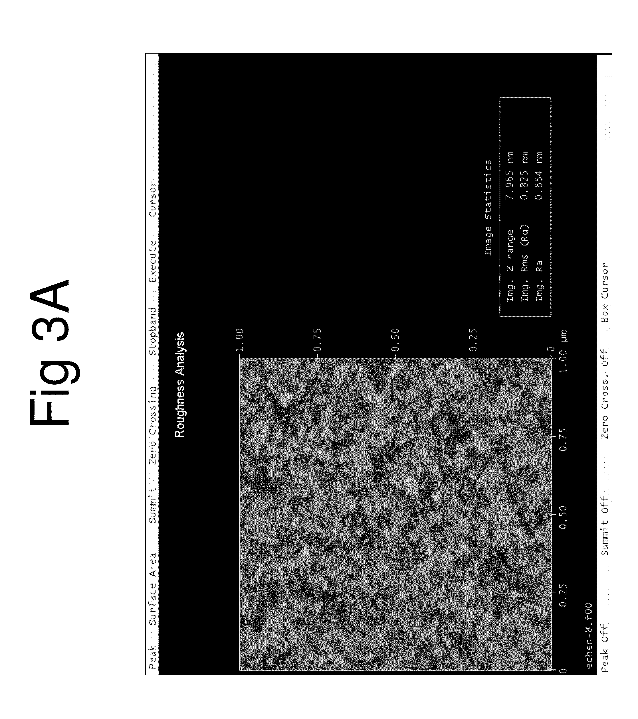
Characterization of the Membrane of AU / SAM

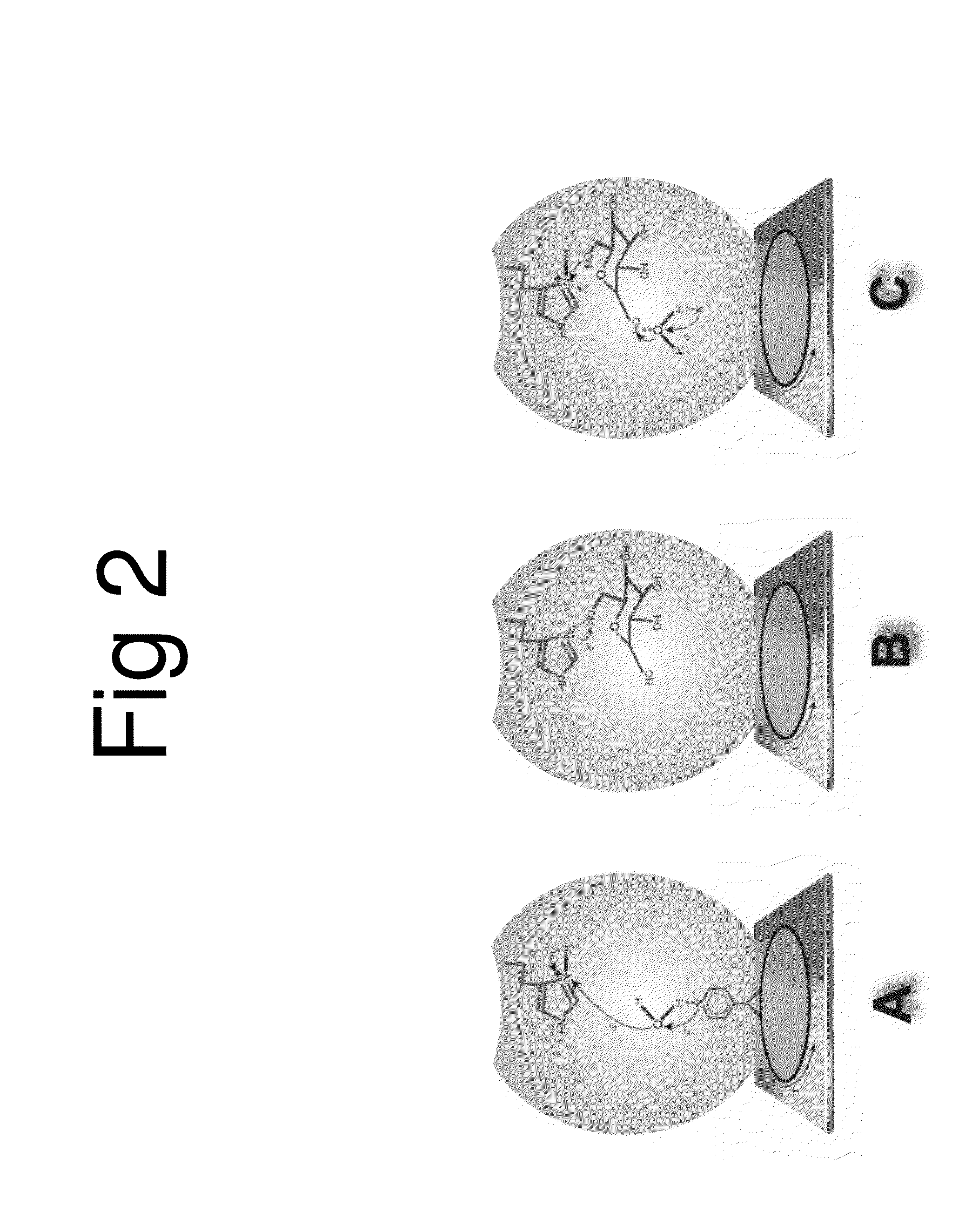
[0027]The morphology of the AU / SAM was characterized using a Dimension 3100 Atomic Force Microscope (AFM) (Bruker Nano, CA.). FIG. 1 is an art illustration of the model used to construct the Au-nanopored sensor cross linked with polymers and modified cyclodextrins by SAM method. FIG. 1 illustrates an art work for the model used to construct the Au-nanopored His 516 receptor-CD SAM electrode. The moiety of the receptor-CD was cross linked with polyethylene glycol diglycidyl ether (PEG) and poly(4-vinylpyridine) (PVP) and self-assembled a nanopore structured SAM through hydrogen bonding. Possible driving forces to form such a nanopore could be the changes in the heat of formation in the active site and the change of free energies of solvation that are favorable to the electron-relay processing. The pKa value difference between the receptor His 516 and the pyridine, and the difference of hydrophobicity between the internal cavity of CD and the ...

example 3

Human Cancer Cell Line MDA-MB-231 and the Glioblastoma Brain Cancer Line SNB-19

[0029]Breast cancer cell samples are human adenocarcinoma cells line MDA-MB-231 as shown in FIG. 4A taken from breast cancer tissue. The glioblastoma brain cancer cells samples are human neuro blastoma line SNB-19 as shown in FIG. 4B. The cell cultures are held in a base growing medium of DMEM (Dulbecco / Vogt Modified Eagle's minimal essential Medium—a common growth culture medium used for human cell incubation) (Invitrogen, CA infused with a 10% concentration of FBS (fetal bovine serum), 10 mM HEPES, 100 units / mL penicillin / Streptomycin and 2 mM L-glutamine. It was kept in a normal atmosphere at a temperature of 37.0° C. with 10% CO2 and humidified air. The cancer cells in the DMEM media were incubated for 24 hrs. Before test the cancer cells, dilution procedures were conducted.


PUM


Abstract

The present invention provides a novel electrochemical device and a cancer cell heat release map method to visually display the cancer progresses. The novel device comprises an electrode having a nanopore biomimetic electron-relay network with imidazolium-ATP of cancer cell-water-pyridine at the active sites that mimic the electron-relaying between His 516 and N(5)-FAD of GOx for the purpose to selectively detect triple-negative breast cancer cell at single cell concentration and it rejects brain cancer cell. The device is fast in millisecond to detect cancers without sample preparation and without interference from other substances, such glucose and proteins under reagent-free conditions. A unique biomarker of the ratio of “Action/Resting” cell membrane potential can be used to monitor the cancer progress against the normal cells. A visual contour map of a multiple variable correlation method provided to assess the heat release from the cancer cells against the normal cells is presented. The device for a potential therapeutic application was demonstrated by discharge voltage pulses from the live cancer cells with release extra energy that the cells possessed until it returns to a normal status in terms of normal cell membrane action/resting potential ratio.

Description

CROSS REFERENCE TO RELATED APPLICATIONS[0001]This application is a nonprovisional application of U.S. provisional application No. 61 / 660,072, filed 15 Jun. 2012 and entitled “Nanopore Structured Biomimetic Sensor Device;” is a nonprovisional of U.S. provisional application No. 61 / 660,080, filed 15 Jun. 2012 and entitled “Nanopore Structured Biomimetic Sensor Device;” is a nonprovisional of U.S. provisional application No. 61 / 660,690, filed 16 Jun. 2012 and entitled “Nanopore Biomimetic Device;” and is a nonprovisional of U.S. provisional application No. 61 / 691,632, filed 21 Aug. 2012 and entitled “Nanopore Structured Biomimetic Device.” The disclosure of each aforementioned application is incorporated herein by reference in its entirety.FIELD OF THE INVENTION[0002]The present invention relates to the field of cancer progress monitoring devices and, in particular, to devices handheld comprising a Nanostructured Biomimetic membrane, through functions of spontaneously discharging elect...

Claims


Application Information

Patent Timeline
IPC IPC(8): G01N27/26
CPCG01N27/26G01N15/1031A61B5/0091B82Y30/00G01N27/3278G01N2015/1006G01N2610/00G01N2800/7028
Inventor CHEN, ELLEN T.
Owner CHEN ELLEN T
Why Eureka
Try Eureka