
Prevention and treatment of inflammatory conditions
a technology of retinoic acid receptor and inflammatory conditions, applied in the direction of immunological disorders, drug compositions, peptide/protein ingredients, etc., can solve the problems of liver disease, liver fibrosis, liver fibrosis, etc., to suppress in vitro proliferation, increase the percentage of sulfatide/cd1d-tetramer+, and promote inflammatory respons
- Summary
- Abstract
- Description
- Claims
- Application Information
AI Technical Summary
Benefits of technology
Problems solved by technology
Method used
Image

Examples
example 1
Sulfatide Mediates Inhibition of Type I NKT Cells while Activating Sulfatide / CD1d-Tetramer+Cells
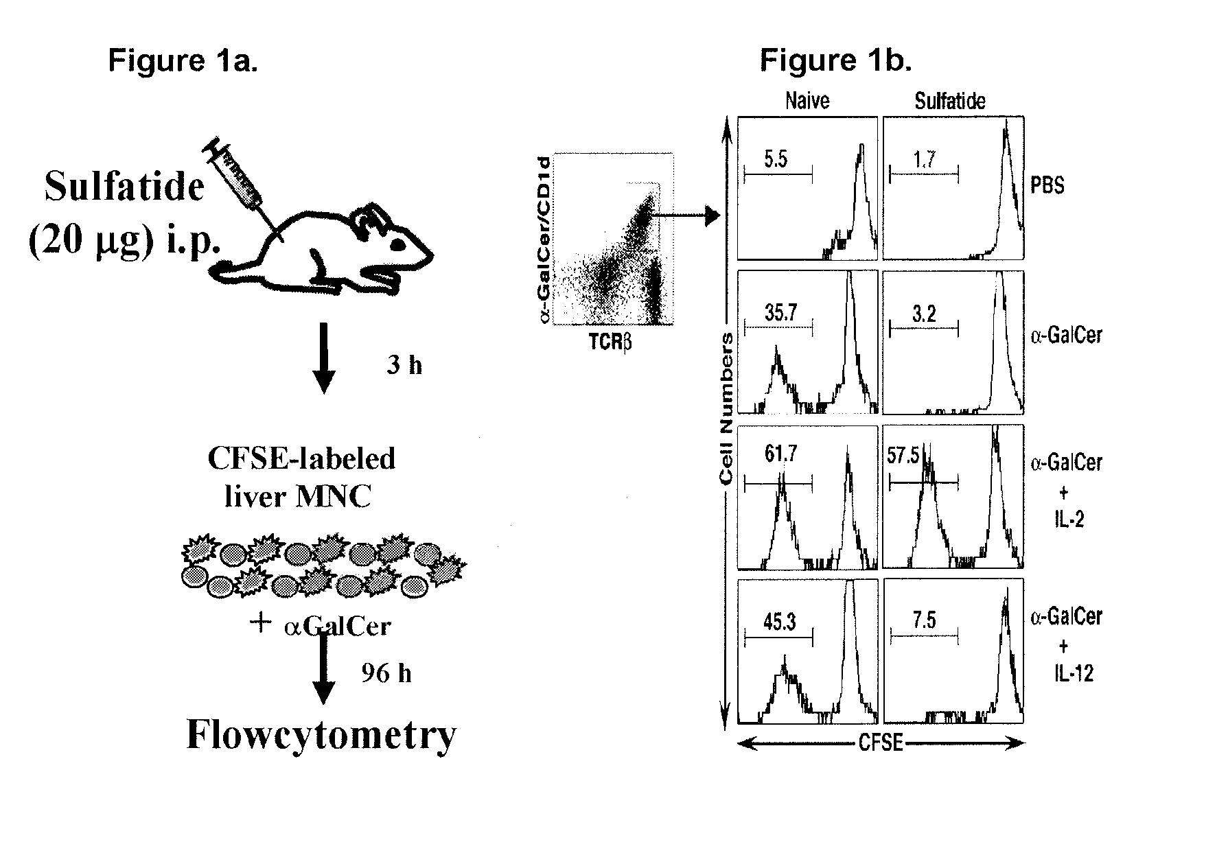
[0110]A dose of 20 μg sulfatide was injected intraperitoneally in mice. Following sulfatide injection, MNCs from liver were isolated and labeled with CFSE. The cells were cultured with αGalCer (10 ng / ml) for 96 hours in the presence or absence of 10 ng / ml cytokine IL-2. Cells were cultured with 10 ng / ml IL-12 alone as a control. Following culture, cells were harvested, stained with anti-TCRβ mAb and αGalCer / CD1d-tetramers, FACs sorted and CFSE dilution analysis was performed on αGalCer / CD1d-tetramer+ (type I NKT) cells. See FIG. 1A. As shown in FIG. 1B, after sulfatide administration, type I NKT cells fail to proliferate in response to an in vitro challenge with αGalCer.
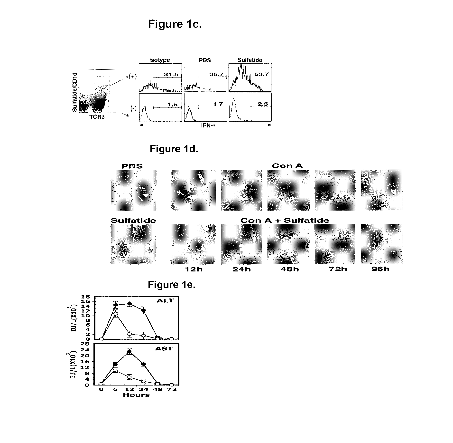
[0111]Liver cells isolated from PBS or 20 μg sulfatide injected mice were sorted into sulfatide / CD1d-tetramer+ and tetramer− populations and stained for intracytoplasmic IFN-γ+. As shown in FIG. 1C, sulfatide-injected mice...
example 2
Sulfatide Administration Protects Against ConA-Induced Hepatitis
[0113]Female C57BL / 6 mice were treated with 8.5 mg / kg of Concavalin A (ConA) (dissolved in pyrogen free phosphate buffer saline, PBS) intravenously (i.v.), which induces liver damage similar to that caused by hepatitis. Immediately after ConA injection, mice were injected intraperitonially (i.p.) with 20 μg (1 mg / kg / m) of bovine brain sulfatide or PBS.
[0114]Damage to the liver gives rise to telltale abnormalities (suggesting liver disease) detectable by liver function tests. For example, viral hepatitis can cause the alanine amino transferase (ALT) and aspartate amino transferase (AST) enzymes in injured liver cells to spill into the blood stream and increase their level in the blood. To test for liver damage, serum was collected and levels of serum enzymes, ALT and AST, were measured at 0, 6, 12, 24, 48 and 72 hours following Con A or Con A+sulfatide injection. Serum enzyme levels were measured with the help of Laborat...
example 3
Analysis of the Effect of the Immune Response to Hepatic Ischemia and Reperfusion Injury on the Composition of the CD11b+Gr-1+Cell Population
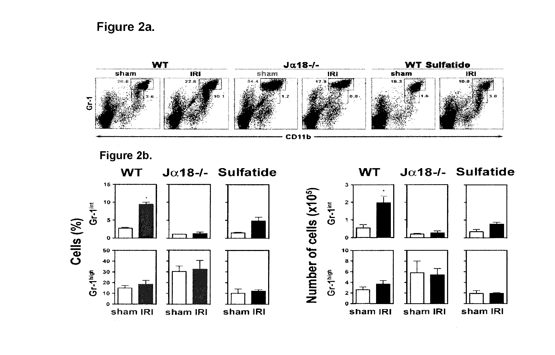
[0118]The hepatic ischemia and reperfusion injury model was established as described in Shen X D, Ke B, Zhai Y, et al., CD154-CD40 T-cell costimulation pathway is required in the mechanism of hepatic ischemia / reperfusion injury, and its blockade facilitates and depends on heme oxygenase-1 mediated cytoprotection. Transplantation 2002, 74:315-9, incorporated herein in its entirety, with few modifications.
[0119]WT mice and Jα18− / − mice, which lack type I NKT cells but have normal levels of type II NKT cells, (3-5 mice / group) or WT mice (2-3 mice / group) treated 3 hours prior with 20 μg sulfatide / mouse were anesthetized by intraperitoneal injection of 60 mg / kg sodium pentobarbital. After midline laparotomy, an atraumatic clip was applied to the hepatic triad (hepatic artery, portal vein, bile duct) of the 3 cephalad liver lobes. The caudal lobes re...
PUM
| Property | Measurement | Unit |
|---|---|---|
| concentrations | aaaaa | aaaaa |
| concentrations | aaaaa | aaaaa |
| concentrations | aaaaa | aaaaa |
Abstract
Description
Claims
Application Information