Implants for Soft and Hard Tissue Regeneration

a soft and hard tissue regeneration and implant technology, applied in the field of implants, can solve the problems of difficult to distinguish a single initiating change in either tissue, further degeneration, etc., and achieve the effect of enhancing osteointegration

Active Publication Date: 2014-07-17
TEPHA INC
View PDF23 Cites 10 Cited by
  • Summary
  • Abstract
  • Description
  • Claims
  • Application Information

AI Technical Summary

Benefits of technology

[0010]It is still another object of the invention to provide methods for manufacturing biocompatible implants

Problems solved by technology

Nutrition to these tissues is provided by the bone marrow and disruption of the tissues may also compromise nutrition causing further degeneration.
It appears

Method used


Image

  • Implants for Soft and Hard Tissue Regeneration
  • Implants for Soft and Hard Tissue Regeneration
  • Implants for Soft and Hard Tissue Regeneration

Examples


example 1

Manufacture of Knitted Braid Mat

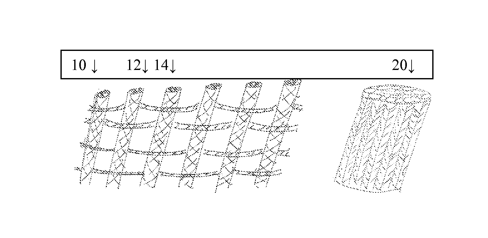
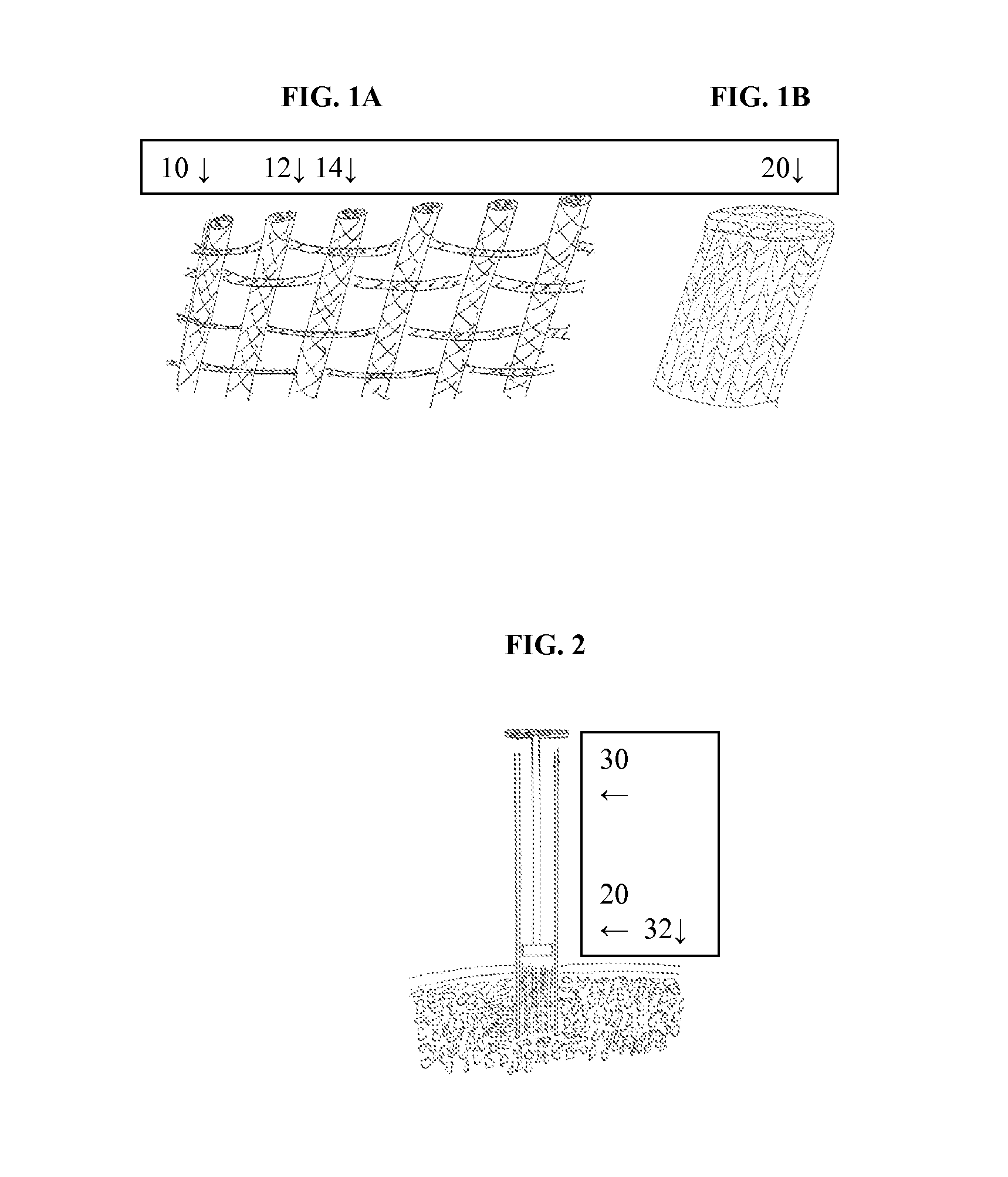
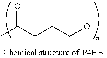
[0073]P4HB (Mw 350 kDa) was melt spun and drawn to produce a monofilament fiber 0.13-0.18 mm in diameter with a tensile strength of 1.8 kg and break elongation of 30%. Sixteen spools of the monofilament were then braided to form a 20 ppi braid that was approximately 1 mm in diameter. The braid was then knitted using a weft insertion knitting machine with a 2 dpf, 120 denier multifilament P4HB warp fiber with an average strength of 6 gm / denier and break elongation of 30% to make a 25 mm wide mat with the braids evenly spaced. There were 6 warp knits, and 20 weft insertions per inch.

[0074]The mat was then cut into pieces 1 cm wide and 35 mm long. These were then rolled to form a cylindrical implant approximately 7 mm in diameter and 1 cm tall.

example 2

Ceramic Coating of the Implant

[0075]A solution preparation formula (for a total aqueous volume of 1 L) is given in Table 1 for preparing implants with ceramic coatings. The first five chemicals given in Table 1 were added, in the order written, to 950 ml of de-ionized water in a glass beaker of 2 L capacity. Before the addition of the next chemical, the previous one was completely dissolved. After all the reagents were dissolved at room temperature, the solution was made up to 1 L by adding additional water and then filtered with a Nalgene MF75 (90 mm) filter unit for sterilization. This stable sterile stock solution of pH value of 4.35-4.40 could be stored at room temperature in a capped bottle for several months without precipitation.

TABLE 1Stock solution preparation recipe, for a total volume of 1 LConcen-AmounttrationSupplier,ReagentOrder(g)(mM)Cat # & Lot#NaCl158.44301000EMD; 7710; 0613B62KCl20.37285Mallinckrodt; 6838;6838Y21610CaCl2•2H2O33.675425EMD; CX0134-1;K92681900MgCl2•6H...

example 3

Implantation Study

[0081]Materials and Methods

[0082]A total of six (6) adult goats were enrolled into a study. Two full thickness osteochondral defects, 7.0 mm in diameter by 10.0 mm deep, were created in the middle lateral trochlear sulcus and in the medial femoral condyle of one stifle of each goat. The medial femoral condyle site was exposed to full weight bearing following cast removal while the middle trochlear sulcus will be a protected, limited loaded site. The devices implanted were either ceramic coated or non-coated, and were prepared according to Examples 1 and 2 above. Three goats had ceramic coated implants and three goats had non-coated implants. At the time of implantation blood immediately wicked up into the implants demonstrating the effectiveness of the design. Postoperatively for all animals, the operated rear limb was placed into a modified-Thomas splint for 14 days.

[0083]On day 84 (12 weeks) after surgery, animals were humanely euthanized. The left and right femo...


PUM

PropertyMeasurementUnit
Diameteraaaaaaaaaa
Diameteraaaaaaaaaa
Porosityaaaaaaaaaa

Abstract

Implants for osteo and osteochondral repair have been developed. These implants include a series of channels between the upper and lower surfaces of the implants, such that when implanted the lower surfaces are situated in an area rich in bone marrow and the channels provide a means for the bone marrow to migrate through the implant. Preferably the implants are made from resorbable polymer fibers, preferably arranged in braids that are knitted or woven together such that the braids are substantially parallel with each other. The implants may be rolled into a bundle of braids with the axis of the braids substantially parallel to the axis of the bundle, to provide channels along the axis of the bundle. A preferred embodiment includes P4HB fibers braided and knitted into a structure that is coated with a ceramic, preferably physiologic calcium phosphate.

Description

CROSS-REFERENCE TO RELATED APPLICATIONS[0001]This application claims the benefit of and priority to U.S. Provisional Application No. 61 / 752,947, filed Jan. 15, 2013.FIELD OF THE INVENTION[0002]The present invention generally relates to compositions and implants comprising resorbable polymer fibers for the regeneration of tissue. The implants can be used in many types of surgery but are most suited to use in osteo and osteochondral defect repair, as well as ligament and tendon repair.BACKGROUND OF THE INVENTION[0003]The tissues that compose and support joint surfaces are collectively called osteochondral tissues. The most superficial of these, the articular cartilage, is essential for absorbing shock and maintaining normal joint environment, and, regardless of the cause, degeneration of articular cartilage can result in irreversible osteoarthritis. While this loss of cartilage is the most noticeable affect of osteoarthritis there are also many changes to the underlying bone caused by...

Claims


Application Information

Patent Timeline
IPC IPC(8): A61L27/58D04C1/02A61L27/40D04B1/14A61F2/28A61F2/08
CPCA61L27/58A61F2/08D04C1/02A61L27/40D04B1/14A61F2/28A61L27/32A61L27/56A61L27/18A61L27/46A61L27/54A61L2300/30A61L2300/414A61L2300/606A61L2430/02A61L2430/06A61L2430/10C08L1/02C08L5/08C08L67/04
Inventor CARTER, ANDREW J.
Owner TEPHA INC
Why Eureka
Try Eureka