Imaging the heterogeneous uptake of radiolabeled molecules in single living cells

a radiotracer and living cell technology, applied in the field of cell imaging, can solve the problems of limited application of fluorescence imaging beyond cell culture imaging and shallow tissue imaging, unsuitable for studying some of the more subtle biological processes, and little is known about the biological behavior of radiotracers at the individual cell level

Inactive Publication Date: 2014-08-28
THE BOARD OF TRUSTEES OF THE LELAND STANFORD JUNIOR UNIV
View PDF11 Cites 13 Cited by
  • Summary
  • Abstract
  • Description
  • Claims
  • Application Information

AI Technical Summary

Benefits of technology

[0025]Accordingly, an aspect of the invention is to image the cellular uptake of radiotracer labeled biologically active molecules at the single-cell level. The new technique could provide a quantitative determination of biological processes in individual cells under controlled conditions. Furthermore, correlation with biological parameters obtained from fluorescenc

Problems solved by technology

However, fluorescence imaging has limited applicability beyond cell culture imaging and shallow tissue imaging due to the poor ability of the fluorescent light to penetrate biological tissue.
Fluorophores are also relatively large molecules, which makes them unsuitable for studying some of the more subtle biological processes.
Although radionuclide imaging with PET and SPECT is widely used to probe biological processes deep within tissues, little is known about the biological behavior of radiotracers at the individual cell level.
However, film preparation is not compatible with the imaging of live cells.
In addition, film autoradiography requires extremely long exposures due to poor detectio

Method used


Image

  • Imaging the heterogeneous uptake of radiolabeled molecules in single living cells
  • Imaging the heterogeneous uptake of radiolabeled molecules in single living cells
  • Imaging the heterogeneous uptake of radiolabeled molecules in single living cells

Examples


example 1

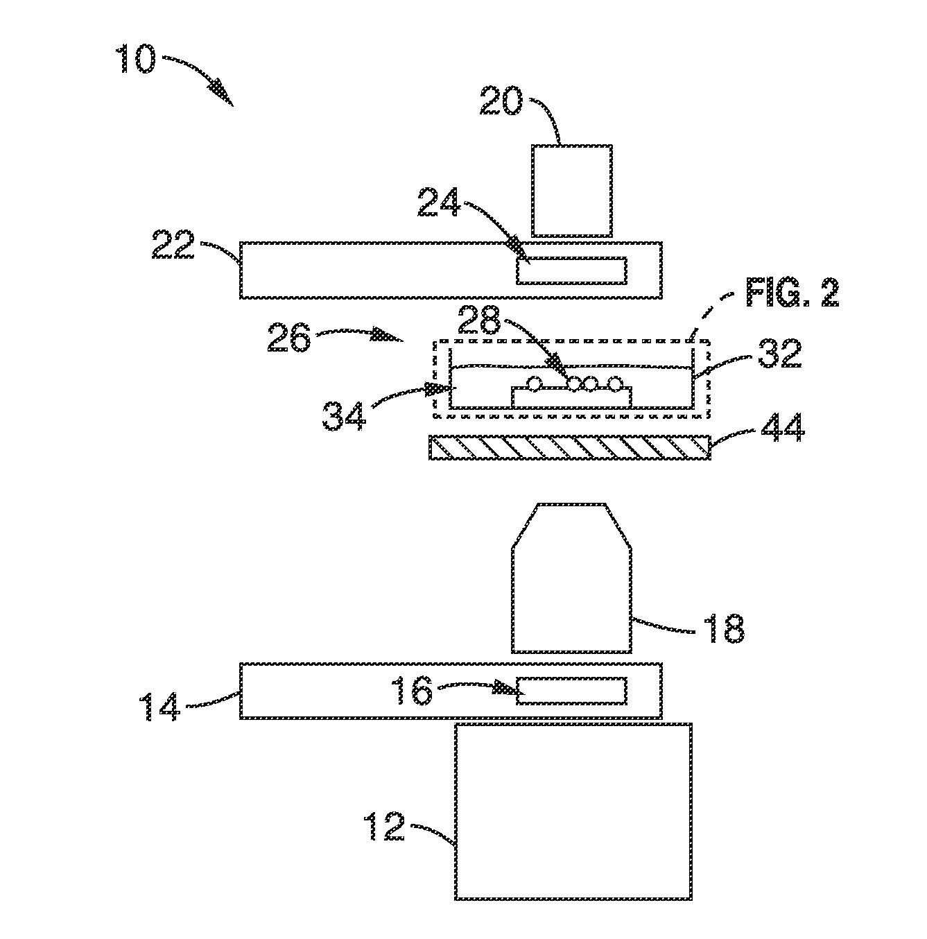
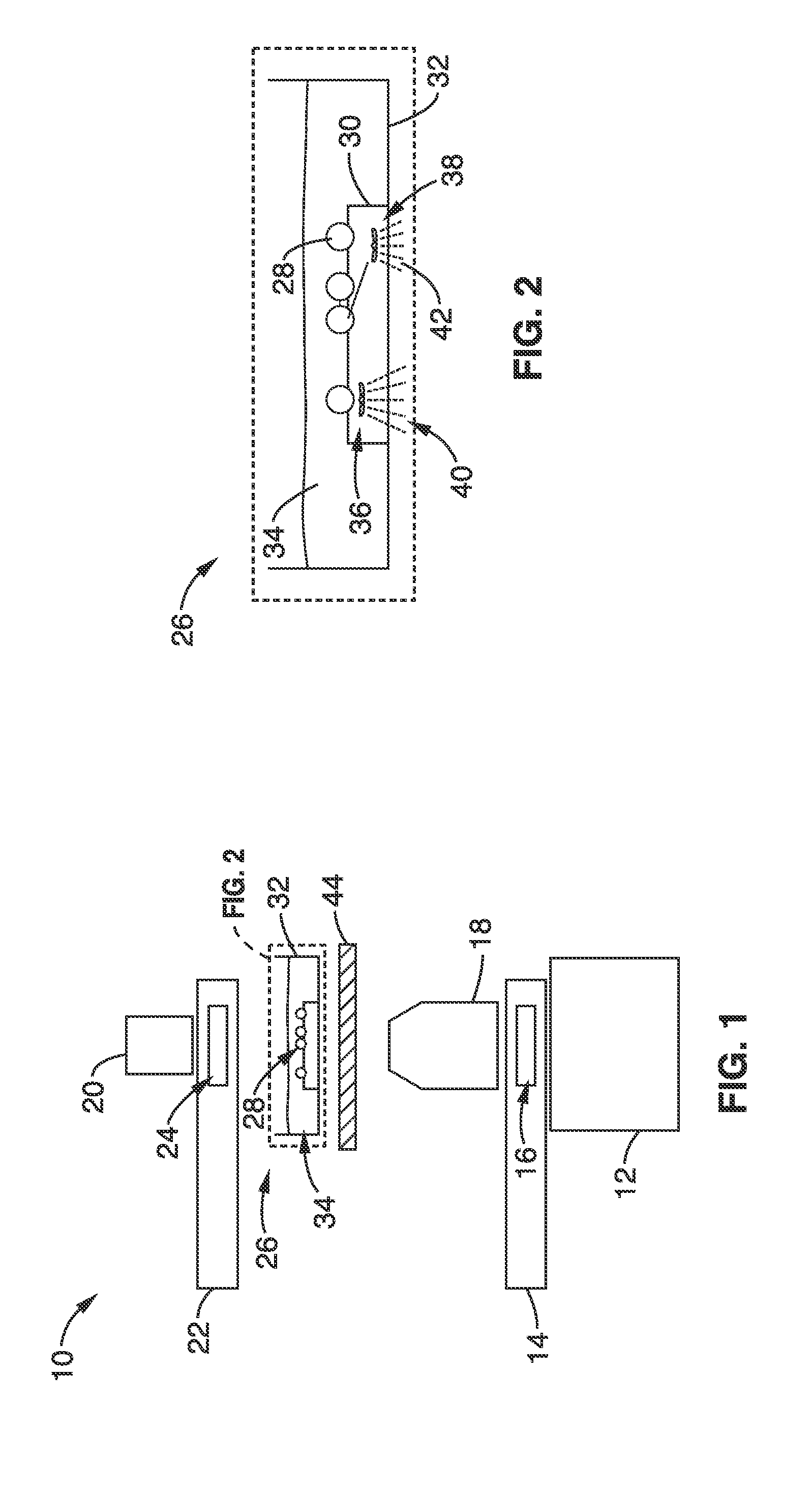
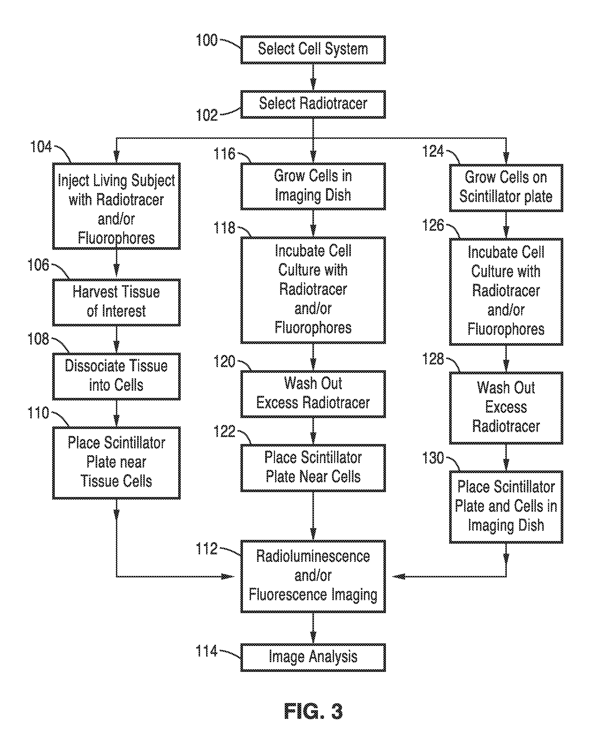
[0094]To visualize the uptake of radiotracer at the microscopic level, cells were cultured directly on a scintillating plate made of a material that converts incident beta radiation into optical photons via radioluminescence. In these experiments, scintillation plates were used with dimensions 10 mm×10 mm×0.5 mm that were made of CdWO4, a dense, high-Z, non-hygroscopic material, with both sides polished to allow for concurrent optical imaging (MTI Corp.). In one experiment, HeLa cells were seeded and cultured on the scintillating plate, immersed in cell culture medium (Dulbecco's Modified Eagle Medium containing 10% fetal calf serum) for 12-18 hours. After the cells had adhered to the surface of the scintillator plate and divided adequately, they were fasted for two hours in glucose-free cell medium and incubated for 20 minutes at 37° C. with 400 μCi of 18F-fluorodeoxyglucose (FDG). The plate, loaded with cells, was then washed thoroughly and placed in a 100 μm-thin glass-bottom mic...

example 2

[0096]To further evaluate the performance of the imaging set-up, a drop of FDG (activity≦2 μCi) was placed between the imaging dish and the scintillating plate. Upon evaporation of the water solvent, FDG precipitated into small solid deposits that could be seen on both brightfield and radioluminescence images. The size of these deposits was measured by fitting them with 2-D Gaussian functions.

[0097]Good correlation (ρ=−0.79) was observed between brightfield and radioluminescence images. A magnified view reveals weaker features present in both imaging modes. A particularly intense deposit was selected and measured by fitting with an isotropic 2-D Gaussian. The full width at half maximum (FWHM) was found to be 5.0 μm for the brightfield image and 6.9 μm for the radioluminescence image, yielding an estimated system resolution of 4.7 μm (FWHM).

example 3

[0098]In another experiment, the sensitivity of the system was evaluated by imaging the decay of 2.6 μCi of FDG over 24 hours. A small drop of FDG was mixed with glycerol and placed on an imaging dish. The mixture was then heated for several hours to allow the water solvent to evaporate, thereby ensuring that no water would evaporate during the acquisition. It was verified that the mixture was uniformly spread between the scintillator plate and the imaging dish. The mixture was imaged every 31 minutes, in 30 minute-long frames, with an EM gain of 251 / 1200. Within a large (370000 pixels) region of interest, pixel values were expressed as a percentage of their value in the first frame. The mean pixel value and the range of pixel-to-pixel fluctuations—defined by one standard deviation—were computed for each frame. For a quantitative assessment of radioluminescence intensity, flat-field and dark image corrections were applied to all acquired images.

[0099]Initially, 69.7 fCi of FDG per C...


PUM

PropertyMeasurementUnit
Thicknessaaaaaaaaaa
Thicknessaaaaaaaaaa
Thicknessaaaaaaaaaa

Abstract

A radioluminescence microscopy system and method for imaging the distribution of radiolabeled molecules in live cell cultures and tissue sections. Cells are grown and incubated with radiolabeled molecules on a scintillator plate or a scintillator plate is placed adjacent to the cells after incubation. Scintillation light produced by decay of radiolabeled molecules inside, bound to, or surrounding the cells, is recorded on an imaging device. Fluorescence microscopy of the same cells with other types of molecules of interest that are labeled with different fluorophores can be conducted concurrently and the biological activity of the labeled molecules can be correlated.

Description

CROSS-REFERENCE TO RELATED APPLICATIONS[0001]This application claims priority from U.S. provisional patent application Ser. No. 61 / 494,568 filed on Jun. 8, 2011, incorporated herein by reference in its entirety.STATEMENT REGARDING FEDERALLY SPONSORED RESEARCH OR DEVELOPMENT[0002]This invention was made with Government support under contract W81XWH-11-1-0087 awarded by the Department of Defense, under contract W81XWH-11-1-0070 awarded by the Department of Defense, under contract W81XWH-10-1-0506 awarded by the Department of Defense. The Government has certain rights in this invention.INCORPORATION-BY-REFERENCE OF MATERIAL SUBMITTED ON A COMPACT DISC[0003]Not ApplicableBACKGROUND OF THE INVENTION[0004]1. Field of the Invention[0005]This invention pertains generally to cellular imaging, and more particularly to imaging radiotracer uptake in single living cells.[0006]2. Description of Related Art[0007]The visualization, characterization, and quantification of biological processes at cel...

Claims


Application Information

Patent Timeline
IPC IPC(8): G01N33/50
CPCG01N33/5005G01N21/6458
Inventor XING, LEICARPENTER, COLINOLCOTT, PETERPRATX, GUILLEMSUN, CONROY
Owner THE BOARD OF TRUSTEES OF THE LELAND STANFORD JUNIOR UNIV
Why Eureka
Try Eureka