

In vivo label-free histology by photoacoustic microscopy of cell nuclei
a cell nucleus and photoacoustic microscopy technology, applied in the field of cell imaging, can solve the problems of complicated histological processes, low contrast in cell nuclei imaging, and inability to achieve label-free imaging techniques with high contrast and spatial resolution
- Summary
- Abstract
- Description
- Claims
- Application Information
AI Technical Summary
Benefits of technology
Problems solved by technology
Method used
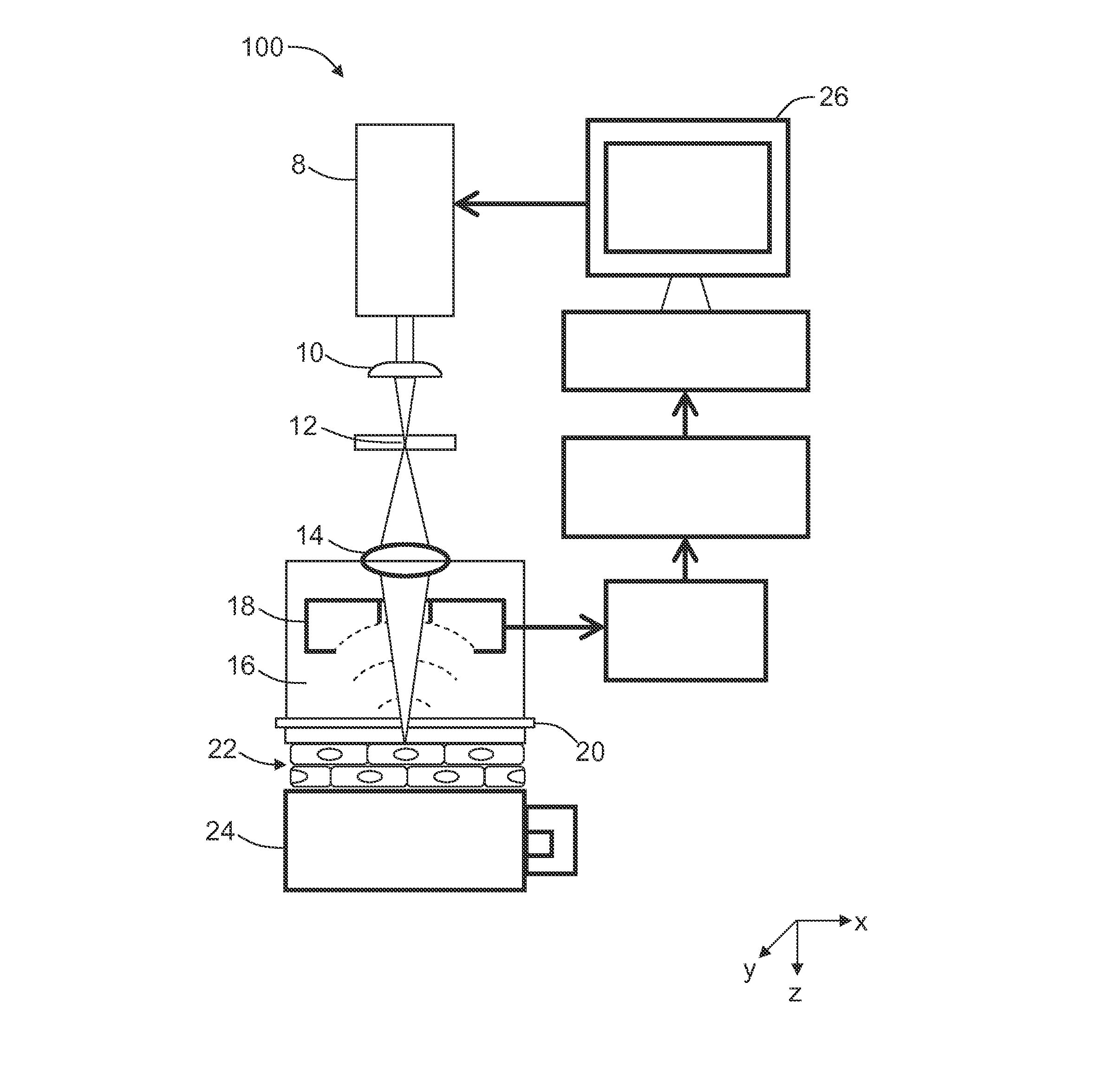
Image

Examples
example 1
[0064]In this Example, spatial resolution of the UV-PAM imaging apparatus was measured.
[0065]Specifically, single submicrometer beads were imaged to measure the spatial resolution of the UV-PAM imaging apparatus. The beads were black polystyrene microspheres 0.2 μm in diameter (Polysciences), immobilized by adsorption on a quartz slide (Chemglass Life Sciences). Bead images were acquired by scanning with a 0.31 μm step size. The lateral FWHM was measured by fitting the Airy pattern to the amplitude profiles of the horizontal cross-sectional images of the single beads and the axial FWHM was measured by Gaussian fitting to the axial amplitude profiles of bead images. By fitting the images of 15 beads, the lateral FWHM was found to be 0.70±0.04 um (mean±standard error) and the axial FWHM to be 28.5±0.8 μm.
example 2
[0066]In this Example, images of cross sections of mouse small intestine obtained using UV-PAM were compared with images of the same tissue after hematoxylin and eosin histological staining.
[0067]Specifically, small intestine was excised from a sacrificed Swiss Webster mouse (Harlan Laboratories), and cut into 6-μm-thick cross sections by a cryostat (CM1850; Leica Microsystems). A UV-PAM image of the cross section of the small intestine was acquired by scanning the tissue section with a 0.62 μm step size (FIG. 3A). After the scanning, the section slide was stained by hematoxylin and eosin (Sigma-Aldrich). Hematoxylin stains the cell nucleus blue, and eosin stains the cytoplasm pink. Next, optical micrographs of the intestine section were obtained using a microscope with a 20× objective (0.45 NA, Nikon).
[0068]FIG. 3B shows the cell nuclei as dark spots, as expected in histology (in a color image of a hematoxylin-eosin stained tissue, the cell nuclei appear as dark-blue spots). A comp...
example 3
[0069]In this Example, cell nuclei in the epithelium of a mouse lip and in the intestinal villi of a mouse small intestine were imaged using UV-PAM ex vivo.
[0070]Fresh specimens were taken from adult Swiss Webster mice and immersed in phosphate buffer solution (PBS, Sigma-Aldrich). After the small intestine was cut longitudinally and unfolded into a sheet, the specimens were mounted on the scanning stage with their inner surfaces in contact with the image window through PBS. The nuclei of the epithelial cells in the mouse lip were imaged by scanning with a 1.25 μm step size for 2.6 min. As shown in FIG. 4A, the image shows a relatively homogeneous distribution of cell nuclei, each approximately 6 μm diameter. The distance between the centers of neighboring cell nuclei ranged from 16 μm to 39 μm, suggesting that the stratified squamous epithelium on the lip was composed of cells with a lateral size of the same range. The nuclei of the epithelial cells on the mouse small intestine wer...
PUM
 Abstract
Description
Claims
Application Information