Photoacoustic microscope and method for monitoring breaking of microvesicles in biological tissue

A photoacoustic microscope and microscope system technology, applied in the field of medicine and medical equipment, can solve the problems of low image resolution, inability to effectively observe changes in tumor vessel diameter, unfavorable long-term monitoring, etc., and achieve low price and easy application and promotion , Ease of use

Pending Publication Date: 2017-07-14
XIAMEN UNIV
View PDF6 Cites 11 Cited by
  • Summary
  • Abstract
  • Description
  • Claims
  • Application Information

AI Technical Summary

Problems solved by technology

The traditional optical microscopic imaging method can observe the blood vessels around the tumor, but it requires tissue sections, which is invasive and not conducive to long-term monitoring; and the use of ultrasound imaging can be used for non-invasive imaging of tumor tissue, but its image resolution is low Cannot effectively observe the change of tumor vessel diameter

Method used


Image

  • Photoacoustic microscope and method for monitoring breaking of microvesicles in biological tissue
  • Photoacoustic microscope and method for monitoring breaking of microvesicles in biological tissue
  • Photoacoustic microscope and method for monitoring breaking of microvesicles in biological tissue

Examples


Embodiment Construction

[0026] The present invention will be further described below in conjunction with accompanying drawing:

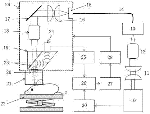
[0027] A photoacoustic microscope, including a microscope system, a signal acquisition control system, and a drive system. The signal acquisition control system controls the drive system and collects the information of the microscope system, and stores and analyzes the collected data; the drive system adjusts the position of the corresponding components , to achieve the adjustment of the microscope.

[0028] The microscope system includes a pulsed laser 10, the pulsed laser 10 generates a short pulsed laser, and the first beam expander combination 11, the first microscope objective lens 12, a fiber coupler 13, and a single-mode optical fiber are sequentially arranged along the advancing direction of the short pulsed laser. 14. Collimator 15, second beam expander combination 16, mirror 17, second microscope objective lens 18, beam splitting element 19, acoustic lens 20, wate...


PUM


Abstract

The invention discloses a photoacoustic microscope. The photoacoustic microscope comprises a microscope system, a signal collection control system and a driving system. The invention further discloses a method for monitoring breaking of microvesicles in biological tissue with the use of the photoacoustic microscope. The photoacoustic imaging is combined with features of high contrast ratio of optical imaging and high resolution ratio of acoustic imaging. Due to low scattering properties of acoustic signal transmission in tissue, deeper penetration degree is obtained compared with a conventional optical imaging method. There is no need to slicing tissue and the method is non-invasive so that long-term monitoring is achieved. Photoacoustic microscope imaging equipment helps achieve imaging of multiple anatomical positions.

Description

technical field [0001] The invention belongs to the technical field of medicine and medical equipment, and in particular relates to a photoacoustic microscope and a method for monitoring the explosion of microbubbles in biological tissues. Background technique [0002] For a long time, microbubbles have been used as an efficient, cheap, and biocompatible diagnostic contrast agent for ultrasound imaging. Because microbubbles have high acoustic scattering properties and nonlinear effects on incident ultrasound, they have many clinical applications, such as the assessment of coronary artery disease, angiogenesis, inflammation and tumor formation, etc. In general, these clinical applications only use the acoustic impedance characteristics of microbubbles for ultrasound imaging, while ignoring other utilization values ​​of this unique structure of microbubbles. [0003] The main way to observe the effect of tumor treatment is to detect it by means of imaging. The traditional op...

Claims


Application Information

Patent Timeline
Patent Type & Authority Applications(China)
IPC IPC(8): A61B5/00A61B5/02A61B8/08
CPCA61B5/0095A61B5/02007A61B8/0891
Inventor 聂立铭吴志友李宏辉赵庆亮
Owner XIAMEN UNIV
Why Eureka
Try Eureka