Method for the quantitative assessment of global and specific DNA repair capacities of at least one biological medium, and the applications therefor

a biological medium and quantitative assessment technology, applied in the field of quantitative assessment of the overall and specific dna repair capacity of a biological medium, can solve the problems of complex repair system, large negative biological consequences of lesions that are taken care of by the ber system, and the formation of base or sugar lesions, etc., to achieve miniaturization, rapid, precise and effective manner

Active Publication Date: 2015-01-29
COMMISSARIAT A LENERGIE ATOMIQUE ET AUX ENERGIES ALTERNATIVES
View PDF1 Cites 1 Cited by
  • Summary
  • Abstract
  • Description
  • Claims
  • Application Information

AI Technical Summary

Benefits of technology

The patent text describes a method to quickly and accurately assess the ability of biological materials to repair DNA damage. This method involves preparing plasmids with specific DNA lesions, depositing them onto a solid support, and then adding repair solutions to study the enzyme activities responsible for DNA repair. The results are measured by measuring the signal produced by a label incorporated into the DNA during the repair reaction. The method is reliable, precise, and can be performed quickly and easily, without the need for control repair solutions. Its technical effects include improved characterization and quantification of DNA repair enzyme activities in biological extracts.

Problems solved by technology

DNA is continuously subjected to endogenous or exogenous attacks that result in the formation of base or sugar lesions.
It should, however, be noted that repair systems, especially in eukaryotes, are very complex and many variants of this simplistic configuration exist (overall repair, repair associated with the DNA transcription, repair associated with DNA replication, etc.).
More than about forty different lesions that are taken care of by the BER system can have considerable and negative biological consequences if they are not repaired.
In particular, it appears that the best repair yield is obtained with whole cell extracts of the type of those used for in vitro transcriptions, whereas cytosolic extracts of the type of those used for promoting plasmid replication from an SV40 origin, and also other crude cell extracts, exhibit nuclease activity, which does not allow correct interpretation of the repair.
The assay from Wood et al. requires the use of radioactive labels, which imposes restrictions that limit the implementation of this method in routine assays; in addition, this assay does not have sufficient qualities of simplicity and of practicality for it to be used routinely.
Cells originating from patients suffering from this disease are very sensitive to ultraviolet radiation and exhibit DNA repair deficiencies.
With this assay, it is therefore impossible to differentiate between breakage repair and the repair of other lesions.
Breakage repair can even mask the repair of other lesions and interfere with the signals attributed to the repair of other DNA lesions.
It is thus limited to characterizing the step of excision of lesions that may be introduced into synthetic oligonucleotides.
Furthermore, although it provides considerable information regarding excision activities, it is not suitable for and does not describe a precise quantification of the enzyme activities for excision / resynthesis of DNA.
The same is true in most of the other variants of the method by Wood et al., described above.Since the assays are relatively laborious to carry out and require the availability of large amounts of biological material, experimenters limit the number of substrates used and the number of biological extracts tested.The lesions introduced into the plasmids are neither measured nor quantified.
The information obtained is very partial and insufficient to precisely define and characterize the repair capacities of a given biological medium.

Method used


Image

  • Method for the quantitative assessment of global and specific DNA repair capacities of at least one biological medium, and the applications therefor
  • Method for the quantitative assessment of global and specific DNA repair capacities of at least one biological medium, and the applications therefor
  • Method for the quantitative assessment of global and specific DNA repair capacities of at least one biological medium, and the applications therefor

Examples


example 1

Preparation of the Range of Plasmids and Assaying of the Lesions

[0108]The plasmid pBluescript II is produced by transformation of XLI-Blue MRF supercompetent cells from Stratagene, according to the protocol provided by Stratagene.

[0109]The plasmid is then purified using the Qiagen plasmid midi kit, according to the recommended protocol.

Additional Purification of the Plasmid

[0110]The plasmid is loaded onto 10 ml of 5-20% sucrose gradient in a 25 mM Tris HCl buffer, pH 7.5; 1M NaCl; 5 mM EDTA and centrifuged in a Beckman ultracentrifuge using an SW-41 rotor, at 4° C. and at 25 000 rpm for 18 hours. 1 ml fractions are then carefully taken and analyzed on an agarose gel. Only the fractions containing at least 90% of coiled form of the plasmid are kept. The plasmid is precipitated with ethanol and dissolved in PBS.

Plasmid Modifications

[0111]UVC Irradiation—Formation of Cyclobutane Pyrimidine Dimers (CPDs) and of (6-4) Photoproducts

[0112]The plasmid, diluted to 20 g / ml in a PBS, is irradi...

example 2

Implementation of the Method According to the Invention with the Range of Plasmids Prepared in Example 1

[0127]Deposition of the Plasmids onto a Support

[0128]The plasmids are diluted to 20 μg / ml in PBS. 500-picoliter deposits are made, using a GESIM robot, on commercial poly-L-lysine-coated glass slides (VWR). The slides are conserved at 4° C.

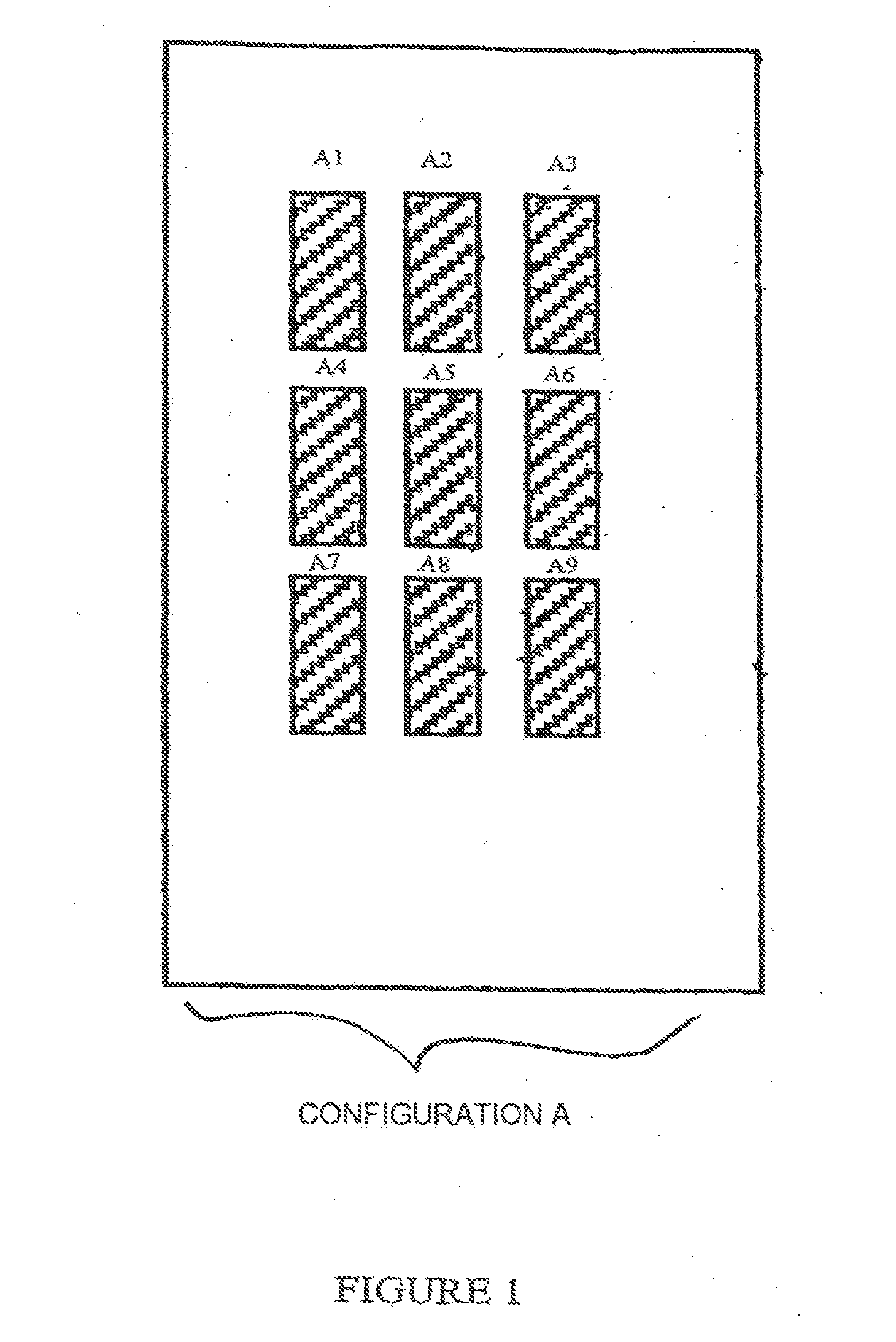
[0129]Each slide (support S) comprises 9 identical zones (A1 to A9) arranged according to configuration A in FIG. 1.

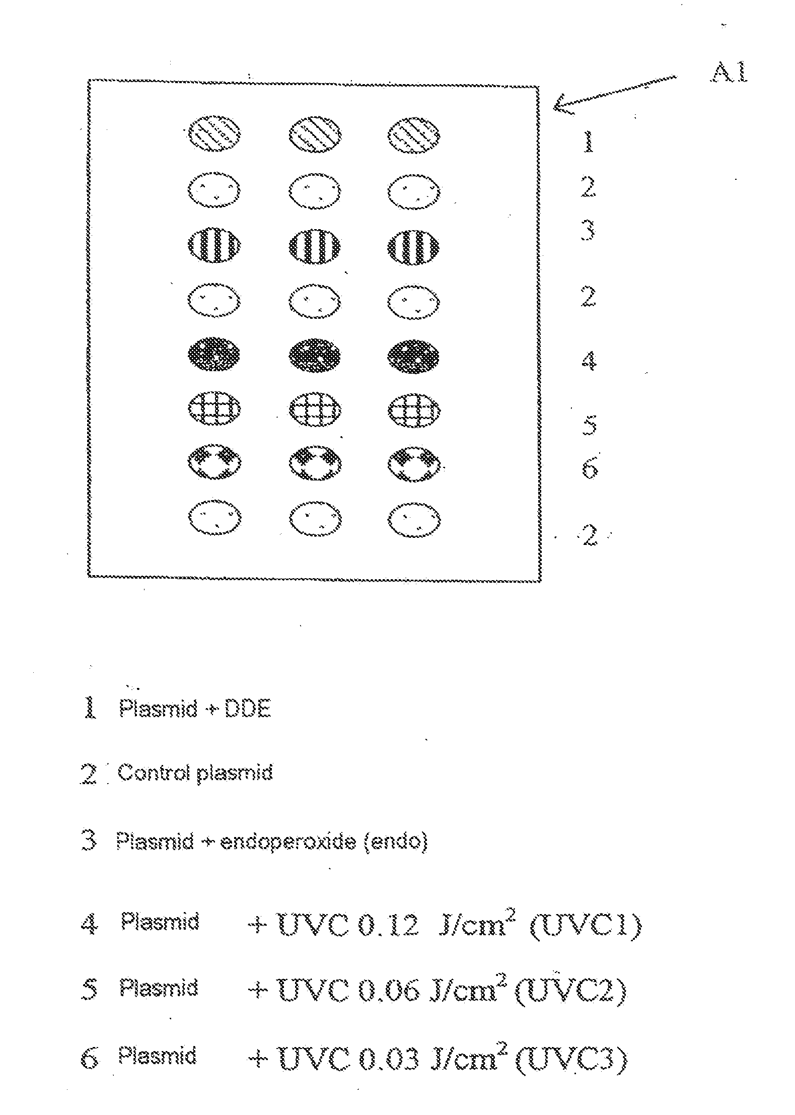
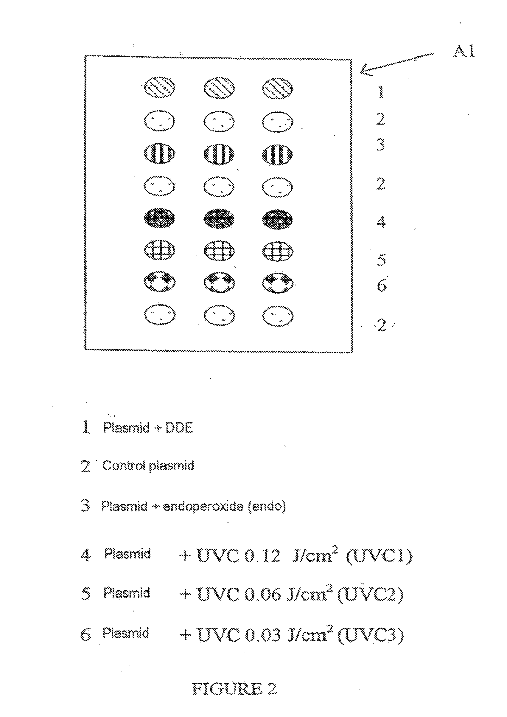
[0130]In each zone, the range of plasmids is deposited in accordance with FIG. 2, which illustrates, for example, zone A1.

[0131]Each zone makes it possible to test a different biological medium.

Repair Reaction

[0132]A solution is prepared containing the biological medium or extract to be tested; for 5 μl of solution, the composition is as follows:

extract0.5μl5x repair buffer1μlCY5-dUTP (Amersham Pharmacia Biotech)0.2μl(0.1 nmol / μl)2M KCl0.2μlATP (Roche - 100 mM)0.1μl

[0133]The volume is made up to 5 μl with H2O2.

Composition of the 5×...


PUM

PropertyMeasurementUnit
concentrationaaaaaaaaaa
volumesaaaaaaaaaa

Abstract

A method for quantitative assessment of base excision repair (BER) and nucleotide excision repair (NER) DNA repair capacities of at least one cellular extract, which method entails: a) preparing a range of plasmids with at least one physical or chemical treatment or both and recovering a supercoiled fraction of each of said plasmids, b) characterizing the lesions present on each of the plasmids of the range of plasmids; c) depositing the plasmids of the range of plasmids, and at least one supercoiled control plasmid without lesions onto a single solid support, according to a pre-established configuration A, so as to form a functionalized support divided into different zones A1 to Ax, corresponding to an integer equal to the number of cellular extracts to be simultaneously tested, each zone containing supercoiled plasmids treated with at least one physical or chemical treatment that induces a lesion acted upon by BER mechanisms, and at least one other zone containing supercoiled plasmids treated with at least one physical or chemical treatment that induces a lesion acted upon by NER mechanisms; d) incubating the functionalized support obtained in step c) with various repair solutions, each of which contains at least one cellular extract from a subject, and labeled nucleotide phosphates; e) washing the functionalized support at least once; f) directly or indirectly measuring the signal produced by the labeled nucleotide triphosphate incorporated into the DNA during the repair reaction in step d), in each of the different and pre-established zones A.sub.1 to A.sub.x; g) recording and quantifying the signal corresponding to each deposit of plasmid in each zone A.sub.1 to A.sub.x; and h) determining the ratio of the signals of the plasmids containing the lesions relative to the control plasmid jointly deposited.

Description

TECHNICAL FIELD[0001]The present invention relates to a method for the quantitative assessment of the overall and specific DNA repair capacities of a biological medium, by assessing the excision / resynthesis capacities of said medium, and also to the applications thereof.BACKGROUND OF THE INVENTION[0002]DNA is continuously subjected to endogenous or exogenous attacks that result in the formation of base or sugar lesions.[0003]The lesions include:[0004]lesions of purine or pyrimidine bases: oxidative lesions induced by the cellular metabolism and by photosensitization; lesions through the formation of chemical adducts, which result from the harmful action of many genotoxic agents, such as polycyclic hydrocarbons, contained in combustion products; lesions through the formation of metheno-bases or of etheno-bases;[0005]lesions of the structure of the DNA double helix: formation of intrastrand (between two adjacent bases of the same strand) or interstrand (between two bases located on th...

Claims


Application Information

Patent Timeline
Patent Type & Authority Applications(United States)
IPC IPC(8): C12Q1/68
CPCC12Q1/68
Inventor SAUVAIGO, SYLVIE
Owner COMMISSARIAT A LENERGIE ATOMIQUE ET AUX ENERGIES ALTERNATIVES
Why Eureka
Try Eureka