Cell Suspension and Use Thereof

a technology of cell suspension and cell suspension, which is applied in the field of cell suspension and use thereof, can solve the problems that the clinicians would otherwise have limited or no access to such autologous epithelial cells, and achieve the effects of preventing clogging of the applicator, and reducing the risk of infection

Inactive Publication Date: 2015-03-19
AVITA MEDICAL LTD
View PDF7 Cites 9 Cited by
  • Summary
  • Abstract
  • Description
  • Claims
  • Application Information

AI Technical Summary

Benefits of technology

[0052]As shown in FIG. 1, skin epidermis contains keratinocytes, melanocytes, Langerhans' cells and Merkel cells. The epidermis forms the surface of the skin. It is made up of several layers of cells called keratinocytes. Keratinocytes in the basal or germinative layer or stratum (also known as keratinocyte basal cells or basal keratinocytes) undergo mitosis and proliferate. Keratinocytes undergoing differentiation progressively move to stratum spinosum, where they become polyhedral in shape and begin to produce tonofilaments. Keratinocytes are the most abundant cell type in the epidermis. Melanocytes locate in the basal layer of epidermis and represent every fifth to every tenth cell therein. Melanocytes produce the pigment melanin, and therefore play an important role in skin and hair follicle pigmentation. The Langerhans' cells reside suprabasally, and represent about 1% of epidermal cells.
[0053]Still referring to FIG. 1, the dermis lies underneath the epidermis and contains skin appendages: hair follicles, sebaceous (oil) glands and sweat glands. Cell types in the dermis include dermal fibroblasts which produce intercellular supportive matrix (e.g. collagens and elastin) and may have a role in wound healing, as well as mast cells, macrophages, melanocytes, Merkel cells, and cells belonging to the immune system and skin appendages.
[0054]Cells capable of reproduction can include skin stem cells and/or fibroblasts. Skin stem cells can include epidermal stem cells, melanocyte stem cells and follicular stem cells. Epidermal stem cells are found in the basal layer of the epidermis and are responsible for everyday regeneration of the different layers of the epidermis. Melanocyte stem cells are responsible for regeneration of melanocytes. Follicular stem cells ensure constant renewal of the hair follicles and can also regene

Problems solved by technology

Clinicians would otherwise have limited or n

Method used


Image

  • Cell Suspension and Use Thereof
  • Cell Suspension and Use Thereof
  • Cell Suspension and Use Thereof

Examples


example 1

Preparation of Recipient Site

[0122]To optimize the success of the epithelium-related procedure, the wound was cleaned and assessed to be of the appropriate depth. Further, blood homeostasis was established and the wound was checked for evidence of surrounding cellulitis or infection. Techniques for preparing the area included, without limitation, sharp dissection, dermabrasion or laser-resurfacing.

Donor Site Biopsy

[0123]The donor site was chosen to appropriately match the recipient site. The donor site was infiltrated with local anesthetic and adrenaline underneath the skin near the subcutaneous tissue. This allowed the donor site to be firm and aided in the taking a biopsy. The dimensions of the biopsy were determined by the size of the surface area of the recipient site to be covered. Typically, the present invention allows for a biopsy size that has an expansion ratio of about 1:10 to 1:80 (donor biopsy size: recipient site size). In one example, a biopsy size of 2 cm×2 cm was ta...

example 2

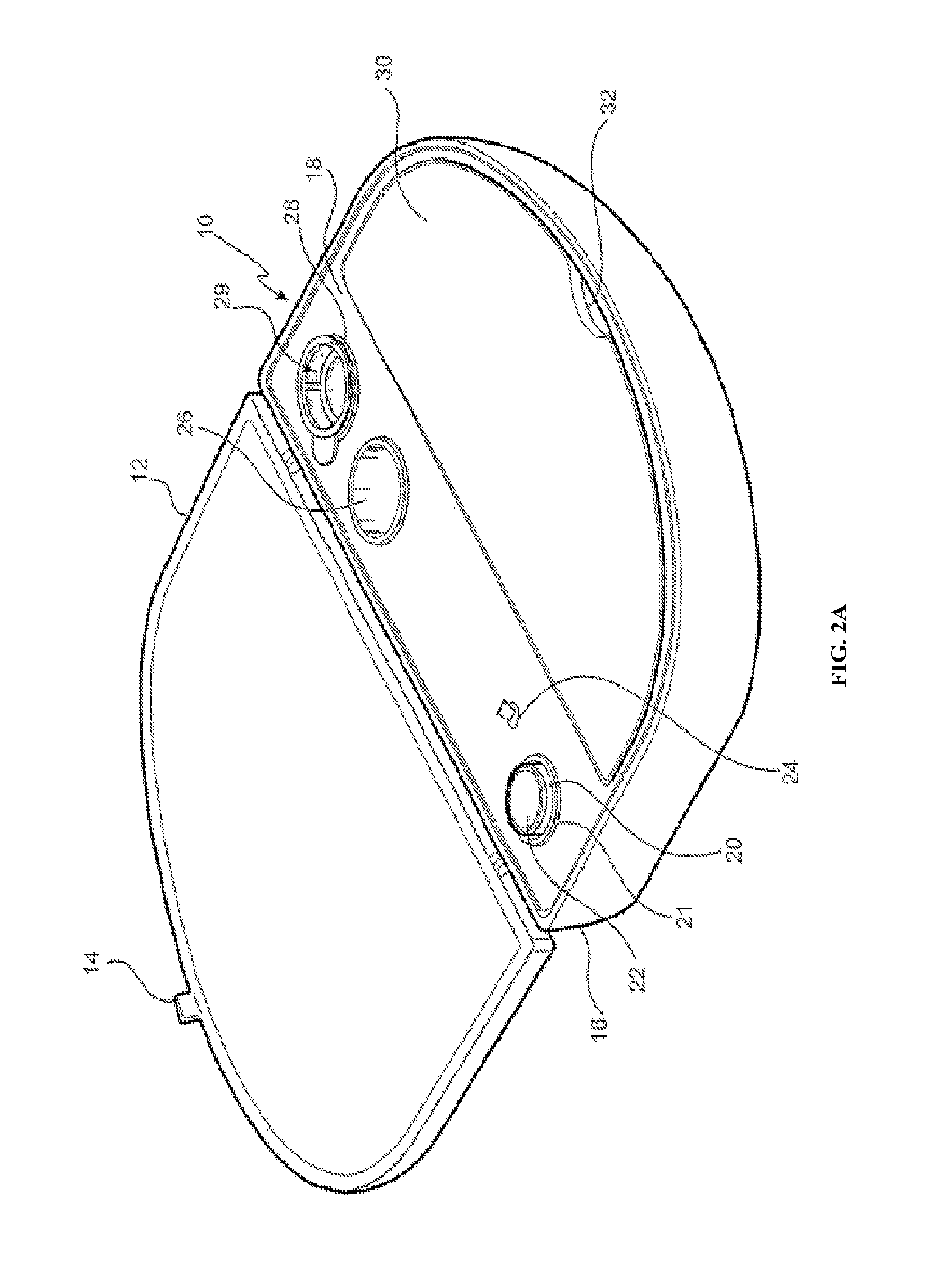
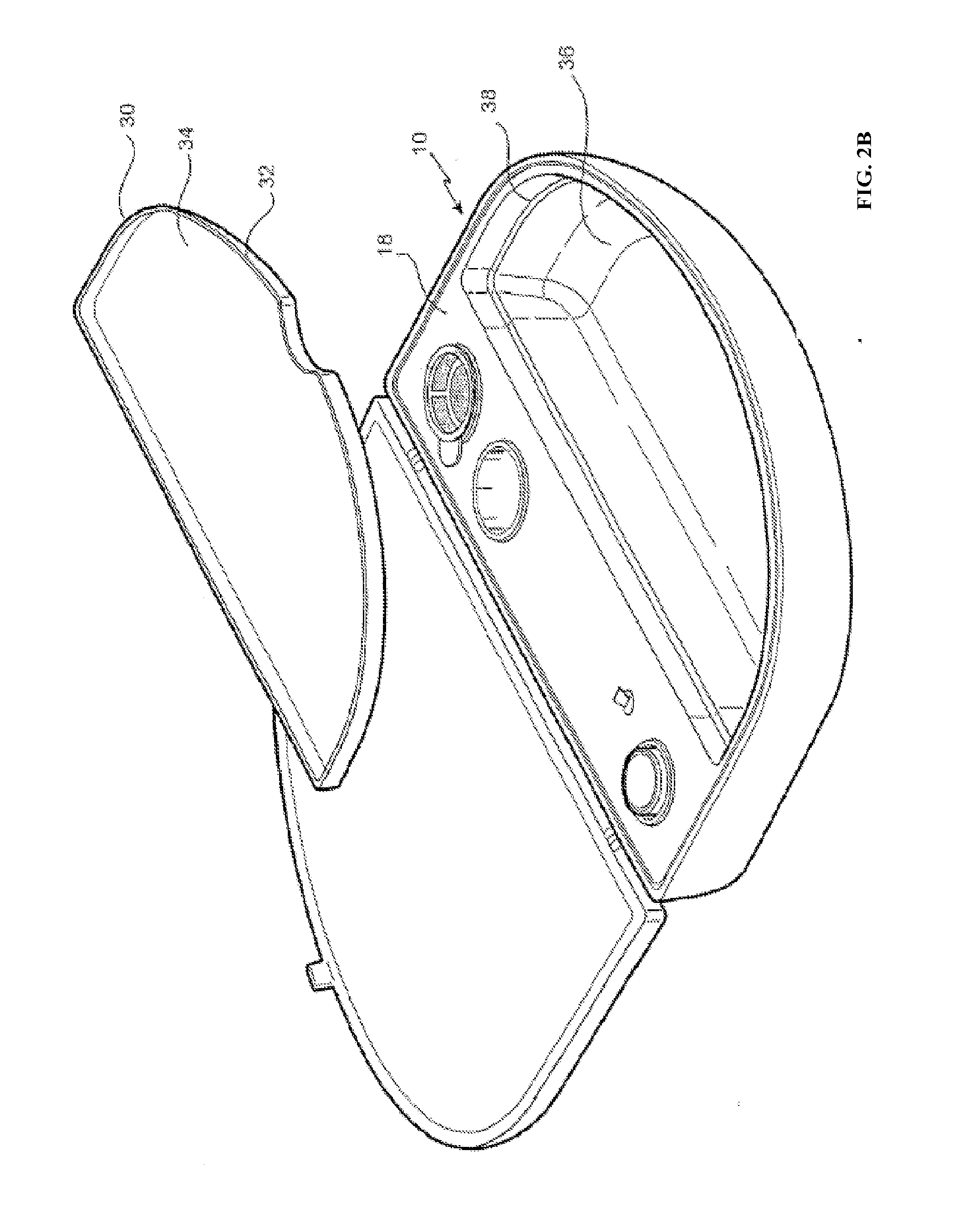
[0134]The embodiment shown in FIG. 2A is directed to a ReCell® Rapid Technique cell harvesting apparatus 10 for use in producing a transplantable cellular suspension of living tissue suitable for applying to a patient in an epithelium-related procedure.

[0135]As illustrated in FIG. 2A the apparatus includes a closure lid 12 possessing a locking mechanism 14 adapted to releasably engage a base portion 16. The locking mechanism 14 provides a means for closing the apparatus 16 when not in use. Located within the base portion 16 is a first member 18 within which there is provided an aperture 20 in which there is located a vial 22 for the enzyme. Adjacent the aperture there is provided an activation switch 24 capable of activating the heating means (not shown). The first member also provides a fluid containment well 26 and a filter recess 28. As presented in this illustration the filter 29 is shown located in the filter recess. Ordinarily the filter is an optional item included as a separ...

example 3

[0143]It was tested whether HA hydrogels increase the viability and migration of human stratum basale epidermal Keratinocytes from adult donors. Various hydrogels were tested using assays including Transwell Cell Migration, CellTiter 96® Aqueous Cell Proliferation Assay and in vitro Scratch Assay described below.

Protocol for Transwell Cell Migration

[0144]1. Maintain stock cultures of Primary Epidermal Keratinocytes (PEK), Normal, Human, Adult cells in Dermal Cell Basal Medium supplemented with Keratinocyte Growth Kit and Gentamicin-Amphotericin B Solution (Complete Media). Subculture the stock cultures of cells to 2.5×10⁴ cells / ml (estimate) and re-feed with media every 2 days or when a density of near confluence is reached.

2. To each well of a 24 well plate add 500 μl of Media. Inserts with an 8 um pore and 0.3 cm2 area will be pre-coated with test material and placed into a well of a 24 well culture dish.

Row A, Columns 1-3: blank, no cells, medium only.

Row B, Columns 1-3: baseline...


PUM


Abstract

The present invention provides for methods and devices suitable for producing a transplantable cellular suspension of living tissue suitable for promoting tissue regeneration in an epithelium-related procedure, as well as compositions produced therefrom. The cellular suspension can include viable and functioning cells at various stages of differentiation, including undifferentiated/progenitor cells and differentiated cells, as well as those in between. In certain embodiments, the cellular suspension can be subjected to a stress to induce a heat shock response therein, or be exposed to an exogenously supplied agent such as heat shock protein or a fragment thereof, hyaluronic acid, platelet-enriched plasma, and/or growth factors. The cellular suspension can be applied directly to a patient's recipient site for in vivo regeneration, or be cultured or seeded to a matrix for in vitro growth/regeneration.

Description

CROSS REFERENCE TO RELATED APPLICATIONS[0001]This application claims priority to and the benefit of U.S. provisional patent application Nos. 61 / 614,112 filed Mar. 22, 2012 and 61 / 614,115 filed Mar. 22, 2012, the disclosures of which are incorporated herein by reference in their entirety.FIELD OF THE INVENTION[0002]This invention relates to a simple, rapid and cost effective method for preparing a cell suspension, particularly a suspension comprising viable epithelial cells such as undifferentiated / progenitor cells and / or differentiated cells. Such suspension is useful for treating a patient in need of an epithelium-related procedure. Device for such preparation and use of the suspension thereof are also provided.BACKGROUND[0003]Tissue regeneration in humans is extremely limited and constitutes a major challenge to the repair of damaged organ function. Wound treatment is a typical area where tissue regeneration is required. Wounds (lacerations or openings) in mammalian tissue can res...

Claims


Application Information

Patent Timeline
IPC IPC(8): A61L15/36A61L15/42C12N5/071
CPCA61L15/36C12N5/0625A61L2430/40C12N2501/734A61L2430/34A61L15/42A61L27/60C12N2521/00C12N2523/00C12N2527/00A61K35/36A61L27/3813A61L27/3869A61L27/54A61L2300/412
Inventor QUICK, ANDREWDOLPHIN, WILLIAM
Owner AVITA MEDICAL LTD
Why Eureka
Try Eureka