Method for detecting injury to the brain

a brain injury and brain injury technology, applied in the field of brain injury detection, can solve the problems of death and disability, traumatic brain injury (tbi) is considered to be a major health problem, and tbi is particularly devastating among young adults and children, so as to improve the detection and management of secondary brain injury

Active Publication Date: 2015-05-21
TEMPLE UNIVERSITY
View PDF0 Cites 13 Cited by
  • Summary
  • Abstract
  • Description
  • Claims
  • Application Information

AI Technical Summary

Problems solved by technology

Traumatic brain injury (TBI) is considered to be a major health problem in the United States.
TBI is particularly devastating among young adults and children, making it the leading cause of death and disability in the United States.
Despite TBI's major public health issues, resources allocated for TBI research (from basic science to clinical research) at the national level have not been met with the same level of urgency.
Disruption of the BBB leaves the CNS vulnerable to neuronal damage.
Once leakage occurs, secondary events are triggered that contribute to further injury.
Because of the nature of modern warfare, military personnel engaged in conflict areas are at particular risk of experiencing a BINT event.
Furthermore, abnormal electroencephalogram (EEG) patterns can be observed when there is hyper-permeability of the BBB.
After the onset of stroke, edema formation develops and induces a rise in intracranial pressure which can lead to compression, herniation and damage of brain tissue.
Often the edema that forms worsens during the phase of reperfusion.
Many infectious diseases affecting the brain cause changes to the brain vasculature that often lead to a breach of the BBB.
Under inflammatory conditions, the normal function of the BBB is compromised due to overproduction of pro-inflammatory molecules by inflammatory cells.
Although there have been promising results, these candidate biomarkers have not proven entirely useful for the diagnosis of brain injury.
All of these candidate biomarkers have been poor predictors of long-term outcome after TBI, some lacking specificity or sensitivity.

Method used


Image

  • Method for detecting injury to the brain
  • Method for detecting injury to the brain
  • Method for detecting injury to the brain

Examples


example 1

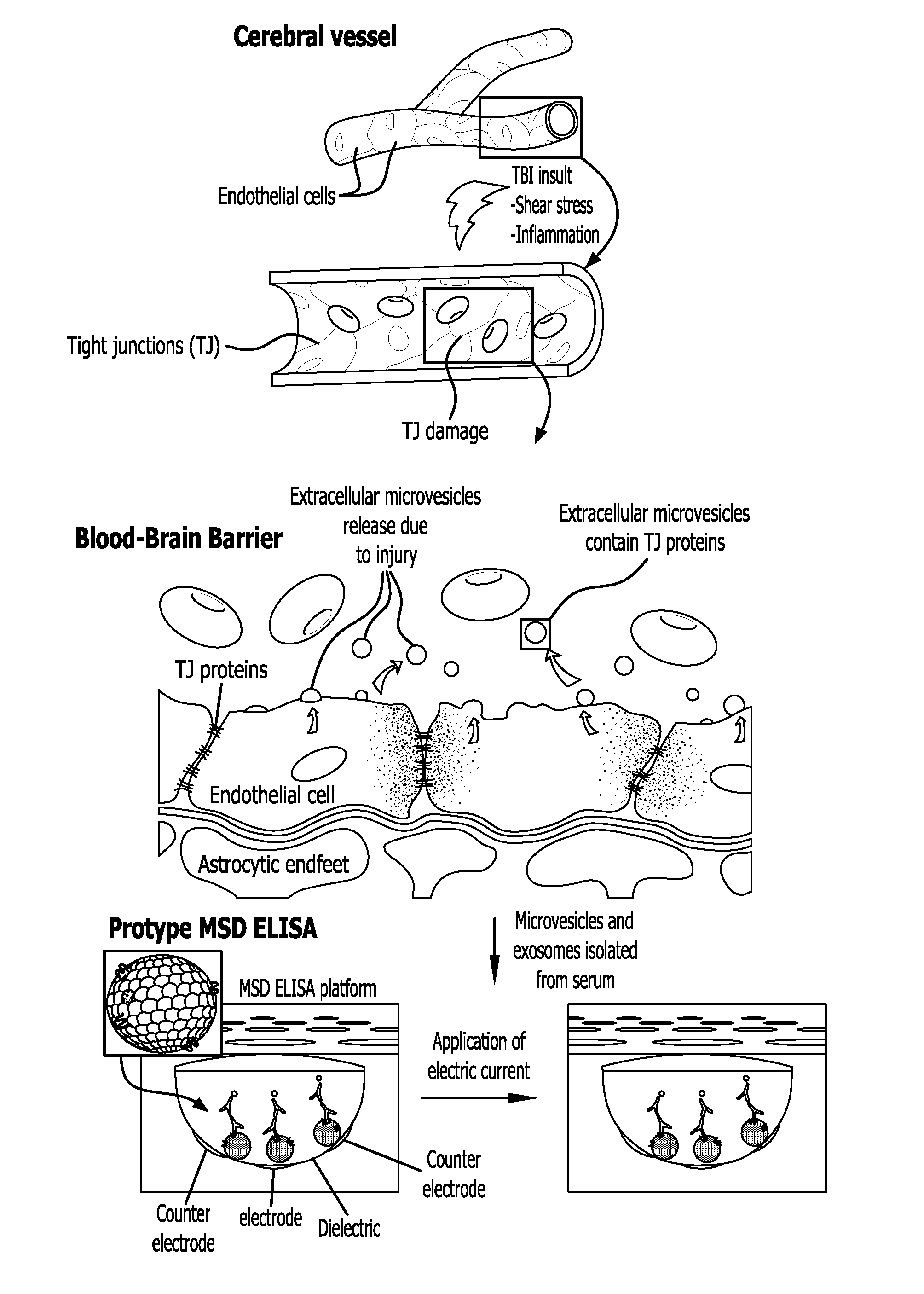
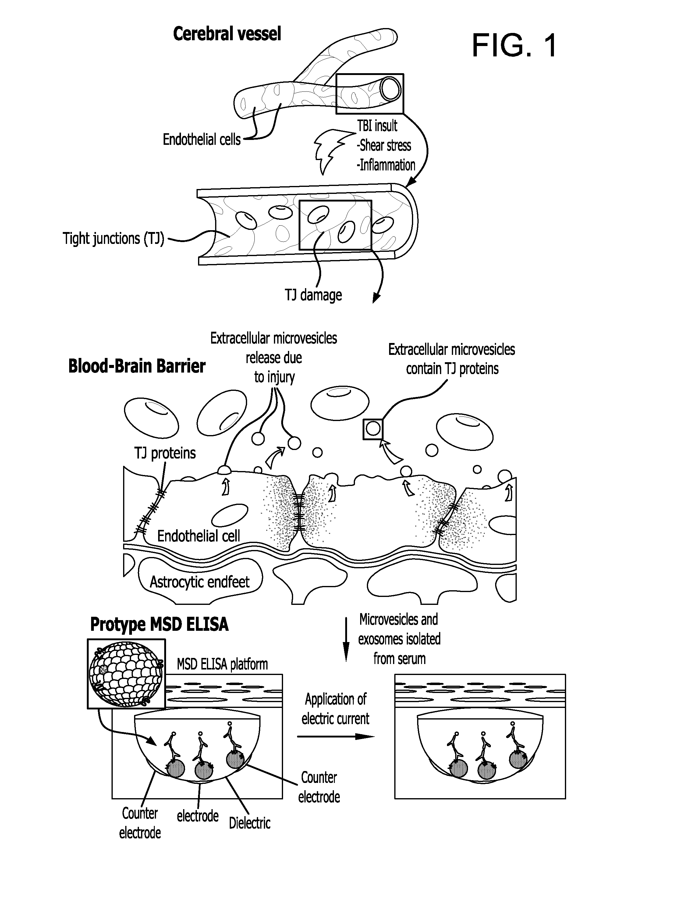
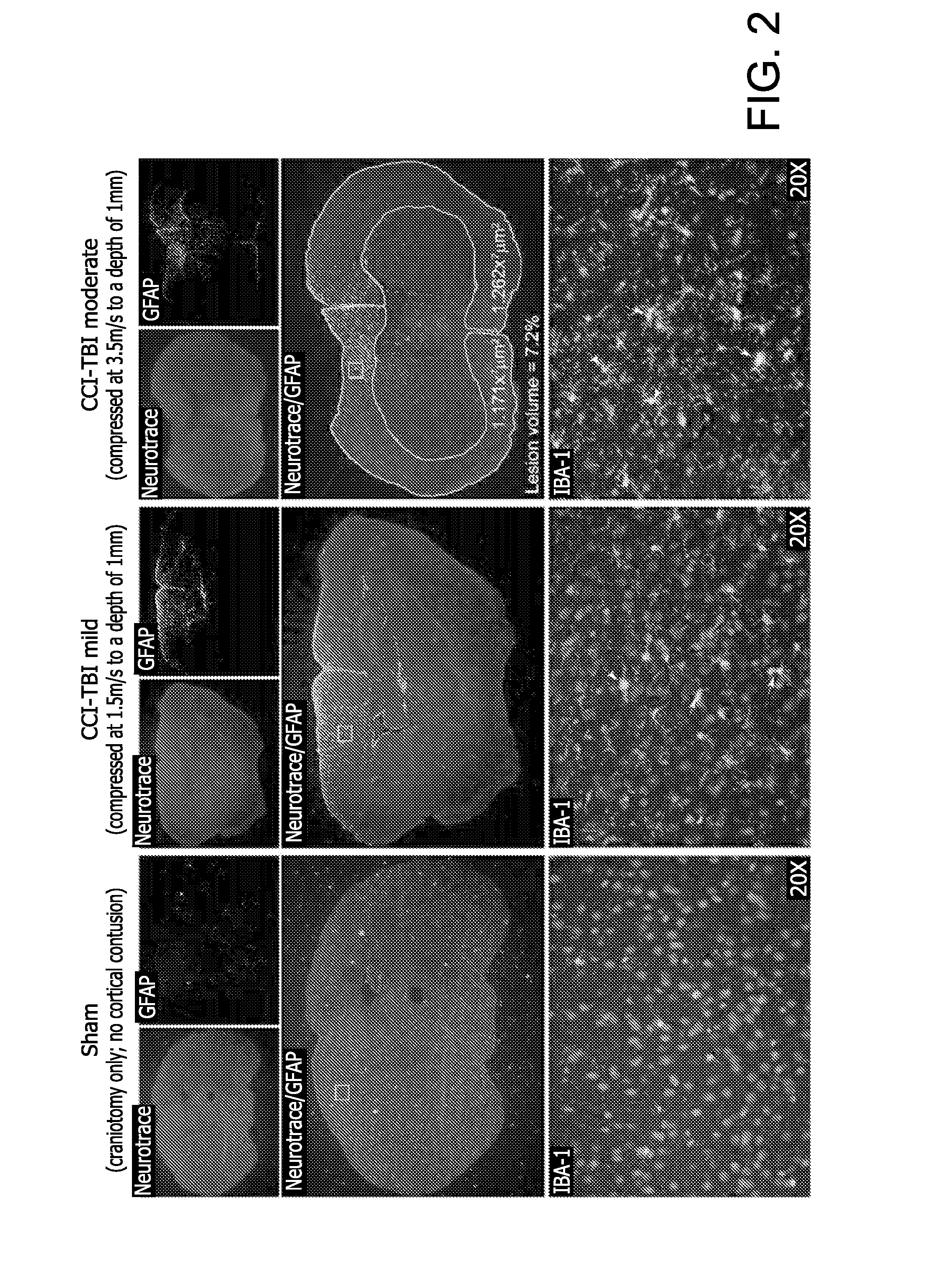
Characterization of the Spectrum of Altered TJ Protein Expression in CNS Vessels after TBI

[0156]The following experiments were carried out to evaluate the status of TJ integrity in the brain following injury using the Controlled Cortical Impact (CCI) model.

Traumatic Brain Injury (TBI) by Controlled Cortical Impact (CCI)

[0157]Using the Controlled Cortical Impact (CCI) experimental mouse model of TBI the status of the T J integrity following injury was evaluated (Elliott M B, Jallo J J, Barbe M F, Tuma R F. Brain Res. 2009. 1305:183-91). The animal protocol used for these studies has undergone full review and approval (#3415, approved on Nov. 9, 2010) by the Temple University School of Medicine Institutional Animal Care and Use Committee (IACUC). A week after arrival, CS7BL / 6 mice (purchased from Taconic Inc, Hudson, N.Y.) were placed into the following groups: untreated (no craniotomy and no TBI), craniotomy only (sham control), mild CCI-TBI, moderate CCI-TBI and severe CCI-TBI. Anes...

example 2

Isolation of Exosomes by Precipitation

[0166]Microvesicles / exosomes were isolated from two sources 1) tissue culture media and 2) from blood plasma collected from either experimental TBI animal samples or human samples. Media from BMVEC, D3 and b-End3 cell cultures exposed to different concentrations of various inflammatory mediators was also used. To induce inflammatory insult the following mediators were used: TNFα (50-100 ng / ml; BD Diagnostics, Franklin Lakes, N.J.), IL1β (100 ng / ml, BD Diagnostics), CCL2 / MCP-1 (Peprotech Inc, Rocky Hill, N.J.), CCL5 / RANTES (Peprotech Inc.), Lipopolysaccharide (LPS) from Escherichia coli 0127:B8 (20 ng / ml, Sigma Aldrich, St Louis, Mo.). After exposure endpoint (4 hrs and 24 hrs), collected conditioned media was centrifuged at 3000×g for 15 minutes in order to remove cells and cell debris. For blood samples, blood was allowed to coagulate for 10-15 minutes and then plasma was separated by centrifugation at 10000×g for 10 minutes. Exosomes were isol...

example 3

Isolation of Exosomes by Ultracentrifugation

[0167]Exosomes can also be isolated by a series of ultra-centrifugations. First the conditioned medium or serum sample (applicable also for CSF) is precleared of cells, dead cells and cellular debris by centrifugation in 50-ml centrifuge tubes for 20 min at 2,000×g in 4° C. The supernatant is then collected and the pellet discarded. In order to minimize contamination, approximately half a centimeter of supernatant is left behind on top of the pellet. The supernatant are then transferred to polyallomer or polycarbonate bottles (appropriate for ultracentrifugation). Next the samples are placed in an ultracentrifuge and spun for 30 min at 10,000×g in 4° C. These supernatants are then transferred to a fresh tube. Caution is taken to ensure that none of the pellet is collected and contaminates the supernatant. At this step the samples are again spun but at higher centrifugation speeds, using at least 70 min at 100,000×g, in 4° C. Note that thes...


PUM

PropertyMeasurementUnit
diameteraaaaaaaaaa
lengthaaaaaaaaaa
pore sizeaaaaaaaaaa

Abstract

Provided is a method for detecting injury to the brain comprising: a) determining the level of a tight junction (TJ) protein in exosomes isolated from a test sample from a subject, wherein the TJ protein is occludin, claudin-3, claudin-5, claudin-12, ZO-1, ZO-2, ZO-3, JAM-A, JAM-B or JAM-C, or any combination thereof; b) comparing the level of the TJ protein in the test sample to the level of the TJ protein in a control sample, wherein an elevated level of the TJ protein in the test sample relative to the level of the TJ protein in the control sample indicates that the subject has an injury to the brain.

Description

CROSS REFERENCE TO RELATED APPLICATION[0001]This application claims benefit of U.S. Provisional Patent Application No. 61 / 664,352, filed Jun. 26, 2012, which is incorporated herein by reference in its entirety.SEQUENCE LISTING[0002]The instant application contains a Sequence Listing which has been submitted in ASCII format via EFS-Web and is hereby incorporated by reference in its entirety. Said ASCII copy, created on Apr. 29, 2013, is named 35926_0452_00_WO_SL.txt and is 1,019 bytes in size.FIELD OF THE INVENTION[0003]The invention relates to a method for detecting injury to the brain.BACKGROUND OF THE INVENTION[0004]Traumatic brain injury (TBI) is considered to be a major health problem in the United States. Approximately every 16 seconds in the United States, a person suffers a TBI. The CDC estimates that 1.5 to 2 million individuals experience TBI annually, resulting in 1.4 million emergency department visits, 235,000 hospitalizations and 52,000 deaths. (Coronado V G, XU L, Basa...

Claims


Application Information

Patent Timeline
Patent Type & Authority Applications(United States)
IPC IPC(8): G01N33/68A61M27/00G01N33/573
CPCG01N33/6896G01N33/573G01N33/6872A61M27/006A61M2210/0693G01N2333/91017G01N2800/56G01N2800/2871G01N2333/70596G01N2333/28
Inventor RAMIREZ, SERVIO H.ROM, SLAVAPERSIDSKY, YURI
Owner TEMPLE UNIVERSITY
Why Eureka
Try Eureka