Interferometric focusing of guide-stars for direct wavefront sensing

a direct wavefront and guide star technology, applied in the field of noninvasive imaging of biological tissues, can solve the problems of limiting the resolution, and achieve the effects of reducing the signal to noise ratio of the wavefront measurement, limiting the resolution, and reducing the penetration depth

Active Publication Date: 2016-01-07
RGT UNIV OF CALIFORNIA
View PDF3 Cites 26 Cited by
  • Summary
  • Abstract
  • Description
  • Claims
  • Application Information

AI Technical Summary

Benefits of technology

[0003]Optical microscopy provides noninvasive imaging of biological tissues at subcellular level. The optical aberrations induced by the inhomogeneous refractive index of biological samples limits the resolution and can decrease the penetration depth. To compensate refractive aberrations, adaptive optics with Shack-Hartmann wavefront sensing has been used in microscopes. Wavefront measurement requires light from a guide-star inside of the sample. The scattering effect limits the intensity of the guide-star, hence r

Problems solved by technology

The optical aberrations induced by the inhomogeneous refractive index of bio

Method used


Image

  • Interferometric focusing of guide-stars for direct wavefront sensing
  • Interferometric focusing of guide-stars for direct wavefront sensing
  • Interferometric focusing of guide-stars for direct wavefront sensing

Examples


Embodiment Construction

Section 1

[0014]To correct the refractive aberration, Adaptive Optics (AO) with different strategies has been applied in optical microscopes. The indirect wavefront sensing method was first applied in an AO system to retrieve the optimal phase by maximizing the detected signal from samples. Numerous iterations are required to find the wavefront, causing photobleaching, while limiting the bandwidth of imaging.

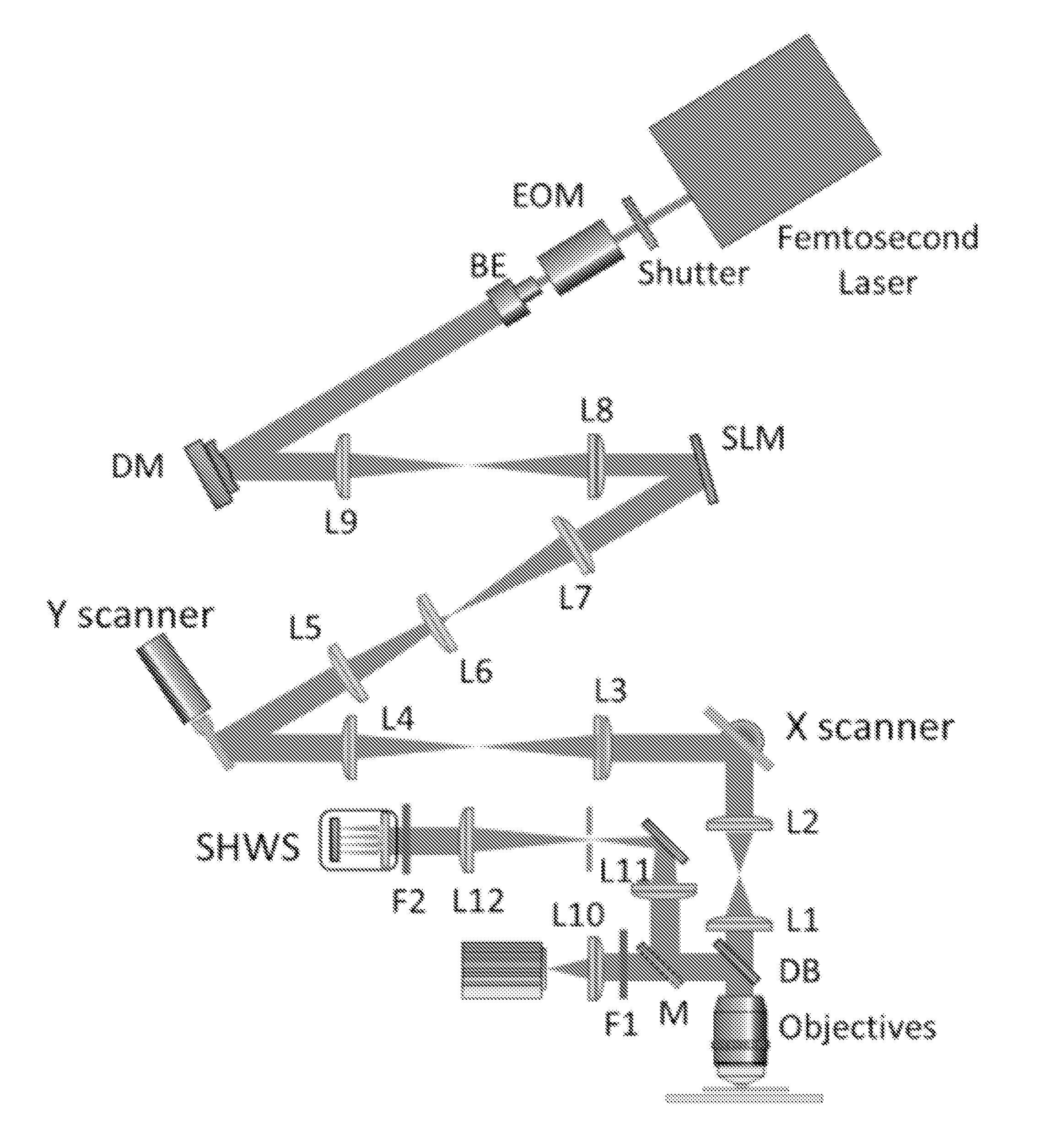
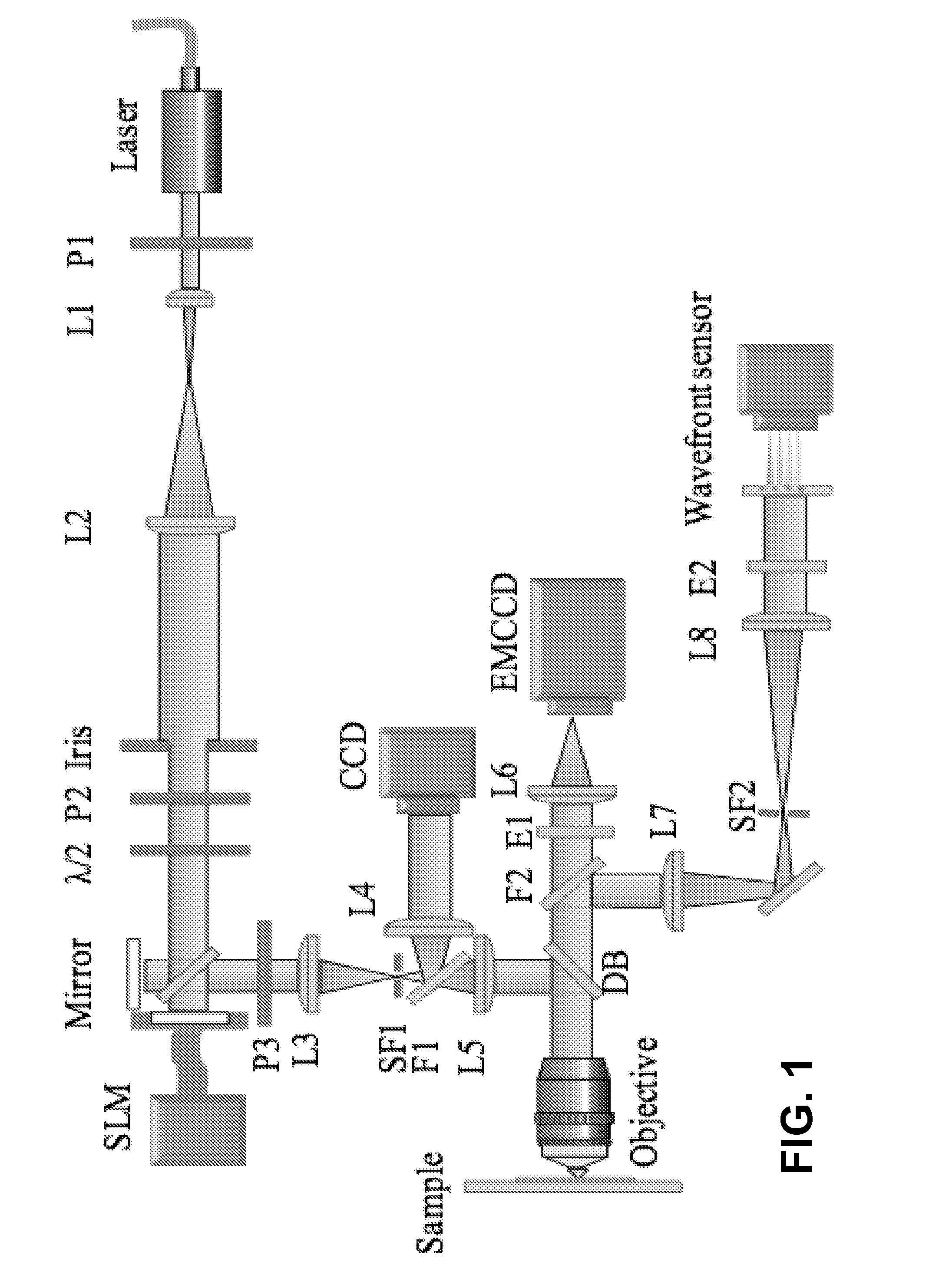
[0015]The direct wavefront sensing method applies a Shack-Hartmann wavefront sensor (SHWS) or interferometer to measure the wavefront directly using the light from a fluorescent microsphere, fluorescent proteins, or backscattered light from the samples. Other approaches use a direct wavefront measurement based on sequential measurements of the wavefront error in each segment of the aperture, or sequential intensity measurements with different trial aberrations.

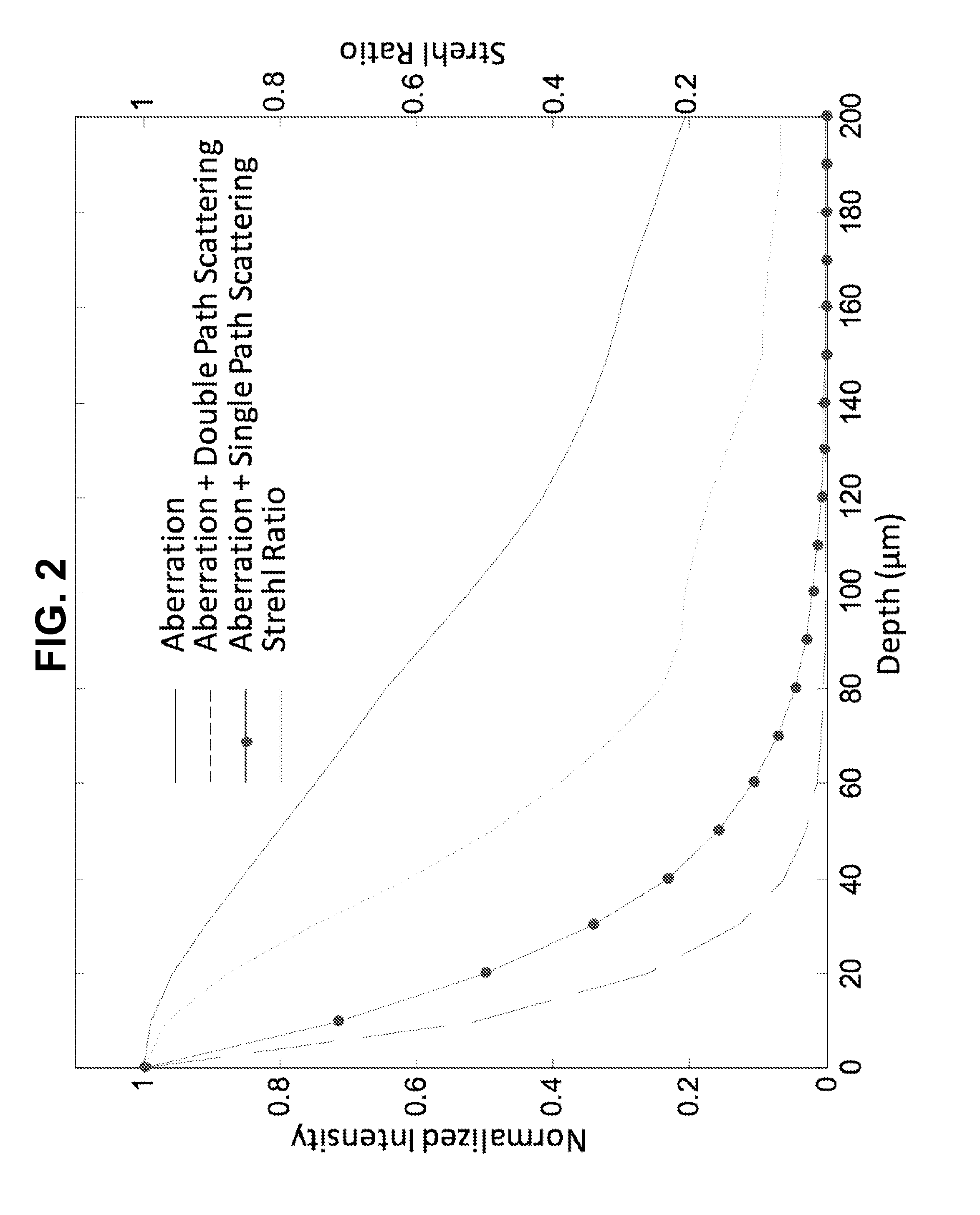
[0016]In the above methods, the performance relies heavily on the intensity of the ballistic light from the samples. In b...


PUM


Abstract

Interferometric focusing (IF), rather than conventional geometric focusing, of excitation light onto a guide-star that is embedded deeply in tissue, increases its fluorescence intensity. The method can extend the depth of wavefront measurement and improve correction inside of tissues because of its ability to suppress both scattering of diffuse light and aberration of ballistic light. The results showed more than two times improvement in SNR and RMS error of the wavefront measurement. Although only ballistic light in the excitation path is corrected, the intensity after wavefront correction increased by 1.5 times. When applying IF to a two-photon microscope with a near infra-red laser, this method would further extend the measurement depth and achieve high SNR for the wavefront sensor.

Description

FIELD OF THE INVENTION[0001]This invention relates to methods and systems for noninvasive imaging of biological tissues at a subcellular level.BACKGROUND OF THE INVENTION[0002]With the advantages of high-resolution and viewing of live organisms, optical microscopy has become an important tool for biological research and continues to open new avenues in its capabilities. In recent years, image resolution and speed has been dramatically improved. However, the improvement of the resolution and penetration depth for optical microscopy is still in its infancy. As light passes through biological tissue, it can be absorbed, refracted and scattered, limiting the resolution and depth of optical imaging in biological tissues. Overcoming these challenges will benefit a wide range of applications from basic biological research to clinical investigations. The present invention addresses these challenges.SUMMARY OF THE INVENTION[0003]Optical microscopy provides noninvasive imaging of biological t...

Claims


Application Information

Patent Timeline
IPC IPC(8): G01N21/45G01N21/64
CPCG01N21/6458G01N2201/0675G01N21/45G01N21/49G02B21/16G02B21/244G02B21/245G02B21/365G01N2021/4709G01J9/00G01N21/6428G01N2021/6439G01N2201/06113G01N2201/121G02B21/361G02B27/0068G02B27/0927
Inventor TAO, XIAODONGKUBBY, JOEL A.
Owner RGT UNIV OF CALIFORNIA
Why Eureka
Try Eureka