
Method and apparatus for percutaneous delivery and deployment of a cardiovascular prosthesis

a technology for cardiovascular prosthesis and percutaneous delivery, applied in the field of cardiovascular valve prosthesis, can solve the problems of valve insufficiency, serious complications, and possible death, and achieve the effects of reducing the number of patients, and improving the quality of li

Active Publication Date: 2016-01-14
CEDARS SINAI MEDICAL CENT
View PDF3 Cites 51 Cited by
  • Summary
  • Abstract
  • Description
  • Claims
  • Application Information

AI Technical Summary

Benefits of technology

The patent text describes a method and system for performing procedures in the heart, specifically repairing the mitral valve. The system includes a catheter with a flexible body and tissue graspers that can be positioned at the mitral valve. The catheter can be advanced through the mitral valve and into the ascending aorta. The tissue graspers can be separated from the catheter and removed from the patient. The technical effects of this system include improved access and repair of the mitral valve with reduced risk of complications.

Problems solved by technology

Valvular insufficiency may result from a variety of problems with the components which make up the valve—for example, the leaflets themselves may degenerate, the tissue cords which tether the leaflets to muscles within the heart may break, or the ring of tissue within which the valve is seated (called the “annulus”) may expand after heart attacks or from congestive heart failure.
Each of these problems leads to a common element in valvular regurgitation: when closed, the edges of the valve leaflets no longer fit snuggly next to each other and allow retrograde flow.
MR is a debilitating disease that can lead to serious complications and possible death.
Although open-heart surgery is a successful method of repairing or replacing faulty heart valves, it poses a significant risk to the wellbeing of the patient, including death, severe injury, and disability.
There is a risk of ischemic or other damage to the heart and other vital organs resulting from the discontinuance of the heart's normal function.
The heart-lung machine may also cause abnormalities of the patient's circulatory, respiratory, hematologic and neurologic systems.
There is a risk of stroke and other consequences from emboli released into the blood during the surgery and during initiation of cardiopulmonary bypass.
There is a risk of heart attack.
Significant damage occurs to the tissues and bone retracted from the patient's chest while gaining access to the heart.
Also, cardiopulmonary bypass carries with it a risk of renal insufficiency, particularly in patients with borderline kidney function.
Such patients may require dialysis after surgery due to the stress on the kidneys during bypass.
Consequently, a patient's recovery can be painful, discomforting, long in duration, and costly.
Moreover, a limited surgical approach or percutaneous approach would decrease or eliminate the tissue trauma that occurs from the extensive incisions of open-heart surgery, sparing patients pain, improving recovery time, and decreasing post-operative complications.
In addition, significant MR and / or TR complicate 30-60% of patients with congestive heart failure, contributing to their impaired cardiac function and causing significant morbidity.
However, because of the significant risks involved in open-heart surgery, many of the patients are unable to undergo valve repair.
No commercial products have been marketed that successfully repair the mitral valve of the human heart with a minimally invasive, beating-heart procedure.
First, the heart and its associated valves are not directly visualized or accessible.
One can use imaging techniques such as fluoroscopy or echocardiography, but these provide a two-dimensional image and a limited field of view.
Three-dimensional imaging technologies, including advanced forms of echocardiography, are available but only in a limited number of medical centers and only to very few clinicians trained to use them.
Second, it is extremely difficult to immobilize the rapidly moving heart valve leaflets for repair purposes while the heart is beating.
Thus, no single device or methodology has successfully been used to repair heart valves in a minimally invasive manner on a beating heart.
Efforts have been made to commercialize catheter based valve clip devices, though such devices are not yet approved for use in the United States.
Although these clip devices can be explained relatively simply, the actual use is not simple.
For example, steering and orienting the clip is a delicate operation that requires skill foreign to most cardiologists.
This intricate control system for orienting the rigid arm is only accurately positioned through the use of complex imaging technologies.
While technically able to repair a mitral valve through peripheral access, these devices and the procedures in which they are used still are very costly to the patient and to the health care system in general.
While these clips devices are described as being able to release, re-approach, and recapture the leaflets, such re-working procedures increase the overall procedure time which is disadvantageous for the patient and the physician.
In practice, the procedure can take two to three times longer than this due to poor initial placement, release and re-grasping efforts.
These and other aspects of the MitraClip design discussed in the EU Heart Journal article of May 23, 2011 make it inconvenient and costly to use.

Method used


Image

  • Method and apparatus for percutaneous delivery and deployment of a cardiovascular prosthesis
  • Method and apparatus for percutaneous delivery and deployment of a cardiovascular prosthesis
  • Method and apparatus for percutaneous delivery and deployment of a cardiovascular prosthesis

Examples


Embodiment Construction

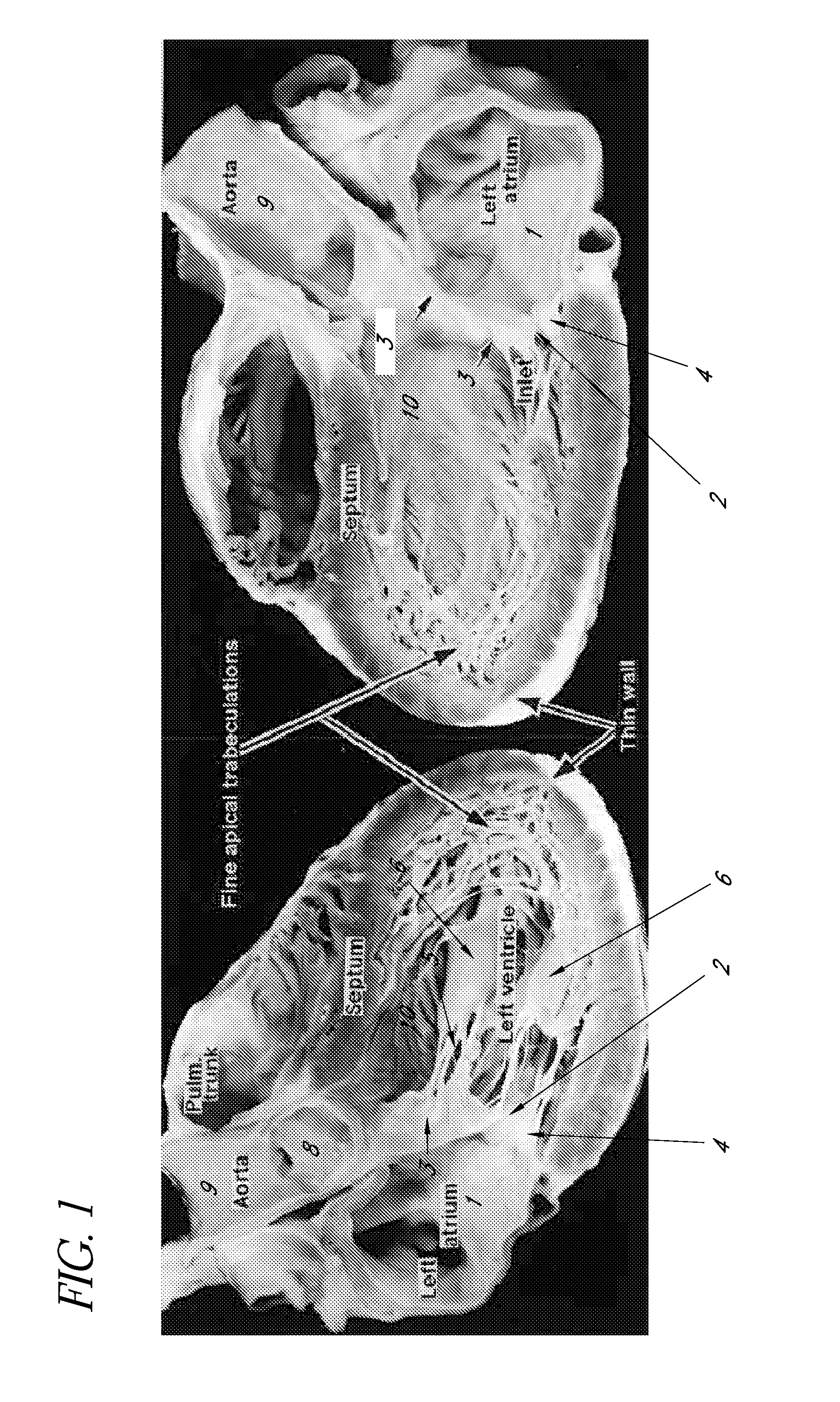
[0046]In FIG. 1, a longitudinal section of the human heart is shown demonstrating the left atrium 1, the mitral valve orifice 2, the anterior leaflet 3 of the mitral valve, and the posterior leaflet 4 of the mitral valve. The subvalvular apparatus consists of the numerous chordae tendinae 5 and the papillary muscles 6. The left ventricular outflow tract (LVOT) 10 is a channel formed by the anterior leaflet 3 of the mitral valve and the interventricular septum.

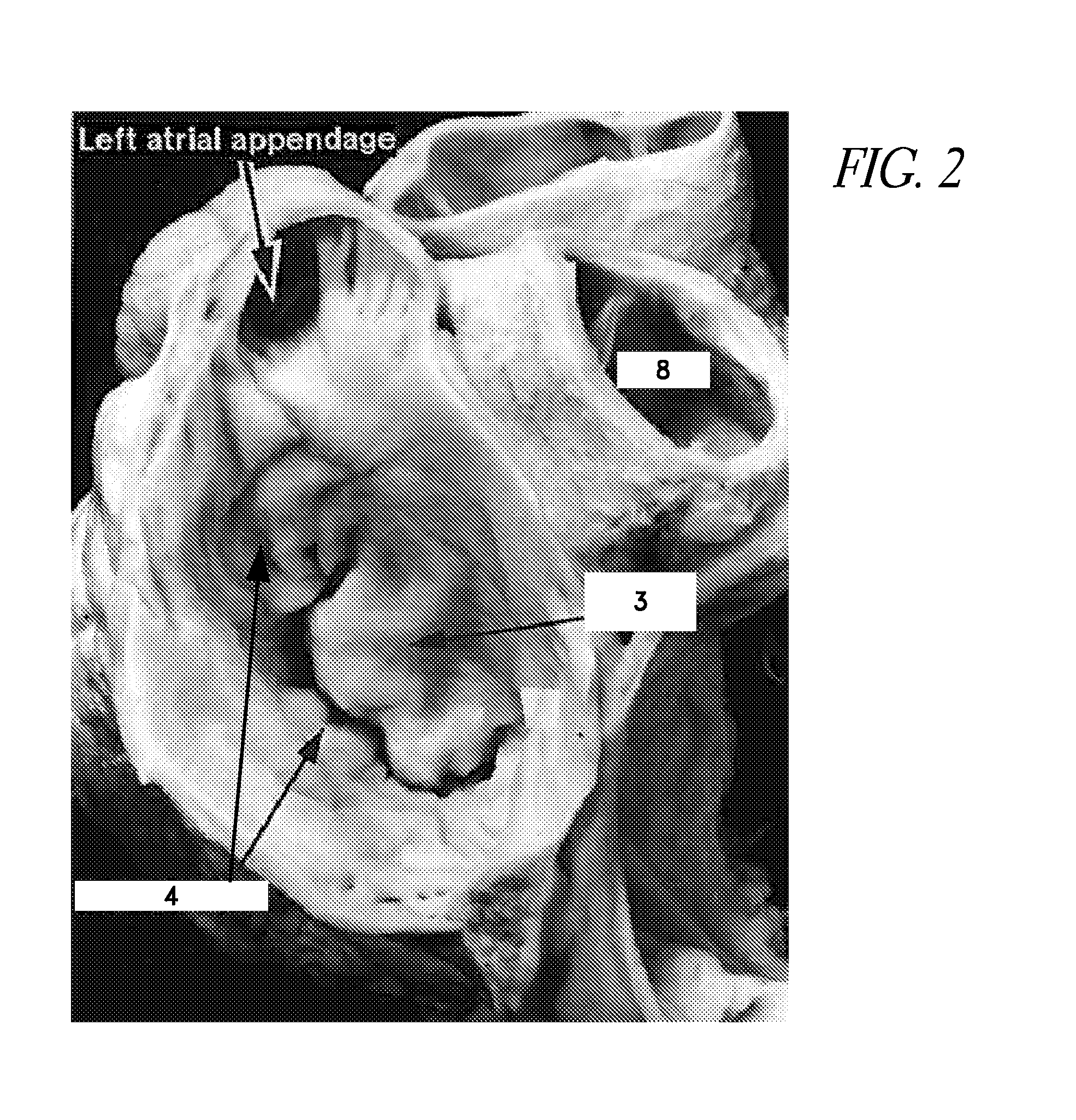
[0047]In FIG. 2, a short axis view of the mitral valve is seen at the level of the left atrium. This demonstrates the asymmetric nature of the mitral valve leaflets. The posterior leaflet 4 has a broad base and of narrow width, while the anterior leaflet 3 has a relatively narrow base and a substantial width. FIG. 2A is a partial dissection of the mitral valve further illustrating the sub-valvular apparatus. These figures illustrate the trajectory along which a catheter device is advanced according to this disclosure to positio...


PUM


Abstract

Catheter apparatuses and methods are provided for repairing heart valves, particularly mitral valves. The method includes providing a catheter having an elongate, flexible body, with a proximal end and a distal end. The distal end can be transluminally advanced from the left atrium through the mitral valve and along the left ventricular outflow tract into the ascending aorta. A valve repair device is deployed to permanently connect leaflets at a mid-section of a mitral valve while permitting medial and lateral portions of the natural leaflets to open and close. The valve repair device detachably connects the distal and proximal ends of the catheter.

Description

BACKGROUND OF THE INVENTION[0001]1. Field of the Invention[0002]This application is directed to cardiac valve prostheses and to apparatuses and methods for deploying such devices in a minimally invasive manner.[0003]2. Description of the Related Art[0004]A major type of heart disease is valvular insufficiency, also called valvular regurgitation, which is characterized by the improper closing of a heart valve. A heart valve consists of a number of leaflets—either two or three—that swing open to allow blood to flow forward (anterograde) out of a heart chamber, and then swing closed to form a tight seal, preventing blood from leaking backwards (retrograde). Valvular insufficiency may result from a variety of problems with the components which make up the valve—for example, the leaflets themselves may degenerate, the tissue cords which tether the leaflets to muscles within the heart may break, or the ring of tissue within which the valve is seated (called the “annulus”) may expand after...

Claims


Application Information

Patent Timeline
IPC IPC(8): A61F2/24A61B17/00
CPCA61F2/2427A61B17/00234A61B2017/00353A61B2017/00292A61B2017/00473A61B2017/00243A61B17/1227A61B17/1285A61F2/246A61F2/2466
Inventor SIEGEL, ROBERT, JAMES
Owner CEDARS SINAI MEDICAL CENT
Learn More