Radiotracer imaging using sodium iodide symporter polypeptides

a radiotracer and sodium iodide technology, applied in the field of radiotracer imaging, can solve the problems of reducing the sensitivity of the nis reporter gene imaging system, sensitivity can be severely compromised, and it is difficult to define, detect, or pinpoint other nis polypeptide-positive cells in the surrounding area, so as to increase the detection sensitivity and resolution of radiotracers, and reduce background signals

Inactive Publication Date: 2016-02-18
MAYO FOUND FOR MEDICAL EDUCATION & RES
View PDF4 Cites 4 Cited by
  • Summary
  • Abstract
  • Description
  • Claims
  • Application Information

AI Technical Summary

Benefits of technology

[0005]This document provides methods and materials involved in radiotracer imaging using NIS polypeptides (e.g., selective radiotracer imaging using NIS polypeptides). For example, this document provides methods and materials for performing imaging techniques that increase the detection sensitivity and resolution of radiotracers localized by NIS reporter gene expression and / or decrease background signals that can be attributed to endogenously expressed NIS polypeptides.
[0007]NIS radiotracers secreted into saliva can be swallowed and can move via the esophagus to the stomach. Considerable amounts of iodide can be transported from the bloodstream into the gastric lumen. NIS radiotracers secreted into the stomach can be reabsorbed after passing into the duodenum and jejunum. Most NIS radiotracers are renally excreted. NIS radiotracer images, therefore, can have high background signals in thyroid, stomach, salivary glands, and bladder, with weaker background signals in esophagus, small intestine, kidneys, and blood. This background radioactivity can decrease the sensitivity of the NIS reporter gene imaging system. For example, sensitivity can be severely compromised for regions in close proximity to the stomach, thyroid, and salivary glands. The current limit of detection in a mouse with pinhole collimation on the X-SPECT instrument in regions with low background activity is approximately 2×10⁵ subcutaneously or intratumorally located NIS-expressing cells (clustered).
[0008]The methods and materials provided herein can be used to decrease background signals in NIS reporter gene imaging, thereby increasing the sensitivity and resolution of the technology. In some cases, background can be reduced by digital subtraction of perfectly co-registered SPECT images obtained by dual isotope imaging using two chemically distinct NIS radiotracers with different gamma emission spectra. The resolution of such a method can be further enhanced through the use of mutant NIS reporter genes encoding mutant NIS polypeptides with selectively diminished ability to concentrate one or other of the two chemically distinct radiotracers.
[0009]In another embodiment, background signals in SPECT and PET imaging can be reduced by co-administering non-radioactive perchlorate anions with a NIS radiotracer, in conjunction with a mutant NIS reporter gene encoding a mutated NIS polypeptide that is relatively resistant to perchlorate inhibition. Methods for isolating a mutated NIS gene encoding a mutated NIS polypeptide having superior performance in the imaging methods are described herein.
[0011]In some cases, an oral CT contrast agent (e.g., barium sulfate, gastrografin, or iodinated contrast) can be used to enhance discrimination of NIS polypeptide expression in areas around the stomach (e.g., the liver, perigastric area, pancreas, spleen, kidneys, and pleural space). The stomach endogenously expresses wild-type NIS polypeptides and uptakes radioisotopes such as radioiodine or pertechnetate, making the stomach highly visible in planar γ-camera, SPECT, or PET images. The strong stomach signal can make it difficult to define, detect, or pinpoint other NIS polypeptide-positive cells in the surrounding area (e.g., the liver, perigastric area, pancreas, spleen, kidneys, and pleural space). Oral administration of an oral CT contrast agent (e.g., barium sulfate, gastrografin, or iodinated contrast) followed by imaging can result in reduced detection of radiotracer signals from the stomach, thereby allowing for detection of radiotracer signals from NIS polypeptide-expressing cells in the area around the stomach.
[0015]In another aspect, this document features an isolated nucleic acid encoding a mutant NIS polypeptide, wherein compared to a non-mutated NIS polypeptide, the ability of the mutant NIS polypeptide to concentrate NIS radiotracers has reduced susceptibility to perchlorate inhibition.

Problems solved by technology

This background radioactivity can decrease the sensitivity of the NIS reporter gene imaging system.
For example, sensitivity can be severely compromised for regions in close proximity to the stomach, thyroid, and salivary glands.
The strong stomach signal can make it difficult to define, detect, or pinpoint other NIS polypeptide-positive cells in the surrounding area (e.g., the liver, perigastric area, pancreas, spleen, kidneys, and pleural space).

Method used


Image

  • Radiotracer imaging using sodium iodide symporter polypeptides
  • Radiotracer imaging using sodium iodide symporter polypeptides
  • Radiotracer imaging using sodium iodide symporter polypeptides

Examples


example 1

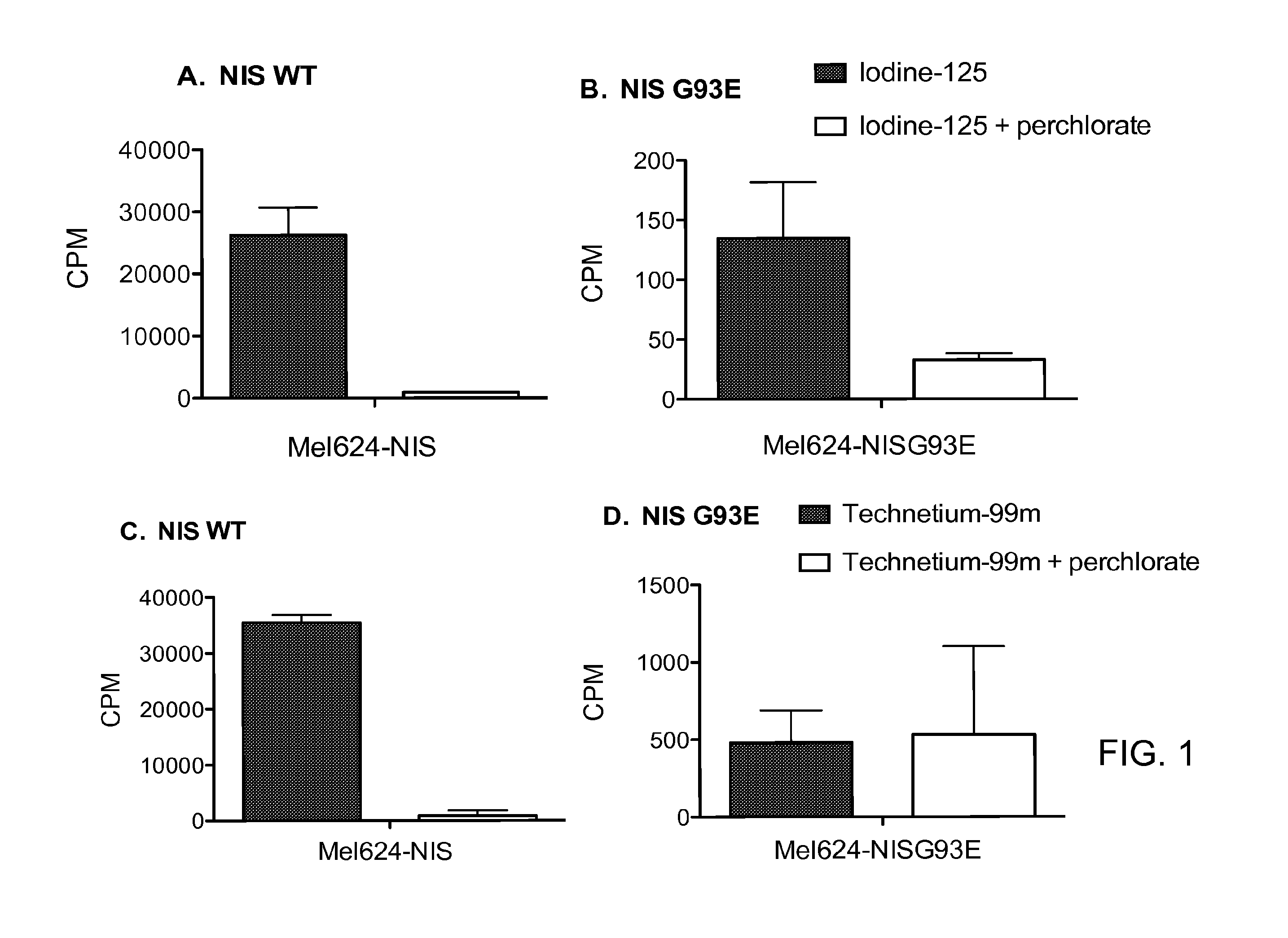
Selective Radiotracer Uptake by a Mutant NIS Polypeptide

[0057]Mel624 cells stably expressing a wild-type human NIS polypeptide (Mel-NIS) or one of two mutant NIS polypeptides (Mel-NIS-93E or Mel-NIS-93Q) were plated in 96-well plates at a density that would lead them to confluency on the day of the uptake assay. 1×Hank's balanced salt solution with calcium, magnesium (HBSS) (Cellgro) and 1 mM HEPES was adjusted to pH 7.3 using KOH (HBBS-HEPES) on the day of the assay. Culture medium was removed from the cells, and 150 μL of HBSS-HEPES+ / −300 μM KClO4 was added to the wells. The cells were incubated for 10 minutes at room temperature prior to the addition of radiotracer. Radiotracers, 125I and 99mTc, were diluted in HBSS-HEPES to about 70,000 to 100,000 CPM / 25 μL (CPM, counts per minute). 25 μL of radiotracer was added to the cells in HBSS-HEPES+ / −KClO4, and the cells were incubated at 37° C. for 1 hour. The cells were then washed twice with cold HBSS-HEPES buffer and lysed to release...

example 2

In Vivo Subtractive Imaging of NIS93E Expressing Tumors to Remove Background Signals from Endogenous NIS Expression in Tissues

[0059]Image subtraction is used to discriminate a tumor that concentrates only one of two NIS isotopes from organs that concentrate both isotopes (e.g., thyroid gland, stomach, and salivary glands). BxPC3 cells (human pancreatic tumor cells) stably expressing either wild-type human NIS polypeptides or human mutant NIS-93E polypeptides are implanted subcutaneously in the flanks of athymic mice. Once tumors grow to a diameter of 5-8 mm, 123I / 99mTc subtraction SPECT is performed using a dual-detector SPECT camera. Four hours after the intraperitoneal administration of 250 μCi 123I sodium iodide and 250 μCi 99mTcO4, SPECT / CT data are acquired. The SPECT emission data are collected in dual-energy windows to separate the 99mTc and 123I counts. The 99mTc window is centered at 140 keV, and the 123I window is placed with a 4% offset above 159 keV to minimize the spill...

example 3

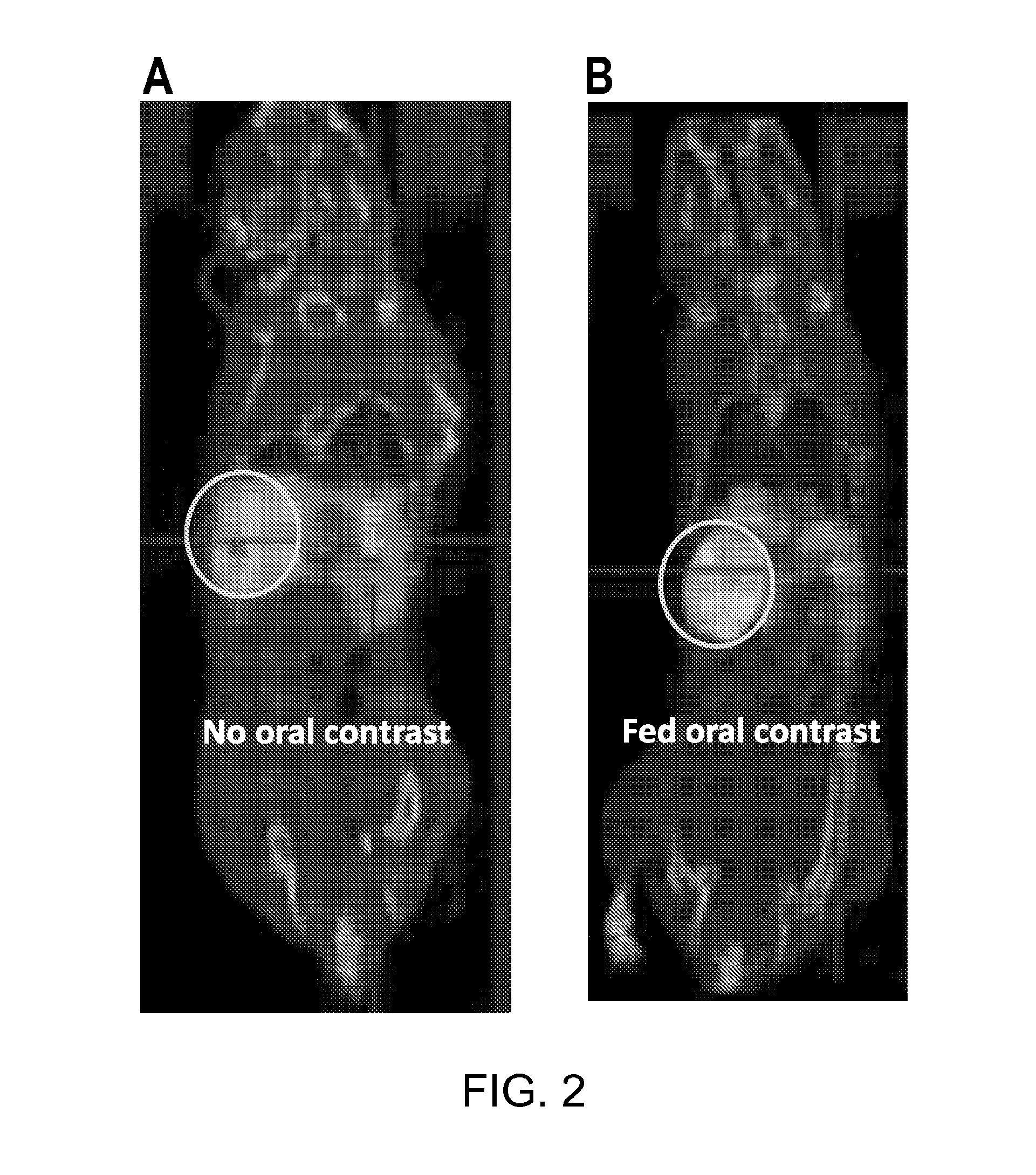
Ingesting Oral CT Contrast Agents Removes Background Signals from the Stomach

[0061]Ingesting oral CT contrast agents (e.g., barium sulfate) removed background signals from the stomach as a result of NIS polypeptide-mediated uptake of radioisotopes, thus enhancing the quality of NIS imaging. In the following example, ingestion of barium sulfate was used to enable and enhance discrimination of NIS expression in the liver. The stomach endogenously expresses wild-type NIS polypeptides and uptakes radioisotopes such as radioiodine or pertechnetate, making the stomach highly visible in planar γ-camera, SPECT, or PET images. The strong stomach signal makes it difficult to define, detect, or pinpoint other NIS-positive cells in the surrounding area (e.g., the liver, perigastric area, pancreas, spleen, kidneys, or pleural space). However, a mouse fed barium sulfate by gavage and then imaged subsequently exhibited reduced isotope signals from the stomach and revealed positive isotope uptake b...


PUM

PropertyMeasurementUnit
pHaaaaaaaaaa
diameteraaaaaaaaaa
diameteraaaaaaaaaa

Abstract

This document provides methods and materials involved in radiotracer imaging using NIS polypeptides. For example, methods and materials for performing imaging techniques that increase the detection sensitivity and resolution of radiotracers localized by NIS reporter gene expression and / or decrease background signals that can be attributed to endogenously expressed NIS polypeptides are provided.

Description

CROSS-REFERENCE TO RELATED APPLICATIONS[0001]This application claims the benefit of U.S. Provisional Application Ser. No. 61 / 788,034, filed Mar. 15, 2013. The disclosure of the prior application is considered part of (and is incorporated by reference in) the disclosure of this application.TECHNICAL FIELD[0002]This document relates to methods and materials involved in radiotracer imaging using sodium iodide symporter (NIS) polypeptides (e.g., selective radiotracer imaging using NIS polypeptides). For example, this document relates to imaging techniques that increase the detection sensitivity and resolution of radiotracers localized by NIS reporter gene expression and / or decrease background signals that can be attributed to endogenously expressed NIS polypeptides.BACKGROUND INFORMATION[0003]NIS polypeptides mediate the uptake and concentration of iodide in the thyroid gland, providing the basis for diagnostic thyroid radioimaging and radioiodine therapy. When nucleic acid encoding a N...

Claims


Application Information

Patent Timeline
Patent Type & Authority Applications(United States)
IPC IPC(8): C07K14/47A61K51/00
CPCA61K51/00C07K14/4702A61K51/02A61K51/12A61K51/025
Inventor RUSSELL, STEPHEN JAMESPENG, KAH-WHYELECH, PATRYCJA
Owner MAYO FOUND FOR MEDICAL EDUCATION & RES
Why Eureka
Try Eureka