3D biomimetic, bi-phasic key featured scaffold for osteochondral repair

a biomimetic and osteochondral technology, applied in the field of 3d biomimetic and biphasic key featured scaffolds for osteochondral repair, can solve the problems of not being able to achieve the robust integration of cartilage and subchondral bone, not being able to achieve the perfect solution for osteochondral regeneration, and not being able to achieve the effect of tissue reconstruction and/or production, excellent interfacial mechanical properties

Inactive Publication Date: 2016-03-10
THE GEORGE WASHINGTON UNIV A CONGRESSIONALLY CHARTERED NOT FOR PROFIT CORP
View PDF7 Cites 38 Cited by
  • Summary
  • Abstract
  • Description
  • Claims
  • Application Information

AI Technical Summary

Benefits of technology

[0013]The present invention relates to a novel method for the creation of 3D biologically inspired tissue engineered scaffolds with both, excellent interfacial mechanical properties, and biocompati

Problems solved by technology

The osteochondral tissue is a nanostructured tissue notoriously difficult to regenerate due to its extremely poor inherent regenerative capacity, complex stratified architecture and disparate biomechanical properties [1, 3].
Although various biomaterials and tissue engineering approaches to treat osteochondral defects have been investigated, it is still very challenging to replicate the robust integration of the cartilage and subchondral bone and the complex stratified cartilage/bone structure.
None of the current available treatment options provides a perfect solution for osteochondral regeneration.
However, the improvements made are still not sufficient to successfully create extremely complex scaffolds that can replicate complex tissues such as cartilage or the bone-cartilage interface.
Current attempts, while producing viable 3D tissue scaffolds, st

Method used


Image

  • 3D biomimetic, bi-phasic key featured scaffold for osteochondral repair
  • 3D biomimetic, bi-phasic key featured scaffold for osteochondral repair
  • 3D biomimetic, bi-phasic key featured scaffold for osteochondral repair

Examples


example 1

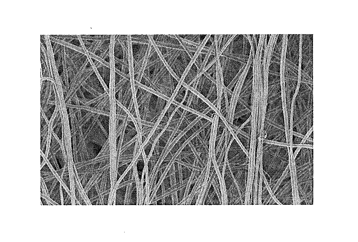
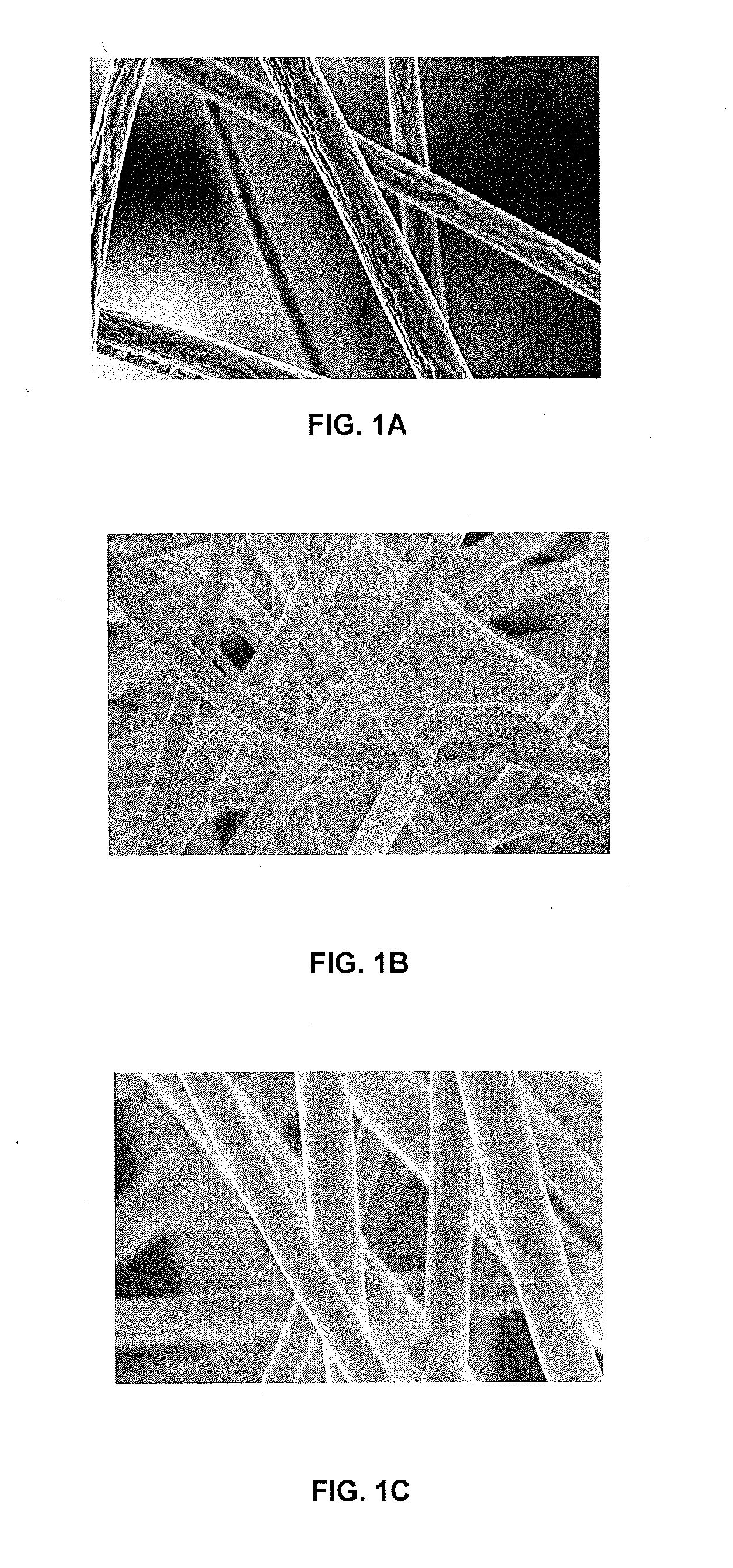
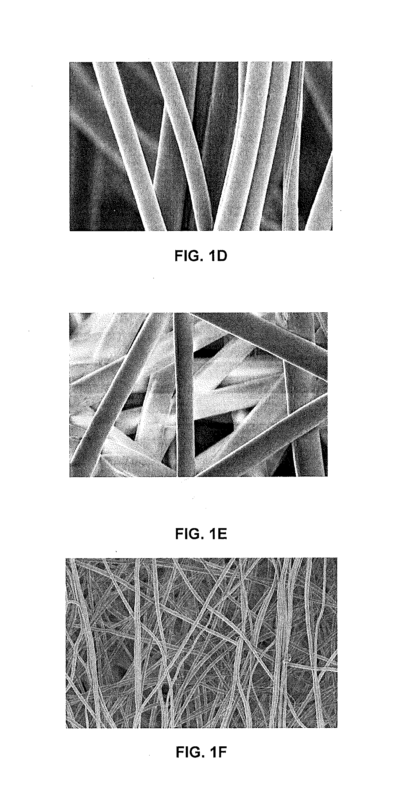
Electrospun Nano / Microscaffold for Cartilage Tissue Engineering

[0073]The purpose of these experiments was to investigate if the mechanical and cytocompatibility properties of electrospun polymer scaffolds for cartilage repair could be enhanced, with the addition of nanomaterials. It was also a goal to evaluate if the nanotubes modified with a cell-favorable molecule can effectively control specific differentiation of stem cells.

[0074]Advances in tissue engineering require more sophisticated materials both to characterize and grow tissues. For this purpose, carbon nanotubes / fibers are emerging candidates. Although the use of carbon nanotubes in tissue engineering is at its infancy, they have been considered exciting alternatives as templates for tissue growth, drug delivery agents and in bio-sensory applications. Carbon nanotubes mimic the dimensions of the constituent components of tissues, where cells are accustomed to interact with nano-fibrous proteins. This property makes them e...

example 2

3D Printed Scaffolds for Osteochondral Regeneration

[0112]Materials and Methods

[0113]3D Osteochondral Scaffold Design and Fabrication

[0114]All 3D osteochondral scaffolds were designed using Rhinoceros 3D modeling package. Scaffolds were then printed in groups of six using a PrinterBot 3D printing system, modified with a 347 μm diameter nozzle, and a spool (or filament fed into the printer) of 1.75 mm diameter biocompatible Polylactic acid (PLA) polymer. PLA comprises an aliphatic polyester of L-lactide units. 3D models were converted into a gcode instruction file using Slic3r, and then used to instruct the printer via the Pronterface software package. The PLA was extruded into a filament using a screw extrusion method. Raw polymer, usually in the form of small beads or pellets, is fed into a hopper attached to a heated, tubular chamber. The chamber has a motor driven screw throughout which turns and moves the pellets down the chamber, and melting them along the way. The chamber termi...

example 3

Additional 3D Printed Osteochondral Devices

[0145]Other embodiments were made with features similar to those in FIG. 9, including: 1) a homogenous cross-hatched structure, with features of 1 to 0.5 mm in size, 2) a bi-phasic structure consisting of a cross hatched pattern and an intersecting rings structure, and 3) biphasic structures but with reinforced key feature in the interface. In addition to above samples printed for cellular study and imaging, a large construct, mimicking the structure and anatomical shape of a human knee with internal bi-phasic and key features was also designed (similar to that shown in FIG. 17). A Stratasys Fortus 250 m 3D printing system was used to fabricate the full large model out of Acrylonitrile butadiene styrene (ABS), a common material used in rapid prototyping 3D printing, for demonstration purpose. Furthermore, the 3D printed cartilage layer of the model was synthesized of biocompatible PLA polymer. This model also had superficial pores on the su...


PUM

PropertyMeasurementUnit
Lengthaaaaaaaaaa
Lengthaaaaaaaaaa
Diameteraaaaaaaaaa

Abstract

This invention describes methods for the creation of 3D biologically inspired tissue engineered scaffolds with both excellent interfacial mechanical properties, and biocompatibility and products created using such methods. In some cases, a combination of nanomaterials, nano/microfabrication methods and 3D printing can be employed to create structures that promote tissue reconstruction and/or production. In other embodiments, electrospinning techniques can be used to create structures made of polymers and nanotubes.

Description

CROSS-REFERENCE TO PRIOR APPLICATIONS[0001]This application is a continuation-in-part of PCT / US2014 / 028914 which was filed on Mar. 14, 2015, which claims priority to U.S. provisional patent application 61 / 799,243 filed on Mar. 15, 2013, the entire contents of each of which are hereby incorporated by reference.U.S. GOVERNMENT SUPPORT[0002]This invention was made with Government support of Grant No. 1DP2EB020549-01, awarded by NIH. The U.S. Government has certain rights in this invention.BACKGROUND OF THE INVENTION[0003]1. Area of the Art[0004]The present invention relates to a novel method for the creation of three dimensional (3D) biologically inspired tissue engineered scaffolds with both excellent interfacial mechanical properties, and biocompatibility. In some embodiments, a combination of nanomaterials, nano / microfabrication methods and 3D printing can be employed to create structures that promote tissue reconstruction and / or production. In other embodiments, electrospinning tec...

Claims


Application Information

Patent Timeline
IPC IPC(8): A61L27/18A61F2/30A61L27/30A61L27/50
CPCA61L27/18A61L27/303A61L27/50A61F2/30942A61F2002/30062A61L2430/24A61L2400/18A61F2002/2835A61L2400/12A61L27/58A61L27/34A61L27/443A61L27/46A61L2420/08D01D5/0046D01F1/10D01F6/625C08L5/04C08L77/04
Inventor HOLMES, BENJAMIN BLAIRZHANG, LIJIE GRACE
Owner THE GEORGE WASHINGTON UNIV A CONGRESSIONALLY CHARTERED NOT FOR PROFIT CORP
Why Eureka
Try Eureka