Tissue interface augmentation device for ligament/tendon reconstruction

a technology of ligament/tendon reconstruction and tissue interface, which is applied in the direction of prosthesis, manufacturing tools, and ligaments, etc., can solve the problems of inferior integration between the tendon graft and the host bone, affecting the resurgence of acl damage, so as to promote osteointegration, promote reestablishment of continuous collagen fibers, and accelerate healing

Active Publication Date: 2016-06-09
NAT UNIV OF SINGAPORE +1
View PDF3 Cites 6 Cited by
  • Summary
  • Abstract
  • Description
  • Claims
  • Application Information

AI Technical Summary

Benefits of technology

[0009]The present invention overcomes some drawbacks of conventional therapies by promoting osteointegration at the bone-(tendon or ligament) graft interface within the surgically prepared bone tunnel such that accelerated healing can be achieved. To achieve this, the inventive device works both as a delivery vessel for osteoconductive and osteoinductive factors and as a scaffold to stimulate and support bone ingrowth. With the presence of osteogenic factors that are optionally incorporated into the tissue interface augmentation (TIA) device according to the present invention, mineralization of this interface tissue will be accelerated, leading to bone ingrowth into the tendon graft. Moreover, the presence of optionally seeded mesenchymal stem cells (MSCs) and / or other biologically active agents will also promote the reestablishment of continuous collagen fibers between the tendon and bone, thereby reestablishing the tendo-osseous junction. The delivery of cytokines and scaffold support provided by the TIA device is particularly essential for the tendon to firmly attach to the bone within the intra-articular environment. With the application of the inventive TIA device, it is anticipated that the bone-graft interface (reconstructed ACL's primary site of weakness) will not exist over extended durations long enough for complications to occur. Complications such as graft failure due to pull-out and micromotion leading to tunnel widening will be avoided with prompt bone-graft integration. Furthermore, firm attachment of the tendon graft to bone will allow earlier and more aggressive rehabilitation, leading to shorter recovery duration thus an earlier return to sports and work.

Problems solved by technology

However, ACL damage is prevalent as it affects 1 in 3000 Americans each year.
This treatment is hampered by the inferior integration between the tendon graft and the host bone.
The native enthesis is usually replaced by a soft fibrous tissue, which does not provide adequate bony anchorage.
Animal studies have demonstrated tendon pullout failure even after 12 weeks of ACL reconstruction.
However, the resultant outcomes are still unsatisfactory.
Yet, delayed healing and poor integration remained outstanding.
It was reasoned that the healing process with the bone-patellar tendon-bone graft was more complicated than normal bone to bone fusion.
This was attributed to the insufficient contact between the bone substratum and the tendon graft.
Unfortunately, the outcomes were compromised by micromotion between the graft tissue and the surrounding bone.
These adverse effects were commonplace with commercially available mechanical implants that only provide temporary anchorage without enhancing biological fusion.
Consequently, the functionality of the transplanted ACL is compromised thus resulting in the need for revision procedures.
Although CaPs have been shown to advance the healing of bone tunnel tissue in animal studies, it does not consist of a biological phase nor allows feasible incorporation of cells to the system.
As such, the method is limited to being an osteoconductive approach, instead of an osteoinductive one.
Nevertheless, the periosteum will have to be autologous, limiting its application as an off-the-shelf product.
At present, the success of tendon or ligament transplantation techniques is severely curtailed by the poor anchorage of the grafts due to insufficient biological integration.

Method used


Image

  • Tissue interface augmentation device for ligament/tendon reconstruction
  • Tissue interface augmentation device for ligament/tendon reconstruction
  • Tissue interface augmentation device for ligament/tendon reconstruction

Examples


example 1

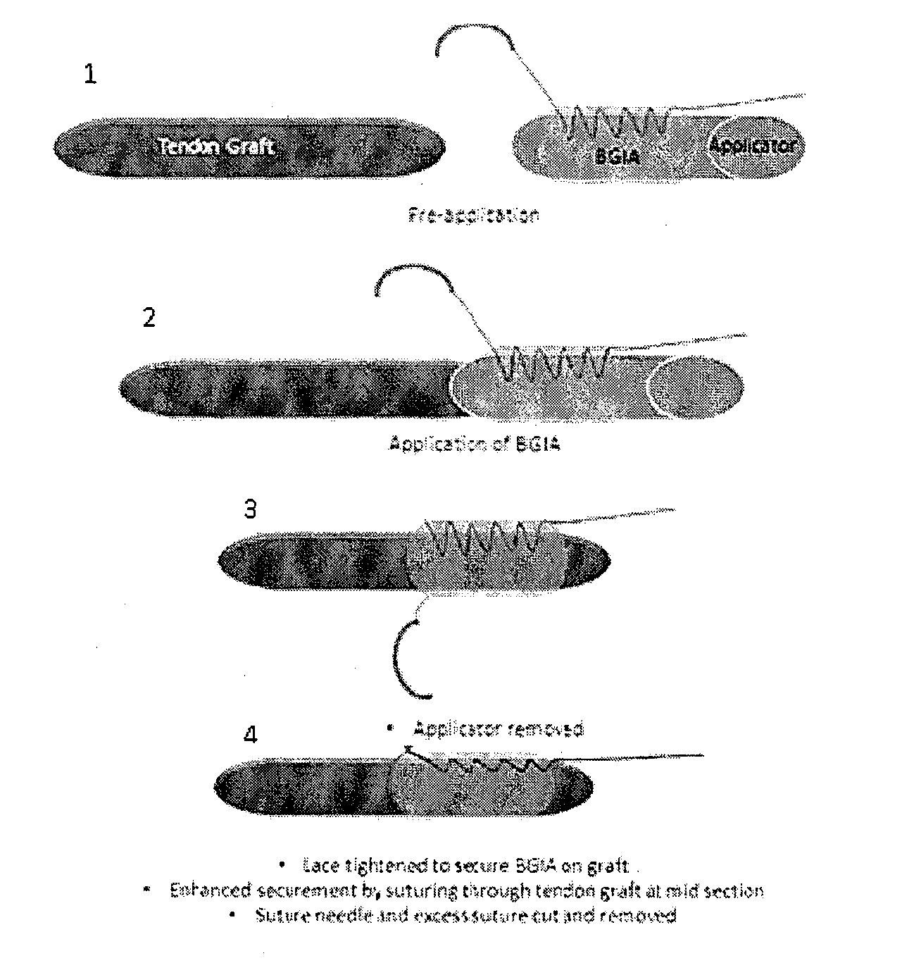
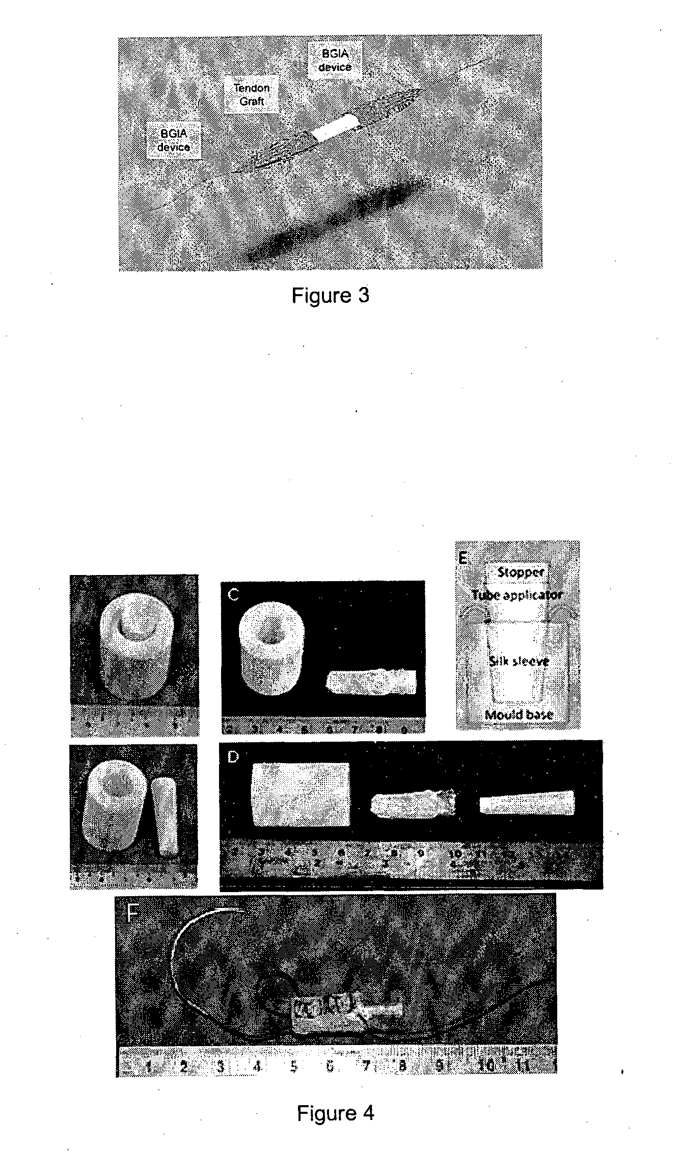
[0105]The TIA device was designed, developed and optimally fabricated using degummed knitted silk sleeve with low crystallinity nano-HA blended in the silk solution to form the sponge.

[0106]The knitted silk sleeve has three components: a knitted silk mesh, a silk sponge and low crystallinity nano hydroxyapatite (LHA). The knitted silk mesh was fabricated by using raw silk fibers with 24 needles on a knitting machine. Then, the knitted raw silk mesh was degummed to remove sericin thoroughly. The silk solution had a concentration of 6 wt.-% of silk fibroin in double distilled water. LHA was prepared by hydrothermal precipitation method: Briefly, calcium chloride, sodium phosphate tribasic and sodium carbonate were dissolved into the water individually at the same concentration. Calcium chloride and sodium phosphate were mixed together, then sodium carbonate was used to modify its crystallinity. The molar ratio of Ca:P:CO3 was 1.67:0.8:0.2. Subsequently, the mixture was agitated at 25°...


PUM

PropertyMeasurementUnit
lengthaaaaaaaaaa
diameteraaaaaaaaaa
diameteraaaaaaaaaa

Abstract

The present invention relates to a device for the interfacial augmentation of tissue grafts for the purpose of tendon and ligament reconstruction. The inventive device works both as a delivery vessel for osteoconductive and osteoinductive factors and as a scaffold to stimulate and support bone ingrowth and comprises a tubular composite silk sheath, said tubular composite silk sheath comprising: a backbone consisting of a tubular silk mesh, and a carrier material consisting of a porous silk sponge, wherein said tubular silk mesh consists of degummed silk fibroin fibers, wherein said porous silk sponge comprises silk fibroin fibers and hydroxyapatite particles and said tubular silk mesh and said porous silk sponge form a composite material. The present invention is also directed to a method for the manufacturing of such augmentation devices, a method for fixation of the thus fabricated tissue interface augmentation devices onto ligament or tendon grafts, a method for applying such devices for ligament and / or tendon reconstruction to tendon grafts, as well as their application in ligament and tendon reconstruction.

Description

FIELD OF THE TECHNOLOGY[0001]The present invention relates to a device for the interfacial augmentation between tissue grafts and bone tissues for the purpose of tendon and ligament reconstruction. Moreover, the present invention is directed to a method for the manufacturing of such augmentation devices, a method for fixation of the thus fabricated tissue interface augmentation devices onto ligament or tendon grafts, a method for applying such devices for ligament and / or tendon reconstruction to tendon grafts, as well as their application in ligament and tendon reconstruction.BACKGROUND ART[0002]In the field of medicine, there has been an increasing need to develop implant materials for correction of biological defects. Particularly in the field of orthopedic medicine, there has been the need to replace or correct bone, ligament and tendon defects or injuries. As a result, there have emerged a number of synthetic implant materials, including, but not limited to, metallic implant mat...

Claims


Application Information

Patent Timeline
Patent Type & Authority Applications(United States)
IPC IPC(8): A61F2/08
CPCA61F2/08A61F2240/001A61F2210/0076A61F2002/087A61L27/3604A61L27/3834A61L27/46A61L27/48A61L27/54A61L27/56A61L2300/414A61L2430/10
Inventor GOH, CHO HONG JAMESTEH, KOK HIONG THOMASSHI, PUJIANGHUI, HOI PO JAMESLI, JUN
Owner NAT UNIV OF SINGAPORE
Why Eureka
Try Eureka