Defined three-dimensional microenvironment for stem cell

a three-dimensional microenvironment and stem cell technology, applied in embryonic cells, non-embryonic pluripotent stem cells, instruments, etc., can solve the problems of low understanding of the effect of cell-matrix interaction in stem cell development, and the current available matrices often lack this level of complexity

Active Publication Date: 2016-12-29
KONKUK UNIV IND COOP CORP +1
View PDF8 Cites 5 Cited by
  • Summary
  • Abstract
  • Description
  • Claims
  • Application Information

AI Technical Summary

Benefits of technology

The disclosure is about a three-dimensional structure made up of a combination of synthetic or natural polymers that can support stem cells. The structure creates a niche which provides a suitable environment for stem cells to grow and differentiate into different types of cells. This invention has potential applications in both regenerative medicine and tissue engineering.

Problems solved by technology

Although biochemical cues including soluble factors such as FGFs, BMPs and Wnts have been well studied for their role in regulating stem cell behavior, the effect of cell-matrix interaction in stem cell development is poorly understood.
While biomaterials technologies are being developed to address these needs, currently available matrices often lack this level of complexity.

Method used


Image

  • Defined three-dimensional microenvironment for stem cell
  • Defined three-dimensional microenvironment for stem cell
  • Defined three-dimensional microenvironment for stem cell

Examples


example 1

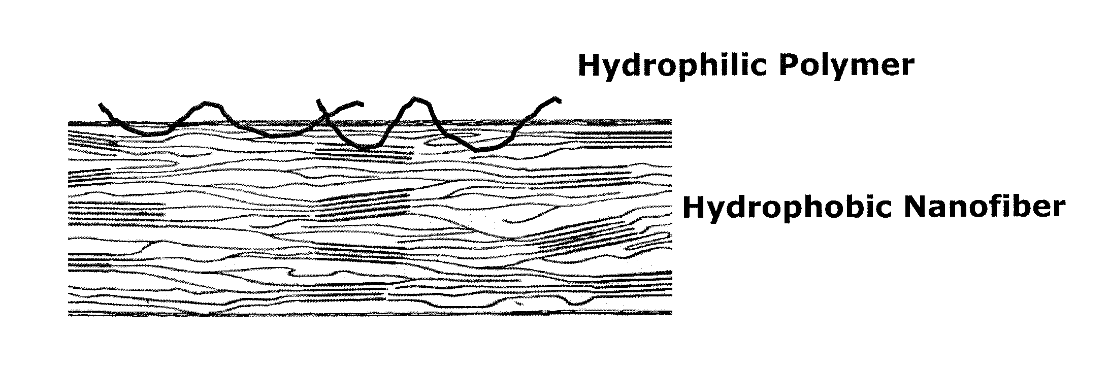
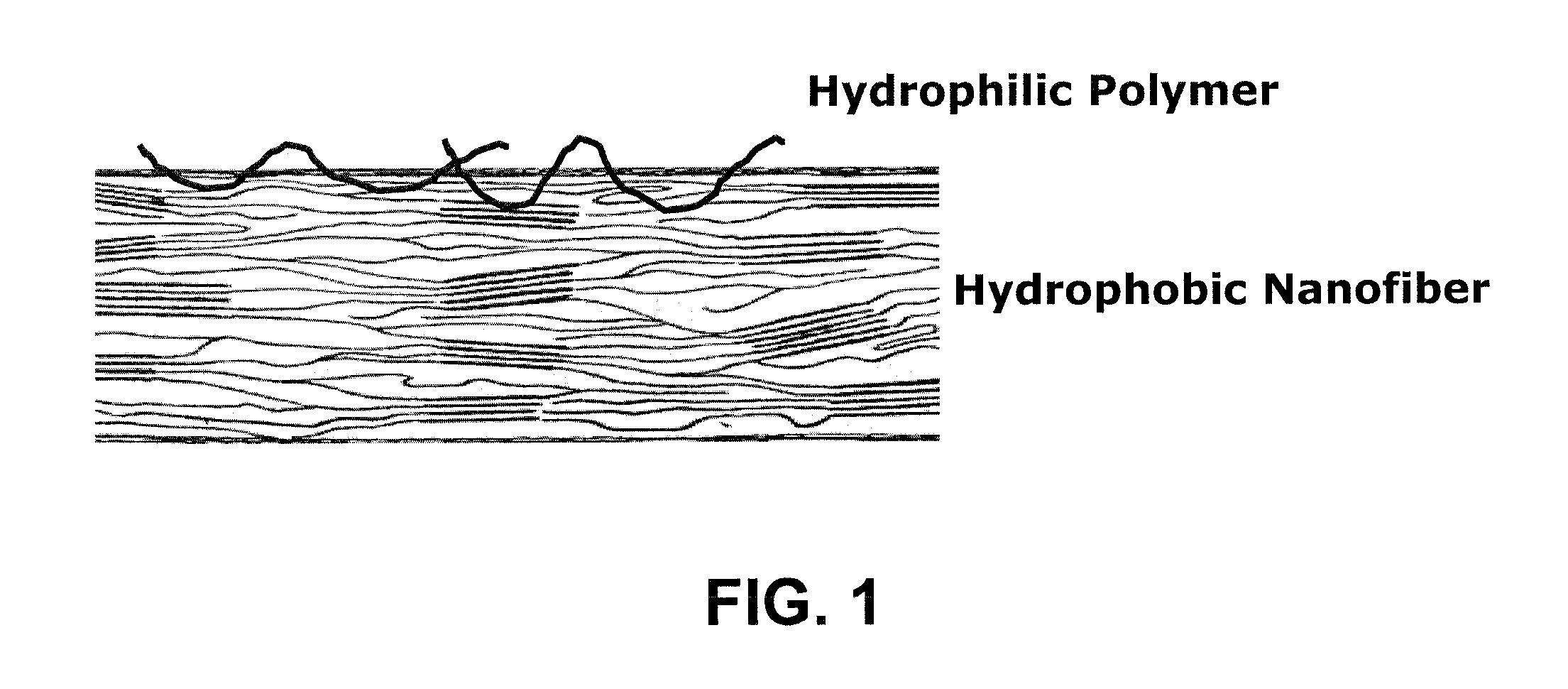
Preparation of Electrospinnable Composition that Provides an Extracellular Microenvironment

[0082]PVDF with an average molecular weight of 400 kDa (Solvay Plastics, Paris), PAN with an average molecular weight of 150 kDa (Sigma-Aldrich, St. Louis) were dissolved in DMAc, respectively, to prepare 20 wt % solution. Each polymer solution was mixed together and the mixture ratio was summarized in Table 2.

[0083]The electrospinnable solution was placed in a plastic syringe fitted with a 27 G needle. A syringe pump (KD Scientific, USA) was used to feed the polymer solution into the needle tip. A high voltage power supply (NanoNC Co. Ltd.) was used to charge the needle tip. The nanofibers were collected onto grounded aluminum foil target located at a certain distance from the needle tip. The fiber meshes were then removed, placed in a vacuum chamber for two days to remove residual solvent, and then stored in a desiccator.

TABLE 2Electrospinnable solution compositionStructural PolymerSolventPV...

example 2

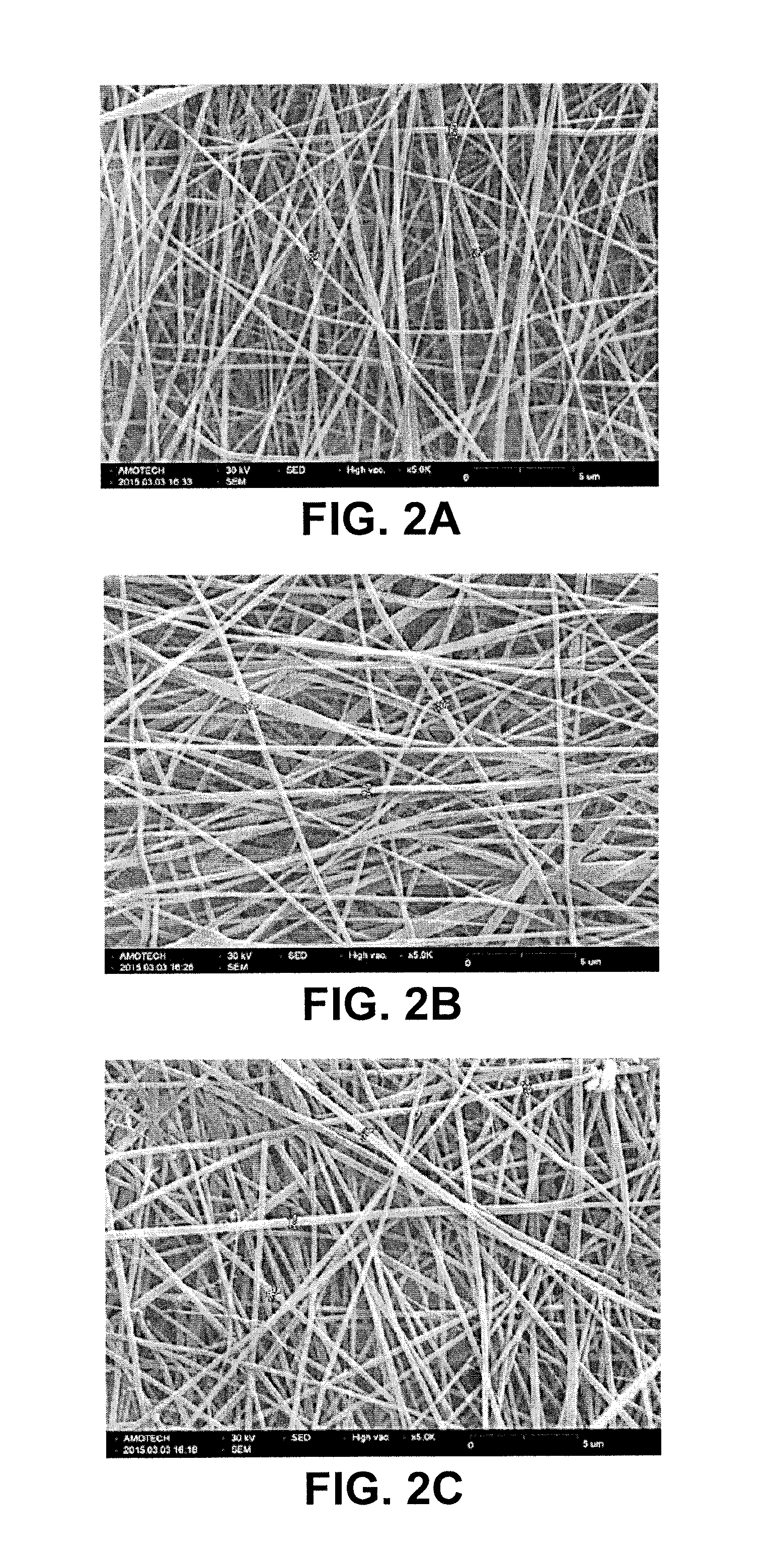
Characterization of an Extracellular Microenvironment

[0084]The nanofiber membranes obtained were observed using a scanning electron microscope. The average diameter indicated is an average of at least three independent measurements, and the average diameter is 400 nm for all samples. The images corresponding to these fibrous microenvironments, obtained by scanning electron microscopy, are shown in FIG. 2A for PVDF / PAN (70 / 30), FIG. 2B for PVDF / PAN (50 / 50), and FIG. 2C for PVDF / PAN (30 / 70).

example 3

Self-Renewal of Murine Embryonic Stem Cell

[0085]The ability of the microenvironment to support self-renewal of mESCs was evaluated by serial passaging of mESCs on the electroprocessed extracellular microenvironment as prepared in EXAMPLE 1.

[0086]For the maintenance of murine embryonic stem cell cultured on poly-D-lysine-coated surface (PDL, Sigma-Aldrich), DMEM Glutamax (GIBCO, Life Technology) containing high glucose 4.5 g / L, Na-pyruvate (0.11 g / L) and L-glutamine was used with L-Glutamine (Invitrogen), 1% non-essential amino acid (Sigma-Aldrich), 50 U / mL Penicillin / streptomycin (GIBCO) and 0.1 mM 2-Mercaptoethanol (GIBCO) as the basal medium, which was added with 20% fetal bovine serum (FBS, Hyclone) and leukemia inhibitory factor (LIF, 1,000 units / mL, Chemicon) at 37° C., 5% CO2 incubator.

[0087]To elucidate the effect of fibrous microenvironment on self-renewal of murine embryonic stem cells, the cells (6×10⁴) were cultured for 48 hours on six different microenvironments placed i...


PUM

PropertyMeasurementUnit
diametersaaaaaaaaaa
diameteraaaaaaaaaa
molecular weightaaaaaaaaaa

Abstract

This disclosure provides for a three-dimensional (3D) microenvironment presenting defined physical or mechanical cues that regulate cellular behavior and use of the matrix. The disclosure also provides for devices and methods for screening for optimal combinations of physical and mechanical cues in order to create a microenvironment that can regulate specific cellular behavior such as cell growth, proliferation, migration or differentiation.

Description

CROSS-REFERENCE TO RELATED APPLICATION[0001]This application claims the benefit of U.S. Provisional Patent Application Ser. No. 62 / 183,663, filed Jun. 23, 2015, the disclosure of which is hereby incorporated herein in its entirety by this reference.TECHNICAL FIELD[0002]This disclosure relates to a defined three-dimensional microenvironment for stem cells.BACKGROUND[0003]Increasing evidence has demonstrated that a local microenvironment, stem cell niche, is important in regulating their self-renewal and differentiation in tissue and organ development process. Stem cell niche provides a complex array of biochemical and physical cues in a spatiotemporally defined fashion, engaging and instructing stem cells to proliferate migrate and differentiate.[0004]Such microenvironment consists of many factors including extracellular matrices (ECMs), growth factors, signaling molecules, etc. Although biochemical cues including soluble factors such as FGFs, BMPs and Wnts have been well studied for...

Claims


Application Information

Patent Timeline
Patent Type & Authority Applications(United States)
IPC IPC(8): G01N33/50C12N5/0735C08J3/28C08L27/16C08L33/20C12N5/00C12N5/074
CPCG01N33/5073C12N5/0075C12N5/0606C12N5/0696C08L27/16C08J2433/20C08J3/28C12N2533/30C08J2327/16C08J2333/20C08J2427/16C08L33/20C12N5/0068C12N2535/00
Inventor CHO, SSANG-GOOBAEK, KYUWONYANG, GWANG-MOKIM, CHANWON, JIHYESEO, IN YONGSEO, DONG-SIKYOO, CHULBAEGOO, HUI-GWANJANG, SEONHOKOO, SONG HEEPARK, SANG-EUN
Owner KONKUK UNIV IND COOP CORP
Why Eureka
Try Eureka