Two-dimensional fluorescence difference spectroscopy characterization of nanoparticles and their interactions

a nanoparticle and fluorescence difference technology, applied in the field of two-dimensional fluorescence difference spectroscopy characterization of nanoparticles and their interactions, nanoparticle complexes, etc., can solve the fundamental, if not disturbing, gap regarding the biochemical and biological activities of these nanomaterials, and the lack of understanding of the effect of many nanomaterials on the structure-function of nucleic acids and proteins, cell metabolism, cell signaling, etc., to achieve the highest fluorescence intensity

Active Publication Date: 2017-05-11
KANSAS STATE UNIV RES FOUND
View PDF16 Cites 6 Cited by
  • Summary
  • Abstract
  • Description
  • Claims
  • Application Information

AI Technical Summary

Benefits of technology

[0011]According to yet another embodiment of the present invention there is provided a method of detecting changes in the structure of a biomolecule associated with attachment of the biomolecule to a nanoparticle thereby forming a nanoparticle complex. The method comprises performing concurrent scans of the fluorescence excitation and emission wavelengths for the biomolecule in comparison to the nanoparticle complex and then determining the intersect between the excitation and emission wavelengths yielding the highest fluorescence intensity. The intersect is compared to one or more known standards observing the change in the biomolecule's

Problems solved by technology

While great numbers of various types of nanoparticles have been synthesized, including elemental nanoparticles and nanoparticle compounds and composites, and the physiochemical properties of these nanoparticles are fairly well understood, there exists a fundamental, if not disturbing, gap regarding the understanding of the biochemical and biological activities of these nanomaterials.
In particular, there is a lack of understanding of the effect that many nanomaterials have on the structure-function of nucleic

Method used


Image

  • Two-dimensional fluorescence difference spectroscopy characterization of nanoparticles and their interactions
  • Two-dimensional fluorescence difference spectroscopy characterization of nanoparticles and their interactions
  • Two-dimensional fluorescence difference spectroscopy characterization of nanoparticles and their interactions

Examples


example 1

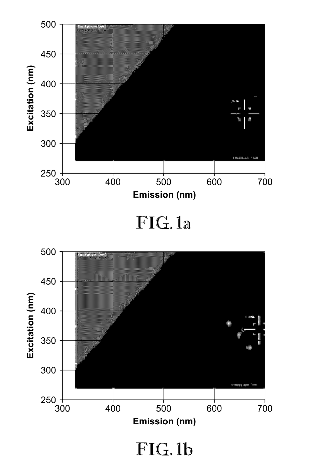
[0046]In this example, 2D-FDS was used to illustrate how two proteins, Luciferase and b-Galactosidase, interact with zinc oxide nanoparticles (ZnO-NP). Particularly, the shift in the excitation and emission wavelength intersect resulting from attachment of the proteins as compared to the uncomplexed nanoparticles was observed.

[0047]Dry powders of nanomaterials were weighed out in milligram quantities and suspended in spectral grade water. Once suspended at the intended concentration (mg / mL), the nanoparticles were ultra-sonicated to evenly disperse the particles throughout the solution. The nanoparticles were transferred to a black 96 well microplate at a volume of 200 μL, and the plate was inserted into a Spectramax i3x 2D-FDS instrument from Molecular Devices. This spectrophotometer (instrument) utilizes Softmax Pro 6.4.2 software to specify the measurement parameters. To generate the 2D-FDS data, the settings were adjusted to detect fluorescence of unknown wavelengths. The spectr...

example 2

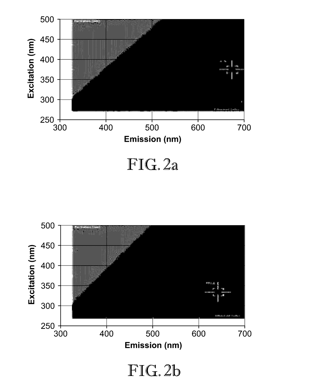
[0050]In this example, 2D-FDS was used to illustrate the shift in the excitation and emission wavelength intersect between cobalt-zinc oxide (CoZnO) composite and CoZnO with polyinosinic-cytidylic acid (poly I:C) RNA attached. FIG. 2a illustrates the 2D-FDS excitation / emission optimum for ZnO—Co composite (375 nm / 675 nm, RFU=1.7K). This optimum value shifts upon poly I:C RNA interaction (335 nm / 685 nm, RFU=491) as shown in FIG. 2b. Thus, a shift in the optimal fluorescence intersect was identified when poly I:C RNA interacts with CoZnO nanoparticle composites. Decrease in fluorescence intensity, from 1.7K relative light units to 491, is another indication of interaction, as the fluorescence emitted from the nanoparticle is quenched by the biomolecule loaded onto the surface.

example 3

[0051]In this example 2D-FDS was used to illustrate the shift in the excitation and emission wavelength intersect between ZnO nanoparticle (FIG. 3a); ZnO nanoparticle in the presence of protamine:poly I:C complex (FIG. 3c); boron carbide (B4C) nanoparticle (FIG. 3b); B4C nanoparticle in the presence of protamine:poly I:C complex (FIG. 3d); MgO nanoparticle (FIG. 3e); and MgO nanoparticle in the presence of protamine:poly I:C complex (FIG. 3f). As can be seen FIGS. 3a-f, the optimum excitation / emission intersect is different between the various material pairs. This example again confirms the utility of 2D-FDS as a rapid and sensitive means of quantifying biomolecular interaction with the nanoparticle. This example further illustrate how the peak position shifts and becomes more intense when ZnO nanoparticle (FIG. 3g) interacts with protein:RNA complex (protamine:poly I:C complex) (FIG. 3h).


PUM

PropertyMeasurementUnit
Nanoscale particle sizeaaaaaaaaaa

Abstract

Methods of characterizing nanoparticles, nanoparticle complexes, and their biomolecular interactions are provided. Concurrent excitation and emission wavelength scans are performed and the optimal fluorescence intensity for the intersect of these wavelengths is determined. The intersect is dependent upon the physical characteristics of the particle and can be used to verify, for example, attachment of biomolecules, amount of biomolecules present, and structure of the attached biomolecule.

Description

CROSS-REFERENCE TO RELATED APPLICATION[0001]The present application claims the benefit of U.S. Provisional Patent Application No. 62 / 251,488, filed Nov. 5, 2015, which is incorporated by reference herein in its entirety.BACKGROUND OF THE INVENTION[0002]Field of the Invention[0003]The present invention is generally directed toward methods of characterizing nanoparticles, particularly nanoparticle complexes comprising an organic species such as a biomolecule or pharmaceutical compound. In certain embodiments, the present invention utilizes two-dimensional fluorescence difference spectroscopy in order to generate a unique identifier or “fingerprint” for the particular nanoparticle or nanoparticle complex that can be used to characterize the nanoparticle or nanoparticle complex.[0004]Description of the Prior Art[0005]In the last 10-15 years, nanotechnology has emerged as an area of great interest in the fields of physics and chemistry. While great numbers of various types of nanoparticl...

Claims


Application Information

Patent Timeline
IPC IPC(8): G01N33/543G01N33/53G01N33/573G01N21/64G01N33/553
CPCG01N33/54346G01N21/6428G01N33/54306G01N33/553G01N33/573G01N2333/938C12Y113/12007C12Y302/01023G01N2021/6421G01N2333/90241G01N33/5308C12Q1/68G01N2021/6419C12Q2563/107C12Q2563/155
Inventor DELONG, ROBERTHURST, MIRANDA
Owner KANSAS STATE UNIV RES FOUND
Why Eureka
Try Eureka