Methods for treating cardiovascular dysfunction and improving fluid homeostasis with a peptide hormone

a peptide hormone and cardiovascular technology, applied in the direction of peptide/protein ingredients, peptide sources, instruments, etc., can solve the problems of patient inconvenience, short-acting peptide limitations, and many of the peptides and proteins have less than optimal pharmacokinetic properties

Active Publication Date: 2017-08-10
UNIV OF MARYLAND BALTIMORE
View PDF1 Cites 3 Cited by
  • Summary
  • Abstract
  • Description
  • Claims
  • Application Information

AI Technical Summary

Benefits of technology

[0006]Methods are provided for treating a subject, preferably a human, that is suffering from a cardiac condition or has a risk factor for developing a cardiac condition. Treatment involves administering to the subject an effective amount of a peptide in a pharmaceutically acceptable form that selected from the group consisting of: (a) a peptide of SEQ ID NO: 1, (ELA-32), (b) a peptide of SEQ ID NO: 2, (ELA

Problems solved by technology

Many of these proteins and peptides have less than optimal pharmacokinetic properties, often because they are smaller than the kidney filtr

Method used


Image

  • Methods for treating cardiovascular dysfunction and improving fluid homeostasis with a peptide hormone
  • Methods for treating cardiovascular dysfunction and improving fluid homeostasis with a peptide hormone
  • Methods for treating cardiovascular dysfunction and improving fluid homeostasis with a peptide hormone

Examples


example 1

Materials and Methods

Chemicals

[0218]The mature form of the 54 amino acid ELA peptide, a 32-amino acid, QRPVNLTMRRKLRKHNCLQRRCMPLHSRVPFP (SEQ ID NO: 1) and apelin-13, QRPRLSHKGPMPG (SEQ ID NO: 3), were purchased by GenScript (Piscataway, N.J.), at more than 98% purity. Other chemicals were from Sigma (St. Louis, Mo.) unless otherwise stated.

Cell culture

[0219]Chinese hamster ovary (CHO) cells were obtained from ATCC (Manassas, Va.) and grown in Ham's F12 medium supplemented with 10% fetal bovine serum (FBS), 100 μg / mL streptomycin and 100 U / mL penicillin. Human umbilical vein endothelial cells (HUVECs) were purchased from Lonza (Walkersville, Md.) and cultured in basal medium supplemented with 0.2% endothelial cell growth supplement (EnGS), 5 ng / mL recombinant human EGF, 50 μg / mL ascorbic acid, 10 mM L-Glutamine, 1 μg / mL hydrocortisone hemisuccinate, 0.75 U / mL heparin sulfate and 2% FBS. HUVECs between passage 3 and 5 were used for all experiments.

Plasmid construction

[0220]The human A...

example 2

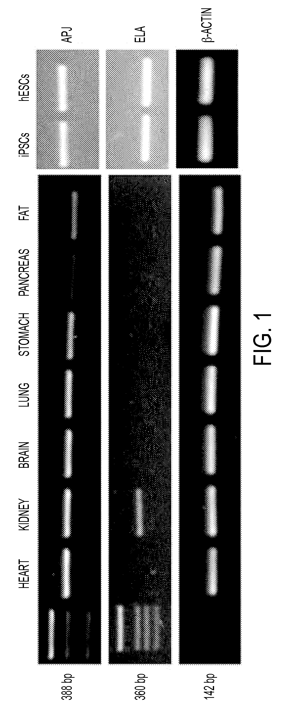
Tissue Distribution of ELA and APJ in Humans

[0232]RT-PCR analyses of ELA and APJ on cDNAs derived from multiple human tissues including the heart, kidney, brain, lung, stomach, pancreas, and fat was conducted with primers specific for ELA or APJ and was conducted to determine ELA tissue distribution in humans, in comparison with APJ. As shown in FIG. 1, APJ is widely distributed and expressed in tissues of heart, brain, kidney, stomach, lung, adipose tissues and pancreas. In contrast, ELA was expressed only in adult kidney tissue and embryonic and induced adult pluripotent stem cells (ESCs and iPSCs).

example 3

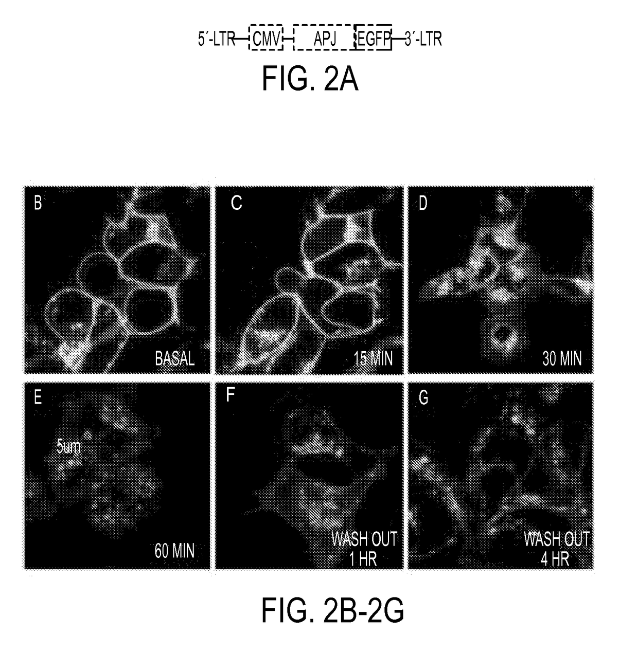
ELA Induction of Human APJ Endocytosis

[0233]To investigate whether ELA would induce APJ internalization, APJ was over-expressed as a fusion protein with enhanced green fluorescent protein (APJ-GFP, FIG. 2B) through lentiviral infection in HEK293 cells. Its intracellular localization in response to ELA treatment was examined. At the basal level, the fusion protein was localized largely at the cell surface, with some perinuclear expression detected (FIG. 2C). Following ELA treatment, intracellular dot-like vesicles appeared by 15 min (FIG. 2C) and became more apparent by 30 min (FIG. 2D). By 60 min (FIG. 2E), large and hollow intracellular vesicles (3-5 p.m in diameter) were clearly visible in the cytoplasm. The fluorescent vesicles remained in the cytoplasm 1 hour (FIG. 2F) and appeared to return to the cell surface 4 hours (FIG. 2G) after the washout in cells previously treated with ELA for 60 minutes.


PUM

PropertyMeasurementUnit
Fractionaaaaaaaaaa
Fractionaaaaaaaaaa
Fractionaaaaaaaaaa

Abstract

The present invention generally to methods of treating subjects suffering from a cardiac condition or having a risk factor for developing a cardiac condition by administering an ELA peptide or fusion protein to a subject in need. The invention relates to fusion proteins of Fc-ELA-32 and Fc-ELA-21 that exhibit improved properties for use as therapeutic agents, e.g. in the treatment of cardiac conditions. In addition, the present invention relates to polynucleotides encoding such fusion proteins, and vectors and host cells comprising such polynucleotides. The invention further relates to methods for producing the fusion proteins of the invention, and to methods of using them in the treatment of disease.

Description

CROSS REFERENCE TO RELATED APPLICATIONS[0001]This application claims the benefit of provisional application 62 / 063,005, entitled “Methods for Treating Cardiovascular Disease and Improving Fluid Homeostasis with a Peptide Hormone,” filed Oct. 13, 2014, the entire contents of which are incorporated herein.STATEMENT OF GOVERNMENT INTEREST[0002]This invention was made with government support under Grant Number DK072488 awarded by the National Institutes of Health. The government has certain rights in the invention.BACKGROUND[0003]Each year, heart disease is one of this country's most serious health problems. The direct and indirect costs of cardiovascular disease and stroke are about $315 billion. This figure is increasing every year. Statistics show that cardiovascular disease is America's leading health problem, and the leading cause of death. Considering the most recent statistics released by the American Heart Association, approximately 84 million people in this country suffer from ...

Claims


Application Information

Patent Timeline
IPC IPC(8): A61K38/22G01N33/68C07K14/575A61K45/06C07K14/47
CPCC07K2319/02C07K2319/30G01N2800/32G01N2800/52G01N33/6893A61K45/06C07K14/4722C07K14/575A61K38/22A61K38/00A61K38/16A61P9/00A61P9/02A61P9/04A61P9/06A61P9/10A61P9/12C07K14/47G01N33/5044C07K14/4705C07K19/00
Inventor GONG, DA-WEIYU, DAOZHANYANG, RONGZESHULDINER, ALANCHEN, LING
Owner UNIV OF MARYLAND BALTIMORE
Why Eureka
Try Eureka