Method for characterisation of perivascular tissue
a perivascular tissue and tissue technology, applied in the field of perivascular tissue, can solve the problems of insufficient blood supply to downstream tissue, weak extracellular matrix separating the lesion from the arterial lumen (also known as the fibrous cap), and often asymptomatic plaques
- Summary
- Abstract
- Description
- Claims
- Application Information
AI Technical Summary
Benefits of technology
Problems solved by technology
Method used
Image
Examples
example 1
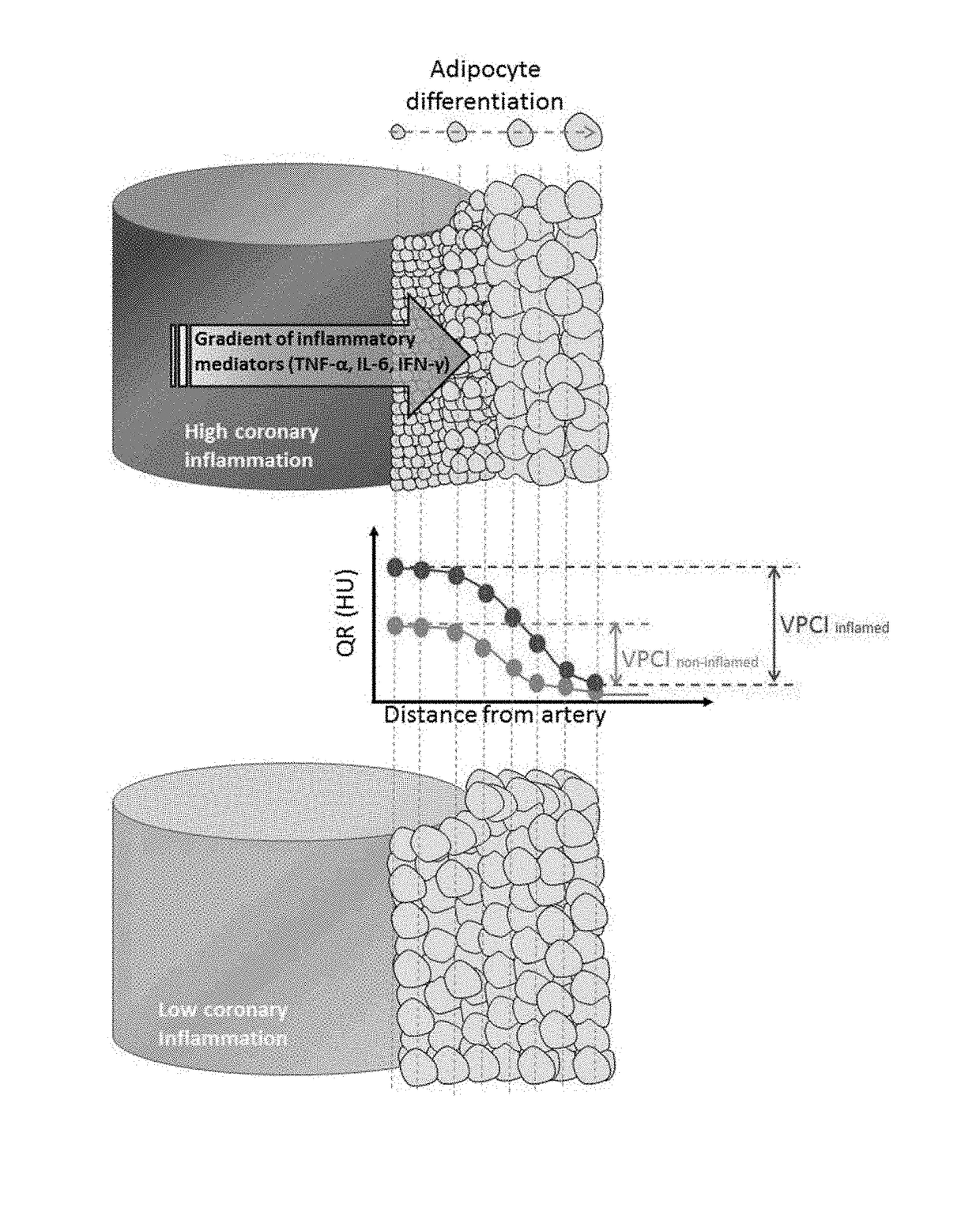
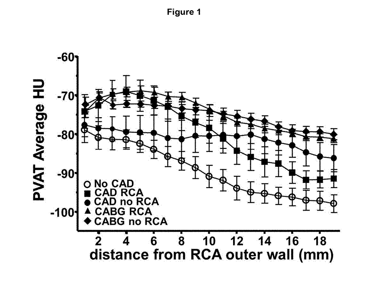
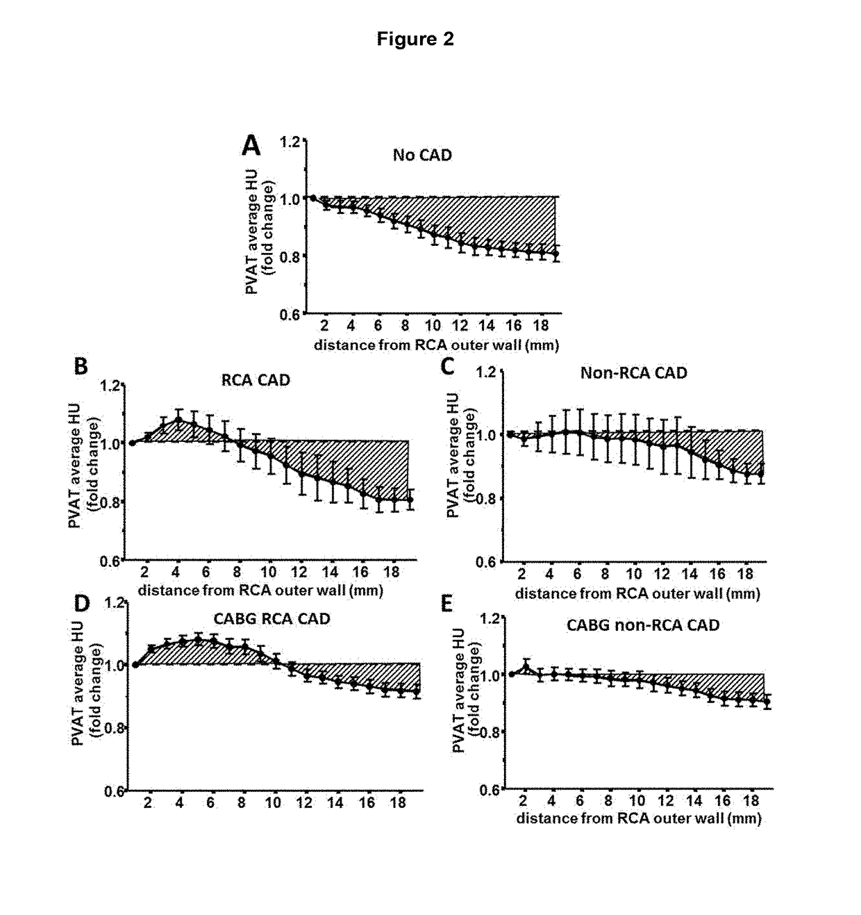
[0077]Available computed tomography (CT) images from a 64-slice scanner (General Electric, LightSpeed Ultra, General Electric, Milwaukee, Wis., USA) were used to analyse perivascular tissue and vascular disease phenotype. Acquisition settings were adjusted according to the local clinical protocols to achieve optimum image quality. The reconstructed contrast-enhanced images were transferred to the Aquarius Workstation® from TeraRecon, Inc. (San Mateo, Calif. V.4.4.11) for volume-rendered analysis. 3D curved multiplanar reconstruction was used to define the vascular segment of interest and to analyse perivascular tissue. The reader was required to manually trace a region of interest and perivascular tissue was then segmented in a semi-automated way into concentric layers around the outer vascular wall. Perivascular tissue characteristics were analysed to determine radiodensity values for voxels corresponding to adipose tissue or water within each concentric perivascular layer for the ...
example 2
[0078]Study Population
[0079]Study arm 1 consisted of 453 patients undergoing cardiac surgery at the Oxford University Hospitals NHS Trust (see Table 1). Exclusion criteria were any inflammatory, infectious, liver / renal disease or malignancy. Patients receiving non-steroidal anti-inflammatory drugs were also excluded. During surgery, samples of adipose tissue were harvested, i.e. subcutaneous (ScAT, from the site of the chest incision), thoracic (ThAT, from the central thoracic area, attached to the pericardium) and epicardial adipose tissue (EpAT, from the site of the right atrioventricular groove, away from any visible vessel). Adipose tissue samples were snap-frozen for gene expression studies, histology and CT imaging as explants, as described below. A subgroup of 105 of these patients underwent also CT angiography (CTA), as described below, with an aim to link the histological and biological characteristics of the adipose tissue biopsies, with the imaging characteristics of the ...
PUM
 Abstract
Description
Claims
Application Information
 - R&D
- Intellectual Property
- Life Sciences
- Materials
- Tech Scout
- Unparalleled Data Quality
- Higher Quality Content
- 60% Fewer Hallucinations
Browse by: Latest US Patents, China's latest patents, Technical Efficacy Thesaurus, Application Domain, Technology Topic, Popular Technical Reports.
© 2025 PatSnap. All rights reserved.Legal|Privacy policy|Modern Slavery Act Transparency Statement|Sitemap|About US| Contact US: help@patsnap.com


