Polymeric Nanoparticles for Enhancing HIFU-Induced Ablation

a technology of polymer nanoparticles and ablation, applied in the field of medical therapy, can solve the problems of limited target organs, limited ultrasound imaging depth, and 10000 nm diameter, and achieve the effect of enhancing the ablative effect of hifu treatment and enhancing the ablation effect of hifu in the focal region

Inactive Publication Date: 2018-07-05
STICHTING KATHOLIEKE UNIV
View PDF3 Cites 0 Cited by
  • Summary
  • Abstract
  • Description
  • Claims
  • Application Information

AI Technical Summary

Benefits of technology

[0012]Contrary to a prejudice in the art, we have found that particles comprising a liquid perfluorocarbon that does not undergo phase change under the influence of high intensity focused ultrasound (HIFU), are very effective in enhancing the ablative effect of HIFU treatment. The invention therefore relates to a polymeric particle comprising a polymer entrapping a liquid perfluorocarbon fo

Problems solved by technology

Ultrasound imaging is potentially quantitative and it is not a whole body imaging modality, and is therefore limited to target organs.
Ultrasound imaging is limited with respect to depth of imaging.
They commonly have a relatively large size (1000-10000 nm diameter) which is generally unsuitable for applications such as cell labeling.
Moreover, they are also unsuitable for imaging outside the blood stream e.g. in tumor imaging.
They also suffer from the additional disadvantage that cell damage, including to blood vessels, may occur as the gas bubbles burst.
Moreover, gas-filled microbubbles can be unstable so that they cannot be stored for a significant amount of time; they typically have to be used soon after hydration.
Finally, such large agents cannot leave the circulation and thus present very limited opportunities for in vivo targeting or drug delivery applications.
Their large size also encourages prompt clearance by the kidneys, which further limits their useful lifetime in vivo.
However, some remaining problems need to be addressed before HIFU can be used extensively in clinica

Method used


Image

  • Polymeric Nanoparticles for Enhancing HIFU-Induced Ablation
  • Polymeric Nanoparticles for Enhancing HIFU-Induced Ablation
  • Polymeric Nanoparticles for Enhancing HIFU-Induced Ablation

Examples


example 1

ncement in a Tissue Model with Polymeric Particles

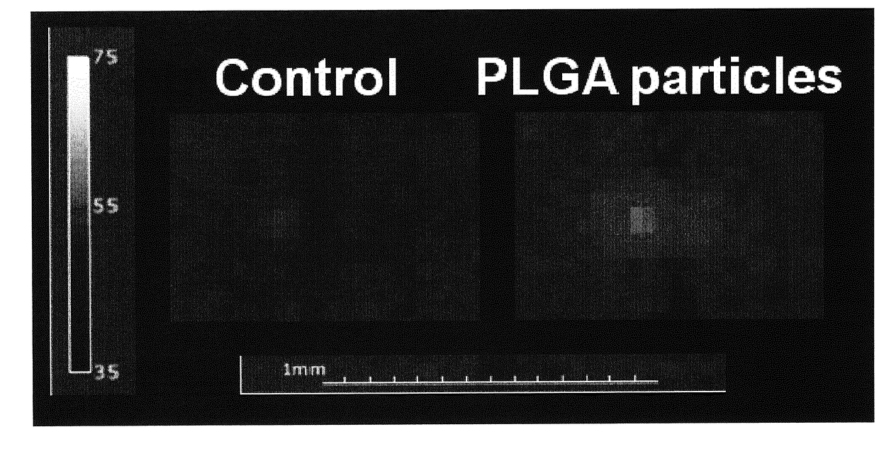
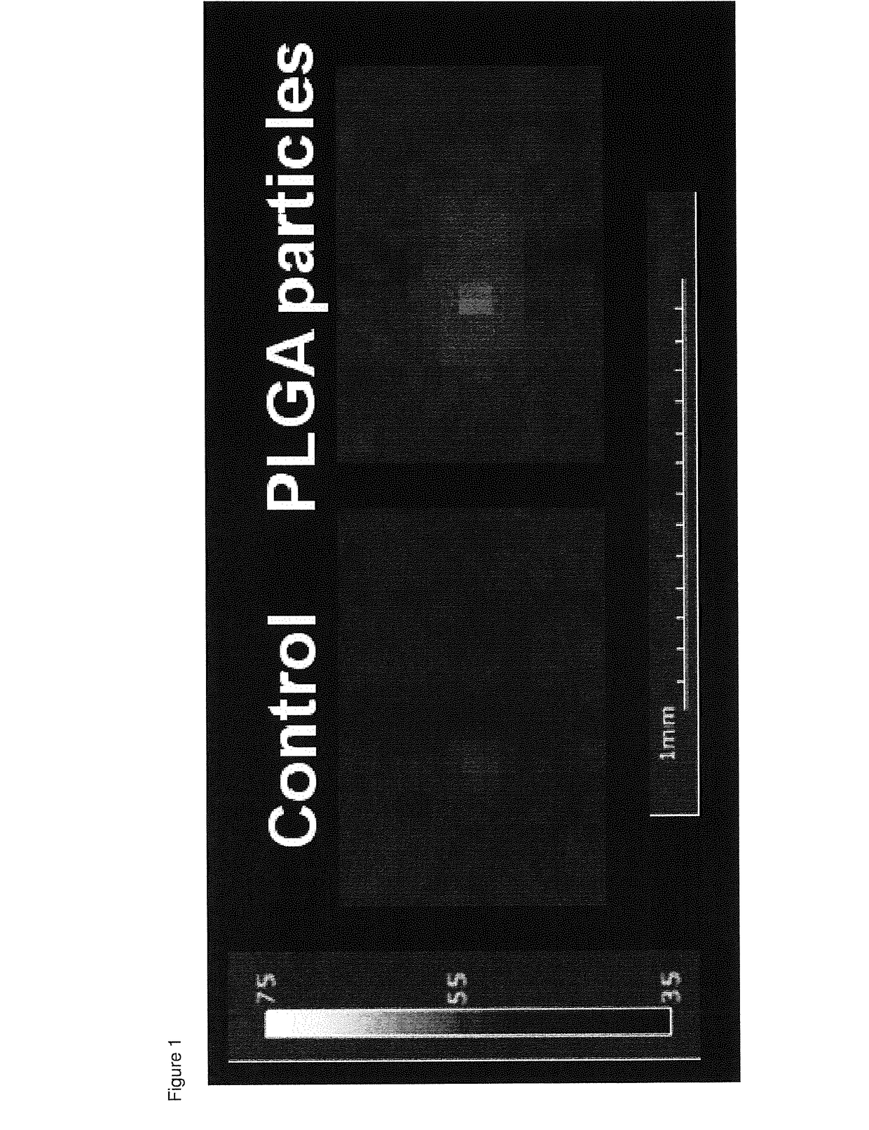
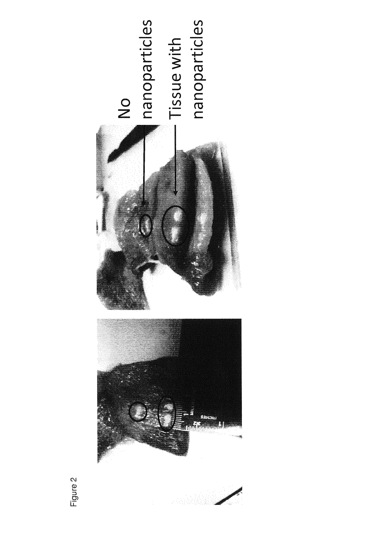
[0042]Particles prepared according to example 2 with a high gadolinium content were injected in a sample of chicken breast that served as a tissue phantom (10 mg / ml). HIFU was carried out at 38 W with a 2 second pulse on a Bruker Clinscan system (7 T horizontal bore). The relevant tissue was then sectioned to directly visualize the ablated zone. Temperature changes were also measured in real time using standard MR thermometry sequences. Comparable results were obtained with the same particles comprising medium and low content of gadolinium. Particles without the gadolinium also showed an enhancement of the ablation effect, although this was less than particles with the low gadolinium content.

example 2

n of Nanoparticles

[0043]PLGA (0.09 gram) was dissolved in 3 ml dichloromethane in a glass tube. Liquid perfluoro-15-crown-5-ether (890 microliter) was added followed by 50 ml of a solution of Prohance (a 3 mg / ml solution of Gadoteridol) diluted in water. Optionally, additional agents, such as a fluorescent dye, may be added to the fluorocarbon at this stage. If a fluorescent particle was required, 1 mg of IcG or IC-Green (Indocyanine Green, Akorn Pharmaceuticals) was added to the solution.

[0044]We prepared particles with a high, medium and low content of gadolinium. For that purpose, the above mentioned solution of Prohance® in water comprised 11.5, 5.75 and 2.85 ml respectively of Prohance® added up with water to 50 ml of solution. The entire mixture was then added dropwise into 25 ml of a solution of polyvinyl alcohol in water (20 gram / liter) under constant sonication (Branson Digital Sonifier 250; 3 minute cycle with 60 sec on and 10 sec off and maximum temperature of 20 degrees ...

example 3

isation of Particles

[0045]We found that particles as prepared above were stable for at least a year when kept at −20 degrees Celsius in the dry form. The particles were also stable in solution at working concentrations for at least 3 months at minus 4 degrees Celsius.

[0046]Diameter of particles prepared according to example 2 was determined using dynamic light scattering (DLS) as previously described (Biomaterials. 2010 September; 31(27):7070-7). The particle size ranged from 80 to 500 nm with a sharp peak at 181 nm.

[0047]The particle diameter distribution remained stable for several months. The particles were lyophilised and frozen for storage. However, particles stored as aliquots in water (frozen) were also stable.

[0048]The particles prepared according to example 2 with high and medium gadolinium content, dissolved in water at a concentration of 1 mg / ml appeared to be exceptionally stable under conditions of ultrasound imaging. We measured particle diameter and count rate (indica...


PUM


Abstract

In the field of medical therapy, more in particular in the field of ablation therapy using ultrasound, such as high intensity focused ultrasound (HIFU), devices and methods are disclosed for enhancing the ablation effect of HIFU. More in particular, a polymeric particle is disclosed, including a polymer entrapping a liquid perfluorocarbon for use in high frequency ultrasound (HIFU) ablation therapy in a human or animal body, wherein the HIFU is focused in a focal region, wherein the ablation effect of the HIFU in the focal region is enhanced by administering the particles to the human or animal body, and the liquid perfluorocarbon does not undergo a phase change from liquid to gas during exposure to the HIFU.

Description

FIELD OF THE INVENTION[0001]The invention is in the field of medical therapy, more in particular in the field of ablation therapy using ultrasound, such as high intensity focused ultrasound (HIFU). The invention provides means and methods for enhancing the ablation effect of HIFU.BACKGROUND OF THE INVENTION[0002]The use of ultrasound in medical imaging procedures is well known in the art. It is the most frequently used clinical imaging modality. Ultrasound is known as an economical, non-invasive, real time technique with a well-established safety record. It can be used for longitudinal studies and repeated use is not harmful for the body.[0003]Ultrasound devices do not produce any ionizing radiation and their operation does not involve the use of radiolabels. The devices for performing ultrasound imaging are portable and already in widespread use. Ultrasound imaging is potentially quantitative and it is not a whole body imaging modality, and is therefore limited to target organs. Ul...

Claims


Application Information

Patent Timeline
IPC IPC(8): A61K41/00A61K49/22A61N7/02
CPCA61K41/0028A61K49/222A61N7/02
Inventor SRINIVAS, MANGALAFIGDOR, CARL GUSTAVDE VRIES, INGRID JOLANDA MONIQUE
Owner STICHTING KATHOLIEKE UNIV
Why Eureka
Try Eureka