Non-human mammal

Inactive Publication Date: 2018-09-13
TOKYO METROPOLITAN INST OF MEDICAL SCI
View PDF1 Cites 0 Cited by
  • Summary
  • Abstract
  • Description
  • Claims
  • Application Information

AI Technical Summary

Benefits of technology

The non-human mammal in this patent can show allergic reactions to things like mite antigen, cedar pollen, egg albumin, and so on. It can also help researchers understand how allergic diseases develop when there are reactions to these substances. This information can be used to develop methods to prevent and treat allergies.

Problems solved by technology

Although the single CD4-positive T cell responses can be analyzed in vivo by transferring an antigen specific-CD4-positive T cell clone that is previously established in vitro into normal mice, in that case all of the CD4-positive T cells in live bodies cannot be antigen-specific.
Although it is possible to use T cell-deficient mice as a recipient side, all the presently existing T cell-deficient mice show some sorts of immunodeficiencies, thereby making it difficult to construct an appropriate experimental system in view of analyzing the immune mechanisms mediated by transferred CD4-positive T cells.

Method used


Image

  • Non-human mammal
  • Non-human mammal
  • Non-human mammal

Examples


example 1

Preparation of Antigen-Specific CD4-Positive T Cells

[0076]Mite allergens (Dermatophagoides farinae (Df) and Dermatophagoides pteronyssinus (Dp)) were mixed with aluminum hydroxide (Alum) or Complete Freund's adjuvant (CFA) in a combination of Table 1, and CD2F1 mice (10-week old, male) were injected with the mixture at tail base. The CD2F1 mice were F1 of BALB / c and DBA2.

[0077]One week after the antigen sensitization, inguinal lymph node was excised, and suspended in a culture medium containing fetal bovine sera to prepare the cells. The CD4-positive T cells were concentrated with a magnetic sorting system using CD4 antibody beads, and thereafter the culture of concentrated cells was started together with splenocytes prepared from the identical mice, of which proliferation ability was made deficient by X-ray irradiation, and specific antigens. The cells were cultured without adding IL-2 for 3 to 4 days from the beginning of culture, in order to reduce the proliferation of the antige...

example 2

Generation of Transnuclear Mice

[0079]Transnuclear mice were generated using the CD4-positive T cells obtained according to Example 1 as a nuclear donor cell. The embryos were generated as prescribed in the methods described in Nature 1998; 394:369-74, and Biology of Reproduction 2012; 86:1-6, and as the culture medium of the embryos, a KSOM medium as prescribed in a method described in Lawitts J. A., and Biggers J. D. 1993, Culture of preimplantation embryos, Methods Enzymol. 225:153-164 was used. Concretely, ova obtained by superovulation of mature CD2F1B6D2F1 female were enucleated in the presence of 5 μg / mL cytochalasin B, and the nuclear donor cell was injected thereto with a piezo-drive micromanupilator. After the injection, the cells were cultured under the environmental conditions of 37.5° C. and 6% CO2 for 1 to 2 hours, and thereafter activated with a KSOM medium in the presence of 50 nM trichostatin A, 5 μM Latrunculin A, and 3 mM strontium. Subsequently, the cells were cul...

example 3

Antigen Reactivities 1 in CD4-Positive T Cells of Cloned Mice

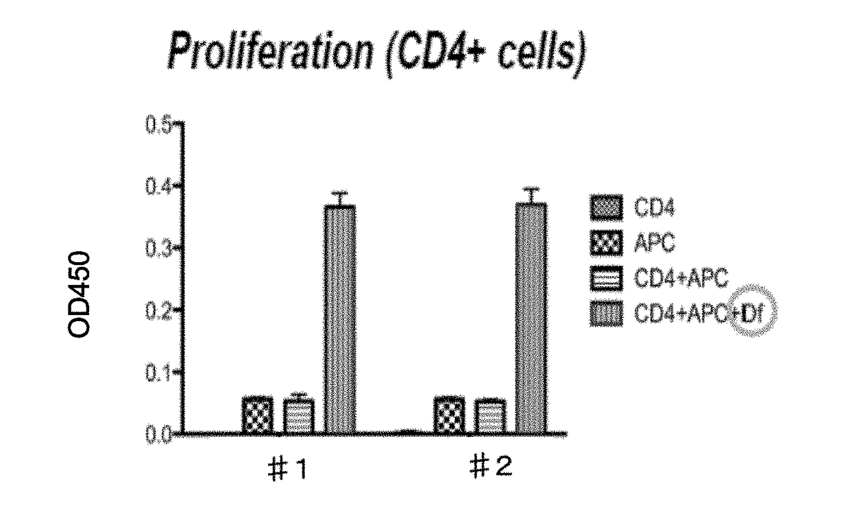
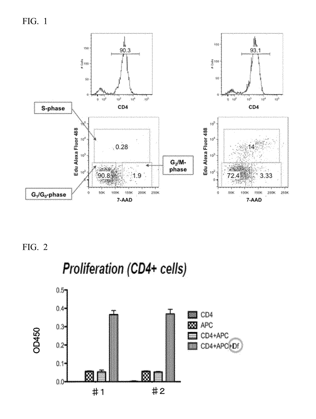
[0080]White blood cells or CD4-positive T cells were prepared from peripheral blood of the Df-specific CD4-positive T cell cloned mice established in Example 2, and the cells were cultured together with X-ray irradiated splenocytes of nolinal mice and a specific antigen (Df). On the fourth to fifth day from the beginning of culture, the cell proliferative responses were analyzed using a Cell proliferation reagent (manufactured by Roche Diagnostics). The results are shown in FIG. 2.

[0081]From FIG. 2, the antigen-specific proliferative responses were found in the mice sensitized with the Df antigen. It was clarified from this matter that the cloned mice possessing the antigen specificity of the donor cell could be established as intended.


PUM


Abstract

A non-human mammal and an offspring thereof, obtainable by a somatic cell nuclear transfer method using a nucleus of a CD4-positive T cell as a nuclear donor. The non-human mammal of the present invention surely and efficiently shows allergic reactions specific to various antigens that are shown to have associations with an immune allergic disease, such as mites and cedar pollens, so that the non-human mammal can be suitably used as the developmental models of allergic diseases for studies of various diseases by studying or pursuing possibilities of applications to the diseases.

Description

TECHNICAL FIELD[0001]The present invention relates to a non-human mammal. More particularly, the present invention relates to a cloned non-human mammal which can be used as an allergy model animal obtainable by a somatic cell nuclear transfer method, and a method for preparing the non-human mammal.BACKGROUND ART[0002]Live bodies have been protected from foreign substances primarily by immune responses, and an immune system which is composed of various cells and factors produced thereby. In particular, leukocytes, especially lymphocytes, play a key role. The lymphocytes are classified in two major types, B lymphocytes (which may be hereinafter referred to as B cells) and T lymphocytes (which may be hereinafter referred to as T cells), both of which specifically recognize an antigen and act to protect the live bodies from the antigen.[0003]Peripheral T cells are mostly composed of CD(Cluster of Differentiation)4-positive cells having a CD4 marker and CD8-positive T cells having a CD8 ...

Claims


Application Information

Patent Timeline
IPC IPC(8): A01K67/027C07K14/725A61K35/17C12N15/877C12N5/0783
CPCA01K67/0275C07K14/7051A61K35/17A01K67/0273C12N15/8775C12N5/0636A01K2227/105A01K2267/0368A01K67/027C12N15/09C12N2517/04A61K39/4632A61K39/464839A61K39/4611A61K39/4644
Inventor KAMINUMA, OSAMUINOUE, KIMIKOKATAYAMA, KAZUFUMIOGURA, ATSUO
Owner TOKYO METROPOLITAN INST OF MEDICAL SCI
Why Eureka
Try Eureka