

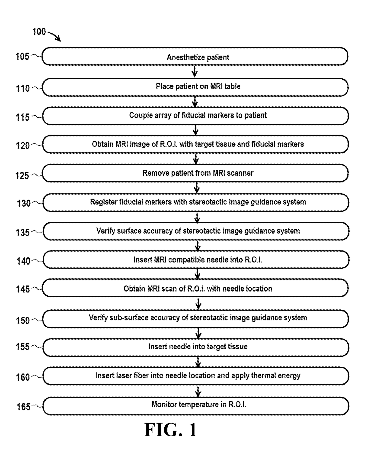
Utilization of laser interstitial thermotherapy guided with real time thermal MRI
a technology of real-time thermal imaging and laser interstitial thermotherapy, which is applied in the field of laser interstitial thermotherapy guided with real-time thermal imaging, can solve the problems of inconvenient visualization techniques, inability to locate and track instruments, and inability to accurately determine the position of instruments, so as to improve the safety and accuracy of a variety of spinal procedures and avoid distortion of surface landmarks
- Summary
- Abstract
- Description
- Claims
- Application Information
AI Technical Summary
Benefits of technology
Problems solved by technology
Method used
Image

Examples
working example
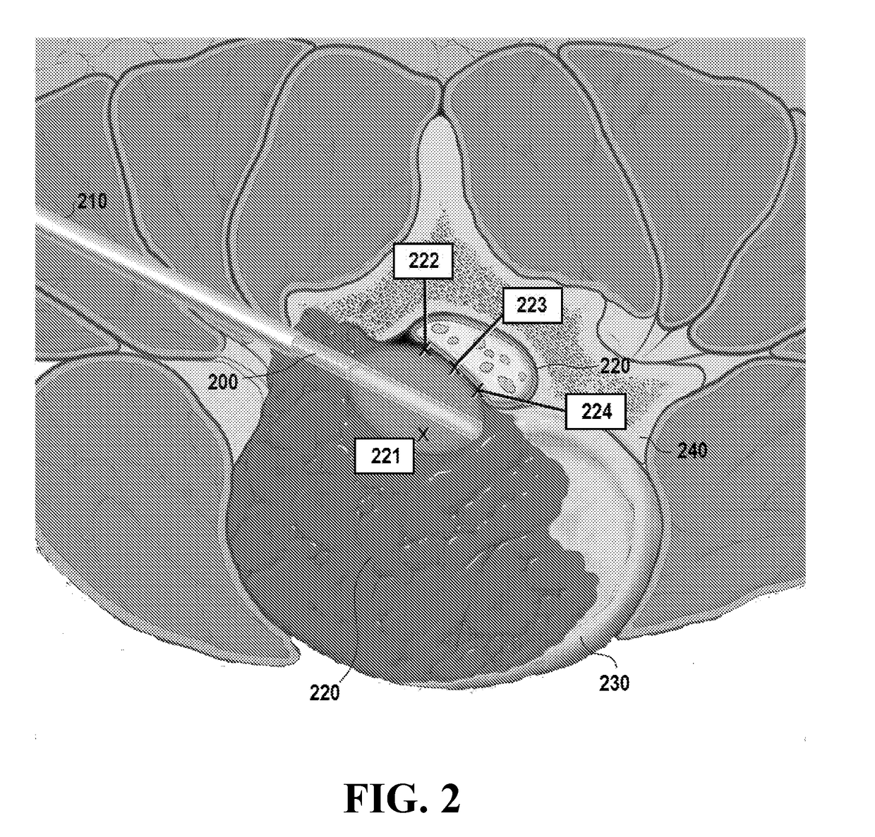
[0069]High grade malignant spinal cord compression is commonly managed with a combination of surgery, aiming removal of the epidural tumor, followed by stereotactic spinal radiosurgery (SSRS), aiming local tumor control. The investigators introduce the use of spinal laser interstitial thermotherapy (SLITT) as an alternative to surgery prior to SSRS.
[0070]Eleven patients with high degree of epidural malignant compression due to radio-resistant tumors were selected. Visual analog pain score (VAS) and quality of life score (QoL) were obtained before, within 30 and within 60 days after the procedure. The investigators performed percutaneous placement of a laser probe in the epidural space. Real time thermal MR was used to monitor the tissue damage in the region of interest. All patients received post-op SSRS. The maximum thickness of the epidural tumor was measured and the degree of the epidural spinal cord compression (ESCC) scored in pre and post procedure MRI as follows: Grade 0: tum...
PUM
 Abstract
Description
Claims
Application Information