Method for generating cell-penetrating stapled peptides that lack nonspecific membrane-lytic properties for therapeutic targeting

a peptide, non-specific technology, applied in the direction of peptides, peptide/protein ingredients, instruments, etc., can solve the problems of inability to definitively know the criteria for generating cell-penetrant peptide analogs, inability to accurately identify and/or optimize promising peptides, and inability to achieve optimal staples. , to achieve the effect of minimizing non-specific cell membrane lytic activity, reducing the propensity for cell uptake, and rapid determination

Inactive Publication Date: 2019-02-14
DANA FARBER CANCER INST INC
View PDF1 Cites 1 Cited by
  • Summary
  • Abstract
  • Description
  • Claims
  • Application Information

AI Technical Summary

Benefits of technology

[0008]We have developed an unbiased statistical method for rapid determination of optimal stapled and / or stitched peptide biophysical design parameters for maximal cell uptake propensity while minimizing nonspecific cell membrane lytic activity (a required feature for stapled and / or stitched peptides to be effective in vivo).
[0012]The method can be applied to enhance the cellular uptake of pro-apoptotic p53 stapled peptides by generating a revised p53 panel bearing E to Q and D to N mutations to optimize the peptides' α-helicity and adjust the overall peptide charge from −2 to 0 and +1. The method can yield cell-penetrant analogs capable of reactivating the p53 pathway through targeted inhibition of HDM24 and HDMX5.
[0013]The method can be applied two or more times to iteratively enhance other biophysical properties of a candidate stapled and / or stitched peptide. For example, the method can be applied to mitigate serum binding, eliminate nonspecific cell lytic activity, and further improve potency6.
[0038]In some embodiments, the HSP has a calculated hydrophobicity that is between 0.5 and 0.9 (e.g., 0.5, 0.6, 0.7, 0.8, 0.9) and has a HPLC retention time of 9.56 or greater at pH7 or pH4 (e.g., a HPLC retention time of 9.56 or greater at pH7, or a HPLC retention time of 11.0 or greater at pH4). In some embodiments, the HSP has a HPLC retention time of 9.56 or greater at pH7 or pH4 (e.g., a HPLC retention time of 9.56 or greater at pH7, or a HPLC retention time of 11.0 or greater at pH4) and a percent α-helicity of 40% to 90% (e.g., 61% to 86%). In some embodiments, the cell-penetrant HSP may have a combination of one or more (e.g., one, two, three, four, or five) of the biophysical properties described above. Table 1 below provides a summary of the key biophysical parameters and values of cell-penetrant hydrocarbon stapled and / or stitched peptides as well as cell-penetrant hydrocarbon stapled and / or stitched peptides that are not non-specifically cell lytic.TABLE 1Biophysical ParametersBiophysicalCell-penetrant hydrocarbon stapled and / or stitchedParameterpeptideTotal Internalizedgreater than 0.5 × 10⁶ (e.g., 0.5 × 10⁶ toFITC Intensity6.0 × 106)(TIFI)Hydrophobicitygreater than 0.5 (e.g., 0.5 to 0.9)HPLC retention9.2 minutes or greater (e.g., 9.2 to 11.2 minutes)time at pH 7 or pH 4α-helicityabout 40% to about 90%**pIless than 9.76**Net charge+2 to −1 (e.g., +4 to −3)**indicates that these values are different for a cell-penetrant hydrocarbon stapled and / or stitched peptide that does not exhibit t non-specific cell lytic activity: α-helicity ranges for the latter are about 21% to about 96%, and pI is less than 9.76 (8.8 to 9.34).+ If both pI is greater than 9.76 and HPLC retention time is greater than 9.78 min, chances of non-specific membrane lysis greatly increase.
[0055]In certain embodiments, the second HSP has increased retention time as compared to the first HSP, and the second HSP has improved cellular uptake as compared to the first HSP. In certain embodiments, the second HSP has an increased net charge as compared to the first HSP, and the second HSP has improved cellular uptake as compared to the first HSP. In certain embodiments, the second HSP has a reduced net charge as compared to the first HSP, and the second HSP has improved cellular uptake as compared to the first HSP. In certain embodiments, the second HSP binds to the same target as the first HSP with the same or greater binding affinity to the target as compared to the first HSP. In certain embodiments, the second HSP binds to the target protein with a binding affinity of less than 100 nM (e.g., less than 90 nM, less than 80 nM, less than 70 nM, less than 60 nM, less than 50 nM, less than 40 nM, about 1 nM to about 10 nM, about 1 nM to about 20 nM, about 1 nM to about 30 nM, about 1 nM to about 40 nM, about 1 nM to about 50 nM, about 1 nM to about 60 nM, about 1 nM to about 70 nM, about 1 nM to about 80 nM, about 1 nM to about 90 nM, about 1 nM to about 100 nM, about 10 nM to about 20 nM, about 10 nM to about 30 nM, about 10 nM to about 40 nM, about 10 nM to about 50 nM, about 10 nM to about 100 nM, about 20 nM to about 100 nM, about 30 nM to about 100 nM, about 40 nM to about 100 nM, or about 50 nM to about 100 nM).
[0056]In some embodiments, the second HSP has the same or reduced effect on non-specific cell lysis.

Problems solved by technology

However, the exact design features that reliably confer cellular penetrance have been elusive.
However, to date, the development of cell-permeable stapled peptides (i.e. cell-penetrant hydrocarbon-stapled and / or stitched peptides) has generally relied on (cumulative) empirical observations and trial-and-error, both of which are inherently inefficient at identifying and / or optimizing promising peptides.
Although some theories have been proposed1,13-15, and specific peptides have been shown to be capable of penetrating cells under certain conditions without lysing them (and causing consequent cell death)4,20,53, the lack of definitive knowledge of the criteria for generating cell-penetrant peptide analogs has presented a significant roadblock to realizing the broader utility of stapled peptides, whether as potential therapeutics in vivo or even only as experimental reagents or tools (e.g., for cellular analysis).
Further, some of the proposed theories have been shown to be inconsistent with experimental evidence.
Additionally, the use of cell-impermeable stapled peptides in cellular studies has led to faulty conclusions about stapled peptide uptake and activity16-19.

Method used


Image

  • Method for generating cell-penetrating stapled peptides that lack nonspecific membrane-lytic properties for therapeutic targeting
  • Method for generating cell-penetrating stapled peptides that lack nonspecific membrane-lytic properties for therapeutic targeting
  • Method for generating cell-penetrating stapled peptides that lack nonspecific membrane-lytic properties for therapeutic targeting

Examples


example 1

and Methods

Stapled Peptide Synthesis and Characterization

[0261]All hydrocarbon-stapled peptides were synthesized, derivatized at the N-terminus with FITC-βAla or acetyl, and purified to >95% homogeneity by LC / MS using methods known in the art and as previously described34. Acetylated peptides were dissolved in 10% (vol / vol) acetonitrile in water for circular dichroism analyses, performed on an Aviv Biomedical spectrophotometer using methods known in the art and as previously described34.

ImageXpress Microscopy Analysis

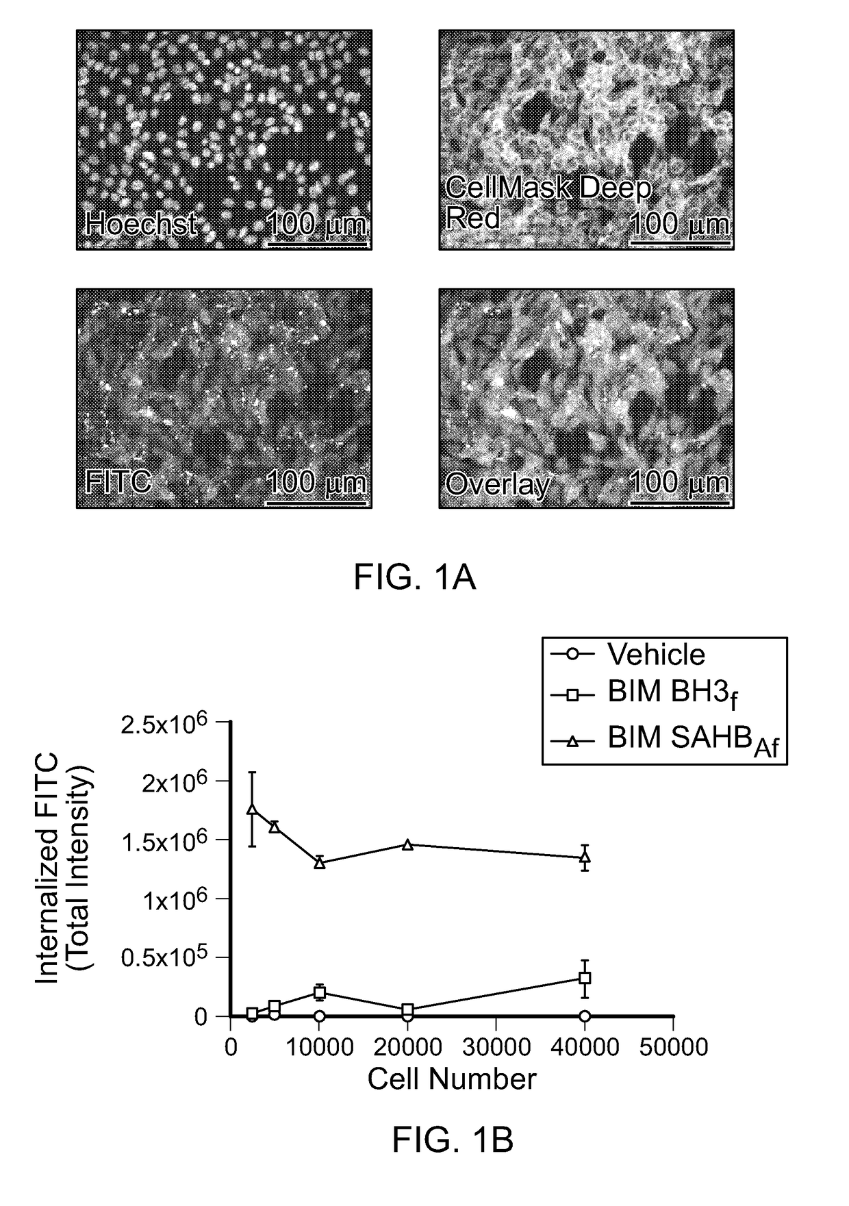
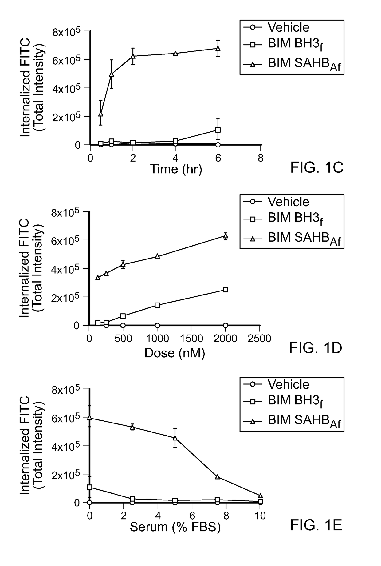
[0262]For high-content fluorescence microscopy analysis, the indicated cell lines were plated in black, clear bottom plates overnight at a density of 2×10⁴ cells per well in DMEM supplemented with 10% (vol / vol) FBS, 1% penicillin / streptomycin, and 1% glutamine. The following day, cells were treated with 0.5 μM FITC-labeled peptides or the equivalent amount of vehicle (0.1% DMSO) for 4 h in serum-free DMEM, and then stained with Hoechst 33342 and CellMask Deep Red (CMDR,...

example 2

nt and Validation of a High-Throughput / High-Stringency Microscopy Assay for Stapled Peptide Internalization

[0271]High-content epifluorescence microscopy and the ImageXpress Micro (IXM) Widefield High Content Analysis System were used to develop a rigorous quantitation platform for measuring cellular uptake of stapled peptides derivatized at the N-terminus with a FITC fluorophore. Custom Module Editor was used to create a custom module (CM) for analysis based on the following principles: (1) define cellular uptake in mouse embryonic fibroblasts (MEFs) based on visualizing cellular and nuclear boundaries using Hoechst 33342 and CellMask Deep Red (CMDR), respectively, thereby excluding extracellular aggregates and autofluorescent debris (FIG. 1A), (2) contract the resultant cellular mask by a defined pixel boundary to avoid quantitation of extracellular, membrane-adherent peptide, (3) exclude FITC signal outliers that reflect out-of-focus fluorescence, (4) acquire images for analysis a...

example 3

nts of Cellular Uptake for a Staple Scanning Library of BIM BH3

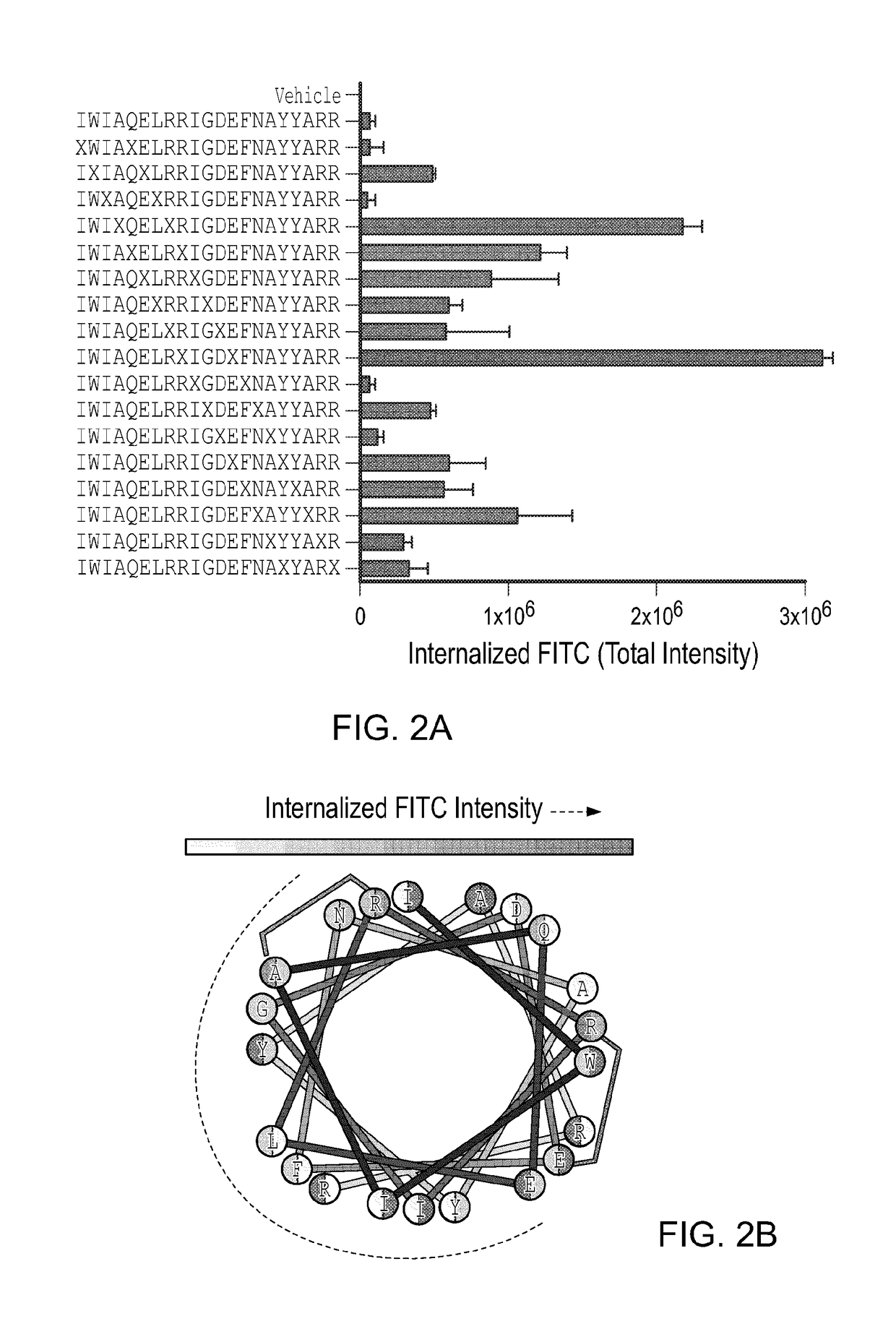
[0274]When designing stapled peptides for biochemical and biological studies, one of the first questions to address is where to install the staple. Here, a library of stapled BIM BH3 peptides was generated by performing a “staple scan” that sequentially places the staple along the length of the template peptide, yielding 17 i, i+4 stapled peptides. Cellular uptake of the FITC-derivatized stapled peptides was monitored by IXM using the previously described CM. Importantly, of the cellular fluorescent dyes measured in the microscopy assay (i.e., Hoechst, CMDR, FITC), only the FITC intensity varied with stapled peptide composition (FIG. 12). Strikingly, our prototype BIM SAHBA1 peptide that bears a staple flanking IGD emerged as the clear winner (TIFI>3.0×10⁶), with four additional constructs containing staples flanking QEL, ELR, AYY, and LRR also demonstrating notable uptake at TIFIs of 2.16×10⁶, 1.21×10⁶, 1.07×10⁶, and 0....


PUM

PropertyMeasurementUnit
pHaaaaaaaaaa
pHaaaaaaaaaa
pHaaaaaaaaaa

Abstract

Methods for generating cell-permeable hydrocarbon-stapled and / or stitched peptides lacking nonspecific membrane lytic properties and methods for using such peptides to target cellular proteins for experimental investigation and / or therapeutic benefit.

Description

CROSS-REFERENCE TO RELATED APPLICATIONS[0001]This application claims the benefit of priority of U.S. Provisional Appl. No. 62 / 298,931 filed Feb. 23, 2016, the contents of which are incorporated by reference in their entirety herein.GOVERNMENT GRANT CLAUSE[0002]This invention was made with government support under grant 1R35CA197583 awarded by the National Institutes of Health (NIH). The government has certain rights in the invention.TECHNICAL FIELD[0003]This disclosure relates to methods for generating cell permeable hydrocarbon-stapled and / or stitched peptides lacking nonspecific membrane lytic properties and methods for using such peptides to target cellular proteins for therapeutic benefit.BACKGROUND[0004]Hydrocarbon-stapled and / or stitched peptides are a class of structured bioactive ligands that have been developed to dissect, target, and / or modulate protein interactions. These peptides comprise a large and diverse array of bioactive α-helices (including evolutionarily-honed mo...

Claims


Application Information

Patent Timeline
Patent Type & Authority Applications(United States)
IPC IPC(8): C07K1/107C07K1/113C07K19/00
CPCC07K1/1077C07K1/113C07K19/00A61K38/00C07K1/006C07K7/08G01N2500/10
Inventor WALENSKY, LOREN D.BIRD, GREGORY H.
Owner DANA FARBER CANCER INST INC
Why Eureka
Try Eureka