Novel protein transduction domain and use thereof
- Summary
- Abstract
- Description
- Claims
- Application Information
AI Technical Summary
Benefits of technology
Problems solved by technology
Method used
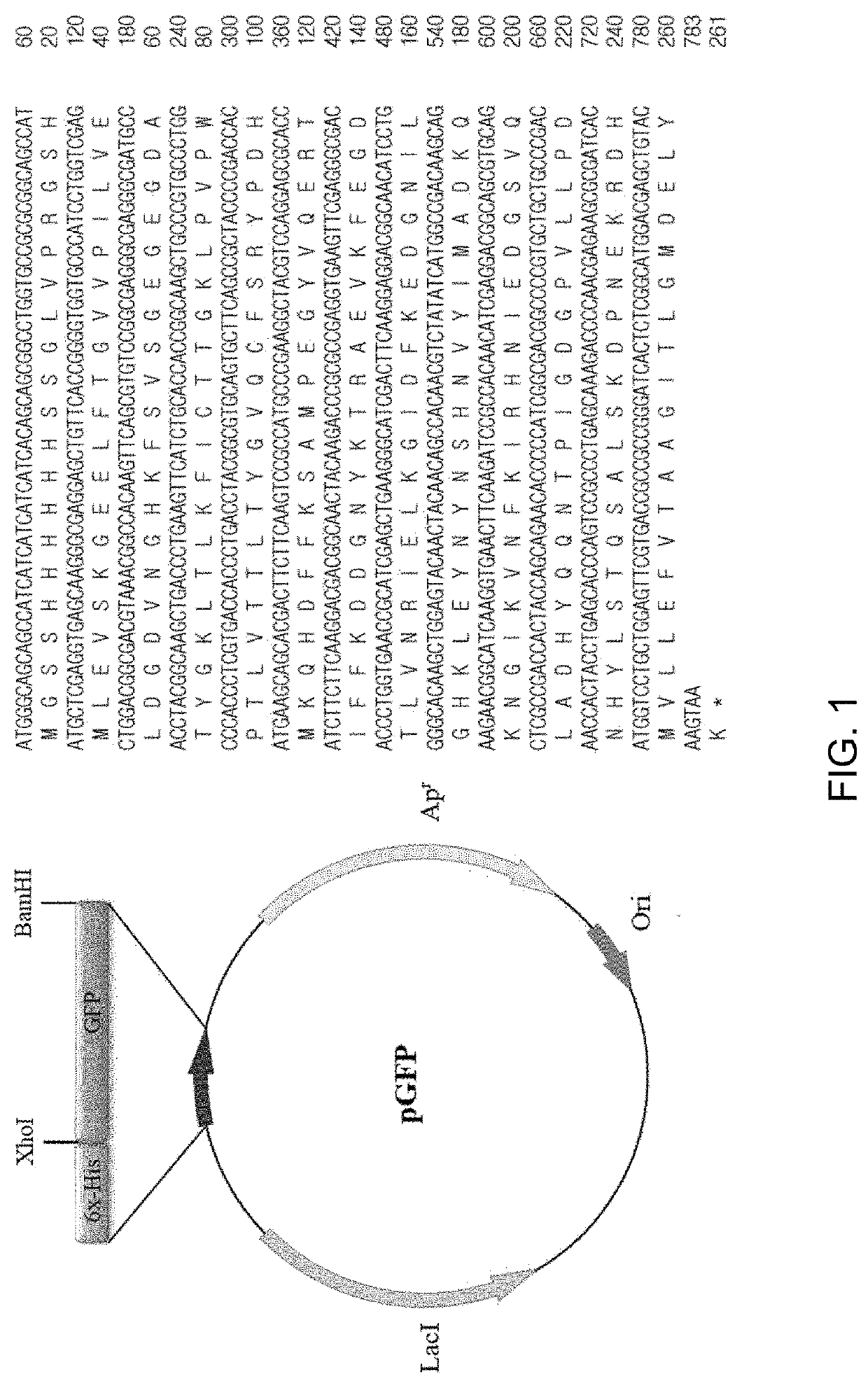
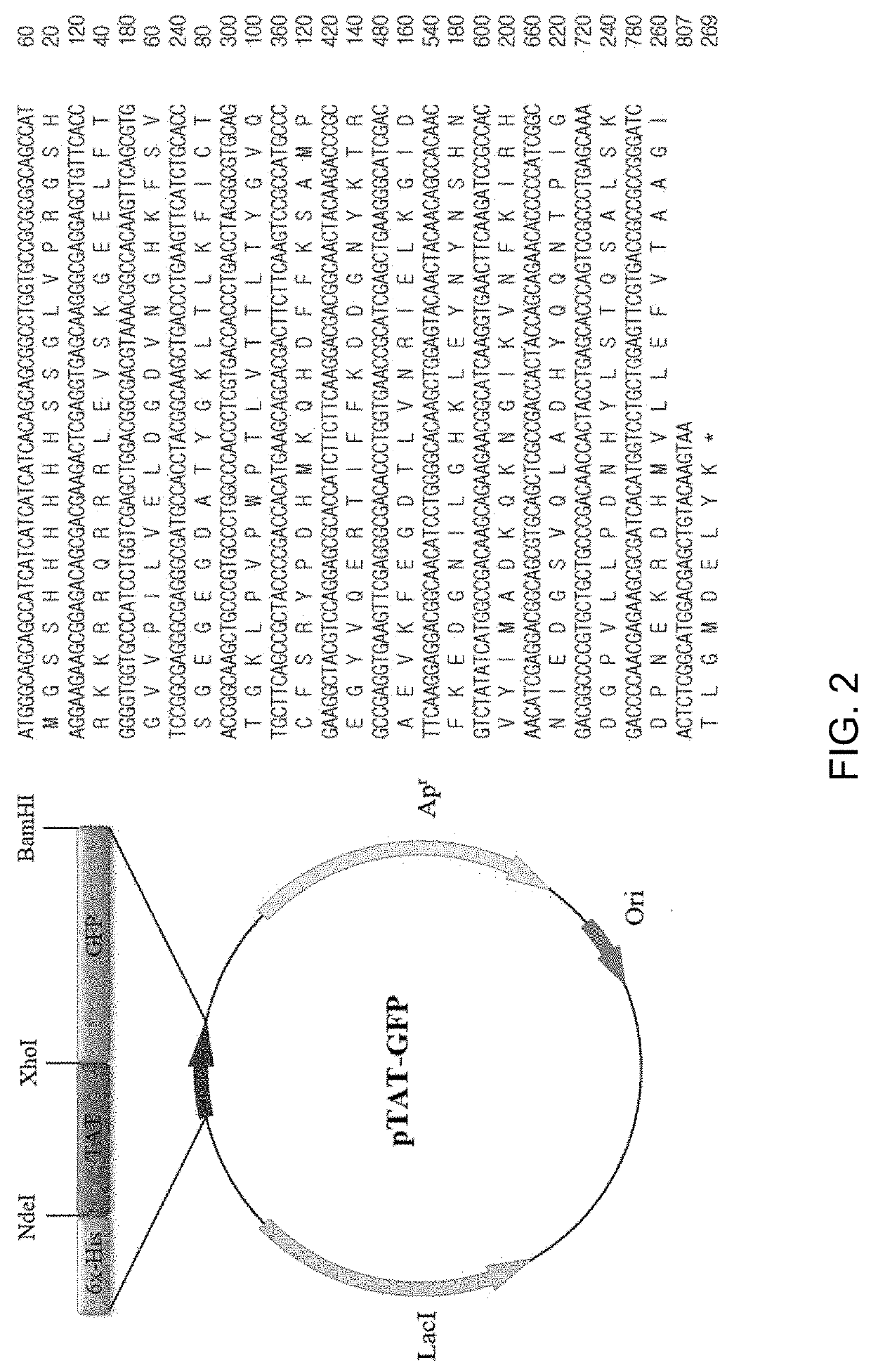
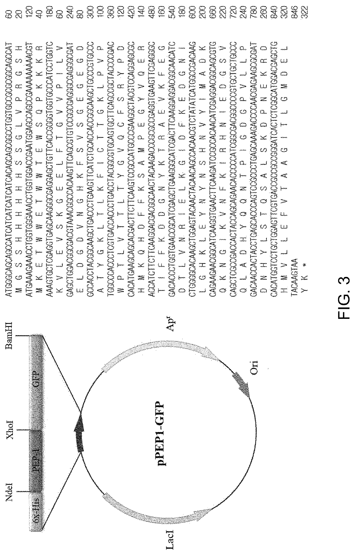
Image

Examples
example 2
n of GFP Recombinant Proteins
[0069]In the present Example, the corresponding proteins were expressed from the recombinant vectors produced in Example above. Four types of expression vectors were each transformed into E. coli Rosetta-gami 2(DE3) cells. The transformed bacterial cells were cultured at 37° C. in 1,000 mL of LB medium containing ampicillin until OD600 reached 0.6 and were further cultured using 0.5 mM IPTG at 22° C. for 24 hours to induce expression of GFP proteins.
[0070]Whether or not respective GFP proteins were expressed was identified by collecting cultured cells before and after inducing expression of target proteins by addition of IPTG, conducting SDS-PAGE electrophoresis and then dyeing the proteins with Coomassie brilliant blue.
[0071]As a result, as can be seen from FIG. 5, GFP expresses 29.35 kDa proteins, TAT-GFP expresses 30.54 kDa proteins, PEP1-GFP expresses 32.18 kDa proteins, and STeP-GFP expresses 32.95 kDa proteins. FIG. 5 shows expression results of GF...
example 3
ion of GFP Recombinant Proteins
[0072]In the present Example, the proteins each expressed in Example were subjected to cell disruption and then only GFP proteins were purely isolated and purified using His tag-affinity columns. Four types of GFP proteins were purified in the same manner as below.
[0073]pET15-GFP, pTAT-GFP, pPEP1-GFP and pSTeP-GFP transformed cells were lysed with a sonicator in 1× native buffer (50 mM NaPO4, 0.5 M NaCl), then centrifuged and separated into an agglomerate (inclusion body) and the cytoplasm (supernatant).
[0074]All four types of GFP proteins were His6-tagged and, using this feature, each supernatant separated by centrifugation after cell disruption was purified with a Ni2+-NTA affinity column (Ni2+-nitrilotriacetic acid Sepharose affinity column, GE Healthcare, USA).
[0075]Equilibrium of columns and bonding with GFP proteins were carried out using 1× native buffer, and washing and elution were carried out using solutions of different concentrations of imi...
example 4
of Cell Penetration of Cell- and Skin-Penetrating GFP Proteins
[0076]In the present Example, in an attempt to compare cell penetration capability of four types of GFP proteins purified in Example, cell penetration was observed using GFP green fluorescence in HaCaT (human keratinocyte) cells.
[0077]HaCaT (human keratinocyte) cells were cultured on a slide for culture for 24 hours. After the culture medium was removed, the cells were washed with HBSS three times and then treated with GFP and cell-penetrating GFP proteins (PEP1-GFP, TAT-GFP, STeP-GFP). Upon treatment of cells, stocks were prepared such that respective proteins reached 10 mM and diluted at 1 / 10 in a medium, and cells were treated such that the final concentration reached 10 μM and then cultured for 2 hours. After 2 hours, the medium was removed, and cells were washed with PBS and then immobilized at −20° C. using cooling methanol for 10 minutes. The cells were thoroughly washed with PBS and dyed with a solution of a nucle...
PUM
| Property | Measurement | Unit |
|---|---|---|
| Hydrophobicity | aaaaa | aaaaa |
| Interaction | aaaaa | aaaaa |
| Bond | aaaaa | aaaaa |
Abstract
Description
Claims
Application Information
 - R&D Engineer
- R&D Manager
- IP Professional
- Industry Leading Data Capabilities
- Powerful AI technology
- Patent DNA Extraction
Browse by: Latest US Patents, China's latest patents, Technical Efficacy Thesaurus, Application Domain, Technology Topic.
© 2024 PatSnap. All rights reserved.Legal|Privacy policy|Modern Slavery Act Transparency Statement|Sitemap