

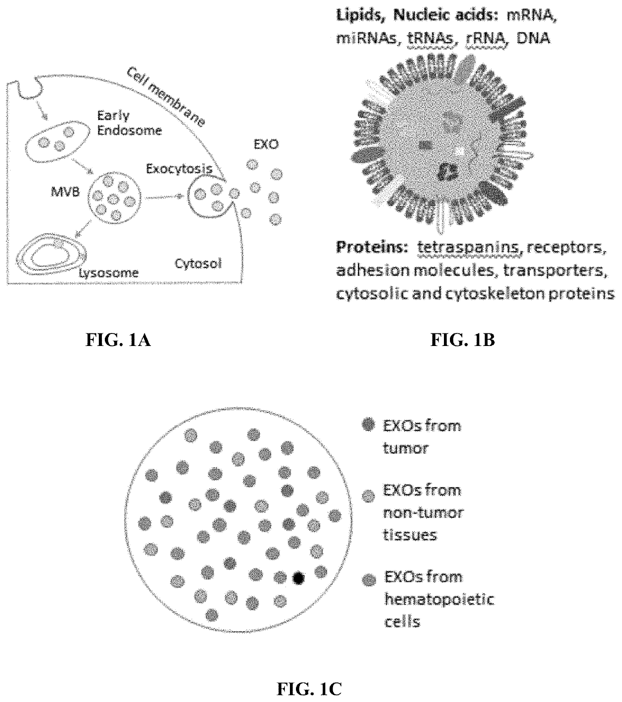
Compositions and methods for the detection and molecular profiling of membrane bound vesicles
a membrane bound vesicle and molecular profiling technology, applied in the direction of fluorescence/phosphorescence, instruments, material analysis, etc., can solve the problems of limited detection sensitivity, impracticality of extracting pure tumor-derived exosomes, and inability to detect pure tumor-derived exosomes
- Summary
- Abstract
- Description
- Claims
- Application Information
AI Technical Summary
Benefits of technology
Problems solved by technology
Method used
Image

Examples
example 1
paration
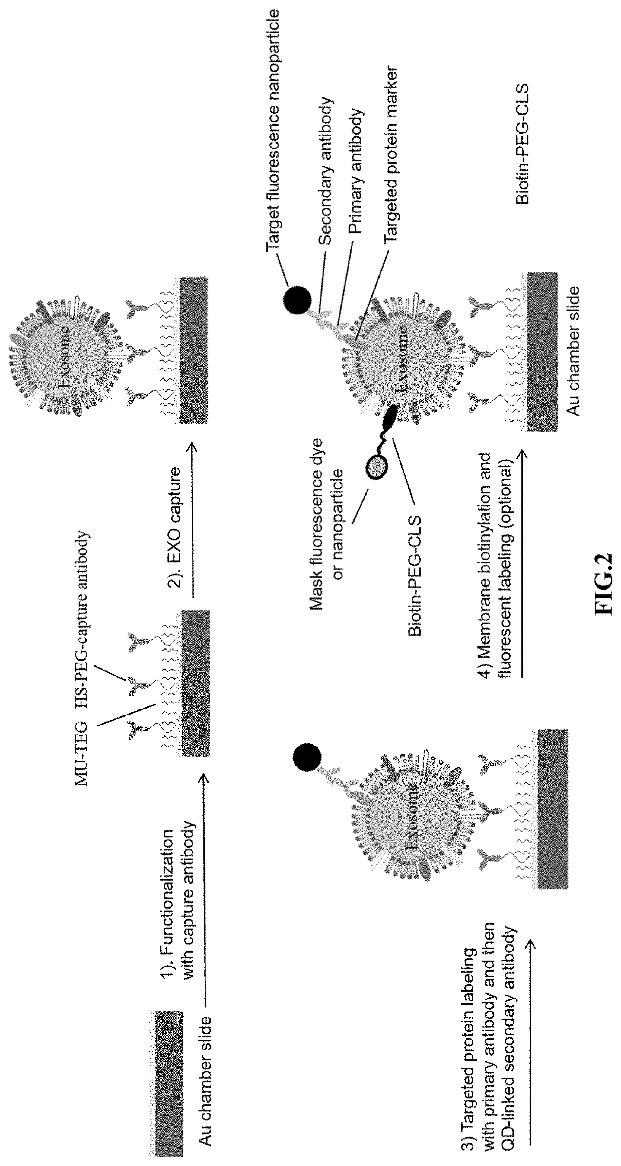
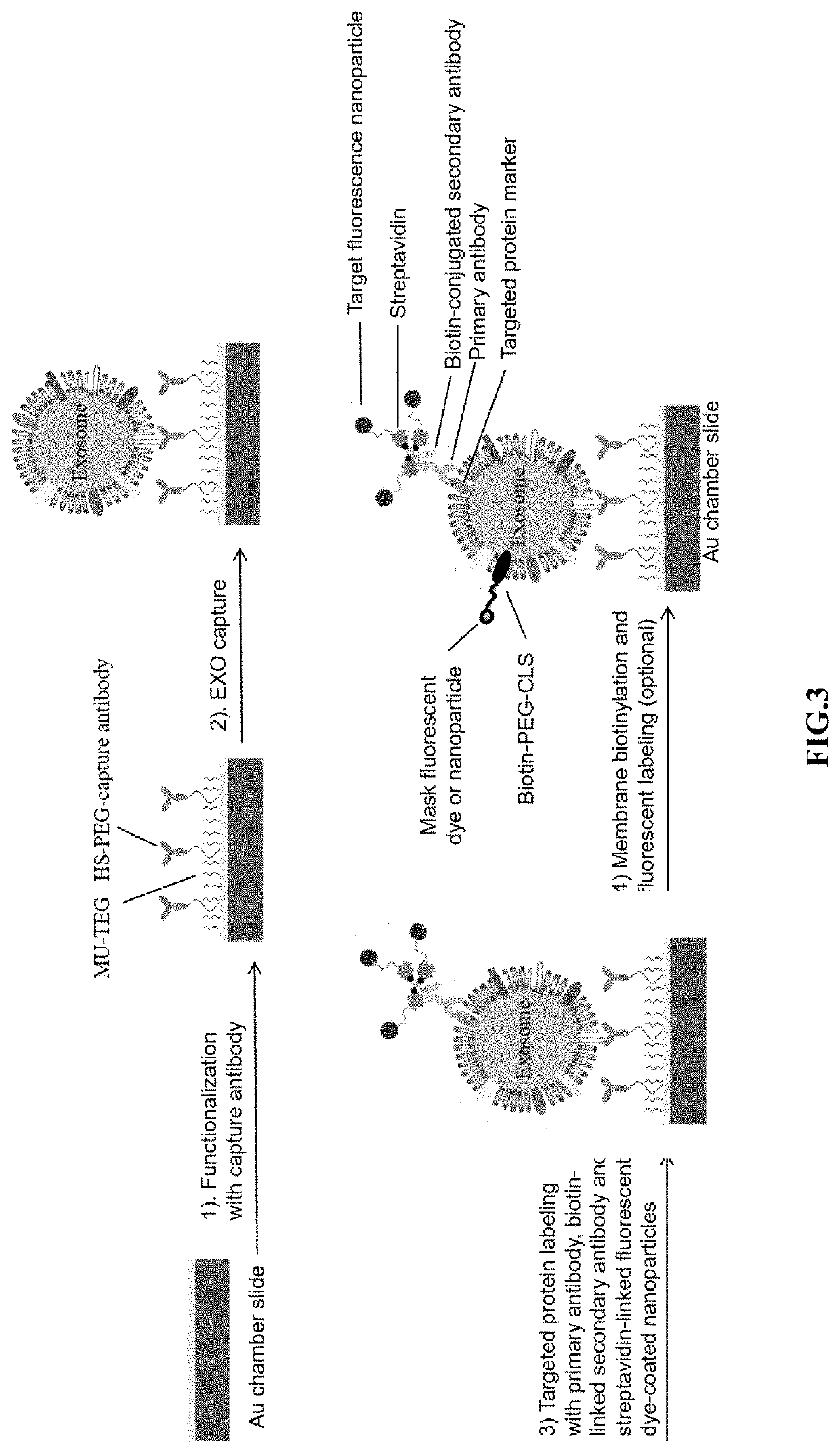
[0111]To detect the presence of extracellular vesicles (exosomes) in a sample, a multi-well cassette was fabricated. Standard microscope glass slides (75×25×1 mm) (FIG. 4A) were coated with an optically transparent gold film approximately 20 nm thick using a magnetron sputter system (AJA International, Inc., North Scituate, Mass.) to facilitate slide surface functionalization. Referring to FIG. 4B and 4C, raised stabilizers on the long edges of the slide enabled fixation of the gold film to the slide, and pressure grease facilitated sealing. 261 wells (9×29) were added to the gold-coated slide using a Formlabs Form 2 3D printer. Each well on the array was about 1.5 mm in diameter and about 0.5 mm in height. Each well had about 0.9 μL capacity and was separated from its nearest neighbors by about 1.0 mm.
[0112]The surfaces of the wells were functionalized with capture molecule by saturating the surface of the slide with 0.1 mM 11-mercaptoundecyl tetra (ethylene glycol) (MU-TEG...
example 2
f Exosomes Comprising CD81
[0113]Array slides were prepared as described in Example 1. Plasma samples from a breast cancer patient were analyzed for the presence of exosomes with a tetraspanin marker (i.e., CD81 primary antibody (approximately 2 μg / ml) (commercially available)) that specifically binds tetraspanin CD81 was added to the array and incubated at room temperature for one hour. DiO dye was used to label exosomes to confirm the success capture of exosomes (50 μM, 30 min). Isotype IgG (commercially available) was used to replace the CD81 capture antibody for a negative control. IgG is a primary antibody that lacks specificity to the target but matches the type and class of the primary antibody used in the cancer marker targeting. IgG was used as the negative control to help differentiate non-specific background signal from specific antibody signal. The assays were then analyzed for fluorescence. FIG. 6A shows the abundant exosomes captured with CD81 antibodies, indicating the...
example 3
abeling with fluorescence nanoparticles
[0114]Exosomes in cell culture media or plasma are diluted over 100× with phosphate buffered saline (PBS) and filtered through a 0.2 μm polyethersulfone (PES) filter to remove debris and larger, non-exosome vesicles. Exosome membranes were then biotinylated with a 50 μM lipophilic cholesterol-PEG-biotin (CLS-PEG-biotin) composition for 30 minutes at room temperature. The PEG moiety conjugated to the cholesterol and biotin moieties has a molecular weight of approximately 2,000 g / mol.
[0115]To detect the presence of a particular exosome surface protein, primary antibody (approximately 2 μg / ml) having affinity for the exosome surface marker was added to the sample and incubated for one hour at room temperature. Secondary antibody labeled with 20 nM QD655 or Magdye 665 (Magnetic nanoparticles coated with Cy5) having an affinity for the primary antibody was then added to the sample and incubated for one hour at room temperature. If dark field imaging...
PUM
| Property | Measurement | Unit |
|---|---|---|
| diameter | aaaaa | aaaaa |
| diameter | aaaaa | aaaaa |
| size | aaaaa | aaaaa |
Abstract
Description
Claims
Application Information