Direct detection of single molecules on microparticles

a technology of single molecules and microparticles, applied in the direction of instruments, particle and sedimentation analysis, measurement devices, etc., can solve the problems of cumbersome detection devices, time-consuming, and additional steps and reagents,

Pending Publication Date: 2020-07-02
ABBOTT LAB INC
View PDF4 Cites 2 Cited by
  • Summary
  • Abstract
  • Description
  • Claims
  • Application Information

AI Technical Summary

Benefits of technology

[0007]The disclosure also provides a method of analyzing two or more analytes of interest in a biological sample in a single assay. The method comprises (a) capturing a first analyte of interest on a surface of a first microparticle, wherein the first microparticle (i) comprises a plurality of specific binding members immobilized on the surface thereof which bind to the first analyte and (ii) is labeled with a first fluorophore; (b) capturing a second analyte of interest on a surface of a second microparticle, wherein (i) the first analyte is different from the second analyte, and (ii) the second microparticle comprises a plurality of specific binding members immobilized on the surface thereof which bind to the second analyte; (c) reacting the captured first analyte of interest with a first conjugate, wherein the first conjugate comprises a specific binding member that is labeled with a second fluorophore and binds to the first analyte; (d) reacting the captured second analyte with a second conjugate, wherein the second conjugate comprises a specific binding member that is labeled with a third fluorophore and binds to the second analyte, and wherein the first, second, and third fluorophores are different; (e) obtaining a transmitted light image of the first and second microparticles; (f) obtaining separate fluorescent images of the first and second microparticles corresponding to the first, second, and third fluorophores, respectively; and (g) analyzing the images using a customized image analysis process.
[0008]The

Problems solved by technology

However, they require additional steps and reagents, cumbersome detection devices, and are time consuming as compared to standard immunoassays.
They are also less adaptable to transitioning between counting (digital) and averaging (analog) modalities, which reduces the detection dynamic range.
Another issue for immunodiagnostic assays is the ability to measure two or more analytes from one sample in a single assay, otherwise known as “mu

Method used


Image

  • Direct detection of single molecules on microparticles
  • Direct detection of single molecules on microparticles
  • Direct detection of single molecules on microparticles

Examples


example 1

[0110]This example demonstrates a method of analyzing an analyte of interest in a biological sample as disclosed herein.

[0111]It has been shown that a single conventional fluorophore (e.g. fluorescein, rhodamime, etc.) can be detected on a low background surface (e.g., specially treated glass or some polymers) using a total internal reflection fluorescence (TIRF) microscope (Skinner, J. P. and S. Y. Tetin, Microsc Res Tech., 78(4): 309-16 (2015)). However, it is difficult to detect a single conventional fluorophore on a microparticle with a conventional fluorescence microscope due to sensitivity issues and fluorescence background of the microparticles. Thus, detecting a single analyte molecule captured by a microparticle with a fluorescent conjugate requires bright signal-generating molecules (e.g., at least 5 times brighter than background fluorescence). As an alternative to nanospheres, streptavidin-phycoerythrin (SAPE) was tested. Phycoerythrin (PE) is known as the brightest fluo...

example 2

[0116]This example describes an immunoassay to detect HCV antigen in accordance with the methods described herein.

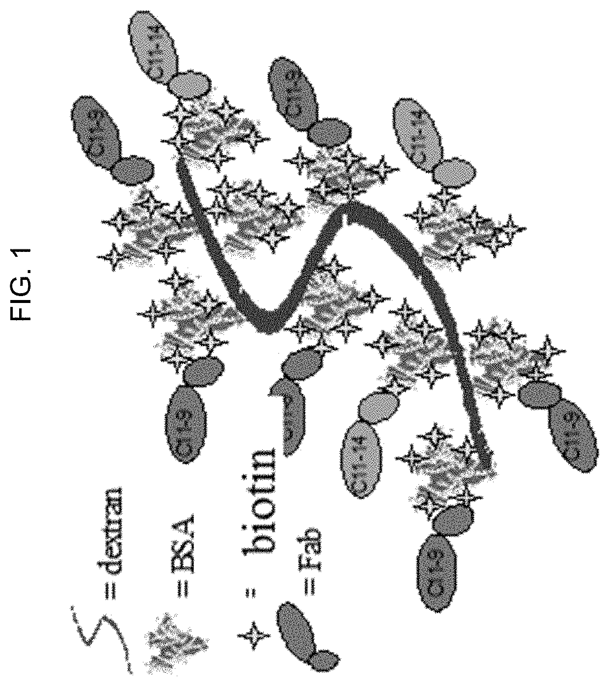
[0117]Microparticles and HCV calibrator from on-market kits (HCV core Ag kit, Ref 6L47-29, and HCV Calibrator set, Ref 6L47-02, respectively, Abbott Laboratories, Abbott Park, Ill.) were diluted with negative human plasma (NHP). Dextran BSA-biotin-Fab clusters were prepared as shown in FIG. 1. Streptavidin phycoerythrin (SAPE, PJR39S) was purchased from Prozyme (Hayward, Calif.).

[0118]The assay was performed in three steps:

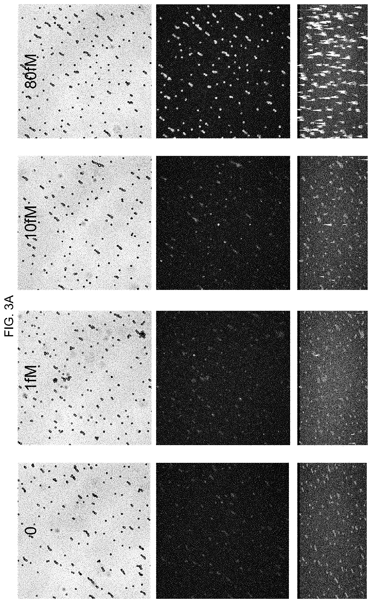
[0119]Step 1: 100 μL of the HCV calibrators ranging from 0-20000 fM were first mixed with microparticles. Microparticles were incubated at 37° C. and then washed.

[0120]Step 2: 100 μL Dextran BSA-biotin-Fab clusters were added to microparticles. Microparticles were incubated and then washed.

[0121]Step 3: 100 μL SA-PE was added. Microparticles were incubated, washed, and then imaged on a microscope.

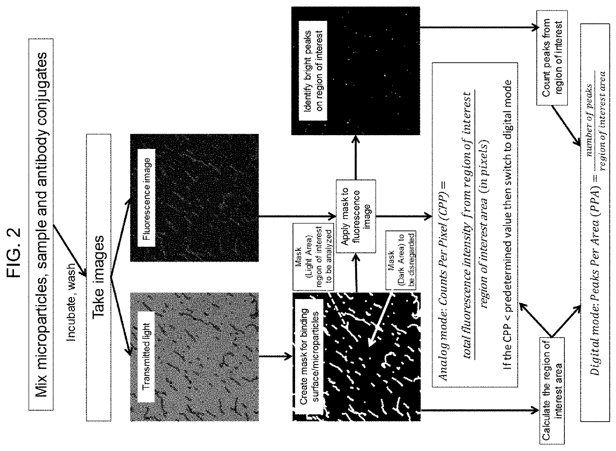
[0122]Imaging and analysis: For each sample, 4-9 sets of...

example 3

[0125]This example describes an immunoassay to detect HIV in accordance with the methods described herein.

[0126]Microparticles used in the assay were coated using an Abbott in-house antibody via EDAC chemistry, and calibrators were prepared from an on-market kit (HIV kit, 25258L100, 65 pg / ml, Abbott Laboratories, Abbott Park, Ill.) and further diluted with negative human plasma (NHP). Dextran BSA-biotin-Fab clusters were prepared. Streptavidin phycoerythrin (SAPE) was purchased from Prozyme (Hayward, Calif.).

[0127]The assay was performed in two steps:

[0128]Step 1: 100 μL of the HIV sample ranging from 0-6.5 pg / ml were first mixed with microparticles, assay specific diluent, and Dextran BSA-biotin-Fab clusters, and microparticles were washed after a 10-minute incubation at 37° C.

[0129]Step 2: 100 μL SAPE was added, microparticles were washed after incubation and then imaged on a microscope with an EM-CCD camera.

[0130]Imaging acquisition and analysis were carried out as described in E...


PUM


Abstract

The disclosure provides methods of analyzing an analyte of interest in a biological sample using fluorescent agents and macroconjugates which comprise a core containing a cross-linked polymer or protein, tags, specific binding members or fragments thereof, and optionally carrier proteins. Also provided are methods of analyzing two or more analytes of interest in a biological sample in a single assay using microparticles and detection conjugates comprising different fluorophore labels, acquiring transmitted light and fluorescent images of the microparticles, and using a customized image analysis process to analyze the acquired images.

Description

CROSS-REFERENCE TO RELATED APPLICATIONS[0001]This patent application claims the benefit of U.S. Provisional Patent Application No. 62 / 785,796, filed Dec. 28, 2018, and U.S. Provisional Patent Application No. 62 / 785,798, filed Dec. 28, 2018, both of which are incorporated by reference herein.BACKGROUND[0002]Conventional immunodiagnostic assays involve ensemble measurements, wherein target molecules are identified in a reaction vial by recording the cumulative signal. More recently, immunoassays have been developed which can count single analyte molecules, which are known in the art as “digital immunoassays.” One such approach involves tagging a detection antibody with an enzyme and trapping individual microparticles in nanowells. It is assumed that at low sample concentration, a single analyte molecule, bound to the antibody-enzyme conjugate and captured by a microparticle confined in the nanowell, can convert enough substrate to a fluorescent state to become detectable in a reasonab...

Claims


Application Information

Patent Timeline
IPC IPC(8): G01N33/58G01N15/14G01N33/543
CPCG01N2015/1018G01N33/54346G01N15/14G01N33/582G01N33/587G01N33/543G01N33/566
Inventor RUAN, QIAOQIAOMACDONALD, PATRICK J.SWIFT, KERRY M.TETIN, SERGEY Y.CALFIN, BRENDALIN, ZHENHAACK, RICHARDPOPE, MARK R.PROSTKO, JOHNQIU, XIAOXINGBOGDAN, FELICIA
Owner ABBOTT LAB INC
Why Eureka
Try Eureka