Infection detection device and method using same

a detection device and technology of infection, applied in the field of test strips, can solve the problems of inability to quickly diagnose wounds and urine infections, inability to quickly and efficiently solve problems such as time-consuming, and rarely heal quickly or without complications, and achieve the effects of facilitating decision-making, rapid operation, and efficient and cheapness

Active Publication Date: 2020-11-12
SALAHSHOOR KORDESTANI SOHEILA
View PDF1 Cites 2 Cited by
  • Summary
  • Abstract
  • Description
  • Claims
  • Application Information

AI Technical Summary

Benefits of technology

[0023]It is the object of the present invention to provide an infection detection system which can operate rapidly, efficiently, and cheaply; thereby, overcoming the disadvantages of currently employed infection detection systems; such as using extra step of culturing or requiring manipulation of dressings or contact layers comprising of many parts. The infection detection ...

Problems solved by technology

Many of these wounds, including those related to chronic venous insufficiency, peripheral arterial disease, and pressure injuries or ulcers, rarely heal quickly or without complications such as infection.
The disadvantages of these new techniques, especially those requiring culturing of biopsies, are that they are time consuming, have to be analyzed in a laboratory, and some tests are expensive or not commonly available.
If however wound healing is delayed, then the pH will oscillate and become increasingly alkaline over time (Sch...

Method used


Image

  • Infection detection device and method using same
  • Infection detection device and method using same

Examples


example 1

Fabrication of a Test Strip

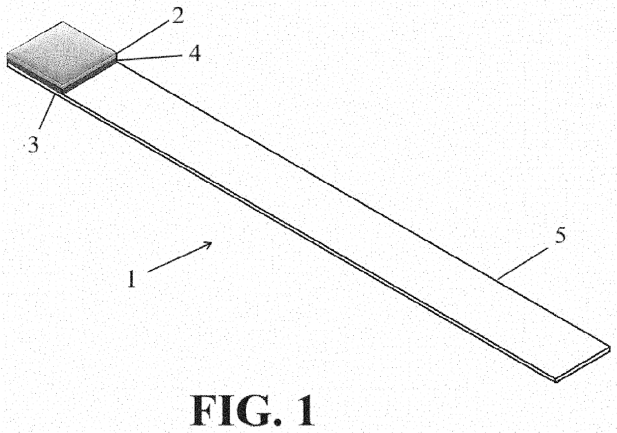
[0042]In accordance with a preferred embodiment shown in FIG. 1, a test strip 1 was manufactured using a detector 2. The detector consisted of a filter paper impregnated with a sensing solution including 3% (w / v) gelatin and 0.1% (w / v) bromethymol blue. A cardboard was used as the base layer 3, and a double sided adhesive as the adhesive layer 4. The filter paper (Whatman® qualitative filter paper, Grade 1) had a thickness preferably of about 0.26 mm, which was cut in shape of a square of about 7 mm length and 7 mm width. The base layer 3 was made of a cardboard with a thickness preferably of about 0.5 to 1 mm, which was cut into a rectangle of 70 mm length and 7 mm width. The adhesive layer 4 (Nano Tape, adhesive type: Cellulose) was a double sided adhesive cut into a rectangle of 7 mm length and 7 mm width. The impregnated filter paper (detector 2) was then attached on the cardboard (base layer 3) using the double sided adhesive (adhesive layer 4). Plura...

example 2

Detection of Infection in Wounds

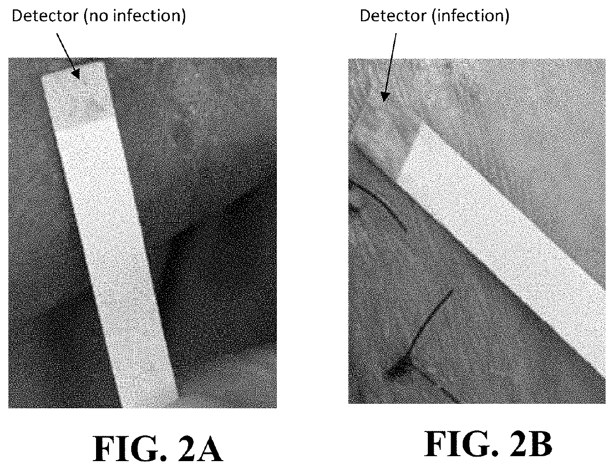
[0043]Fifty-one patients with chronic and acute wounds, not receiving antibiotic treatment within 7 days before the tests, were investigated. Wounds washed with sterile saline and then superficially contacted with the detector of the test strip. In case of an infection, upon contact, the color of the detector start changing rapidly from yellow to green and stabilizes within about 60 seconds. Wound swab cultures were collected and laboratory results were used as control group. Wounds with a growth of ≥105 colony forming unit (CFU) per ml was considered to have a positive culture (Lindsay et al., 2017). Among the 51 patients, 37 were found to have wound infection by both the laboratory and test strip. As was verified by laboratory tests, Staphylococcus aureus and Gram-positive cocci were the most prevalent pathogenic yield from the cultures. Other bacteria detected by cell culture laboratory and were involved with the infections detected by the test str...

example 3

Detection of Infection in Urine

[0046]In this study, urine specimens of 50 patients, with no antibiotic treatment, were examined for the urinary tract infections (UTIs), based on the presence of MMPs activity as an indicator of bacteriuria. The validity of the test strip results of patients with or without clinical symptoms of urinary tract infection (UTIs) was investigated by comparing the test strip results with those of their corresponding urine culture test as a control group.

[0047]Among the 50 patients, 3 were found to have UTIs by both the laboratory and the test strip. The accuracy, sensitivity, and specificity for the strip calculated against the urine culture for the diagnosis of UTIs, were 98%, 100%, and 97.9% respectively. All the results were significant (p≤0.01).

[0048]The bacteria involved with the urine infections that were detected by the strip and cell culture tests were Escherichia coli and Proteus vulgaris, where Escherichia coli was the most prevalent pathogenic yi...


PUM


Abstract

The invention provides device and method for detecting infection in a body fluid, in particular in wound exudates and urine, based on protease activity. The device is founded on a detector consisting of one layer, which is an absorbent layer impregnated with a solution formed by dissolving a pH sensitive dye, such as bromothymol blue, in a gelatin solution. The method uses the device in form of a test strip for quick detection of an infection by observing a change in the color of the detector.

Description

CROSS-REFERENCE TO RELATED APPLICATIONS[0001]Not ApplicableSTATEMENT REGARDING FEDERALLY SPONSORED RESEARCH OR DEVELOPMENT[0002]Not ApplicableREFERENCE TO SEQUENCE LISTING SUBMITTED ELECTRONICALLY[0003]Not ApplicableBACKGROUNDField of the Invention[0004]The present invention relates to a device comprising a detector for diagnosis of infection in body fluids or aqueous body tissues. In particular, the invention relates to a test strip and method for detection of infection in urine and wound fluid exudates.Description of the Prior Art[0005]As the percentage of the older adult population rises, so will the incidence and significance of chronic wounds and wound infections. Many of these wounds, including those related to chronic venous insufficiency, peripheral arterial disease, and pressure injuries or ulcers, rarely heal quickly or without complications such as infection.[0006]The neutrophil population is a well-known marker for the diagnosis of wound infection. This marker increases ...

Claims


Application Information

Patent Timeline
IPC IPC(8): G01N33/52G01N21/78
CPCG01N33/528G01N21/78C12Q1/04C12Q1/37G01N33/523
Inventor SALAHSHOOR KORDESTANI, SOHEILA
Owner SALAHSHOOR KORDESTANI SOHEILA
Why Eureka
Try Eureka