Specific biomarker set for non-invasive diagnosis of liver cancer

a hepatocellular carcinoma and biomarker technology, applied in the field of specific and novel hepatocellular carcinoma tumor biomarkers, can solve the problems of high mortality rate, excessive false positives of afp tests, and inconclusive results, and achieve the effect of facilitating early detection of cancer

Pending Publication Date: 2020-12-10
DRAGON VICTORY DEV
View PDF0 Cites 3 Cited by
  • Summary
  • Abstract
  • Description
  • Claims
  • Application Information

AI Technical Summary

Benefits of technology

[0007]The present invention provides a detection and quantification method for measuring the auto-antibodies against a certain panel of specific tumor HCC biomarkers, which is useful for diagnosing and staging cancers. Comparing t...

Problems solved by technology

Most HCC patients have rapid tumor progressing resulting in high mortality rate.
However, the major problem of the AFP test is excessive false positives.
Despite the fact that AFP test is commonly suggested for diagnosis of liver cancers, its result is not conclusive.
However, conventional detection of ...

Method used


Image

  • Specific biomarker set for non-invasive diagnosis of liver cancer
  • Specific biomarker set for non-invasive diagnosis of liver cancer
  • Specific biomarker set for non-invasive diagnosis of liver cancer

Examples


example 1a

traction from Patients' Biopsies

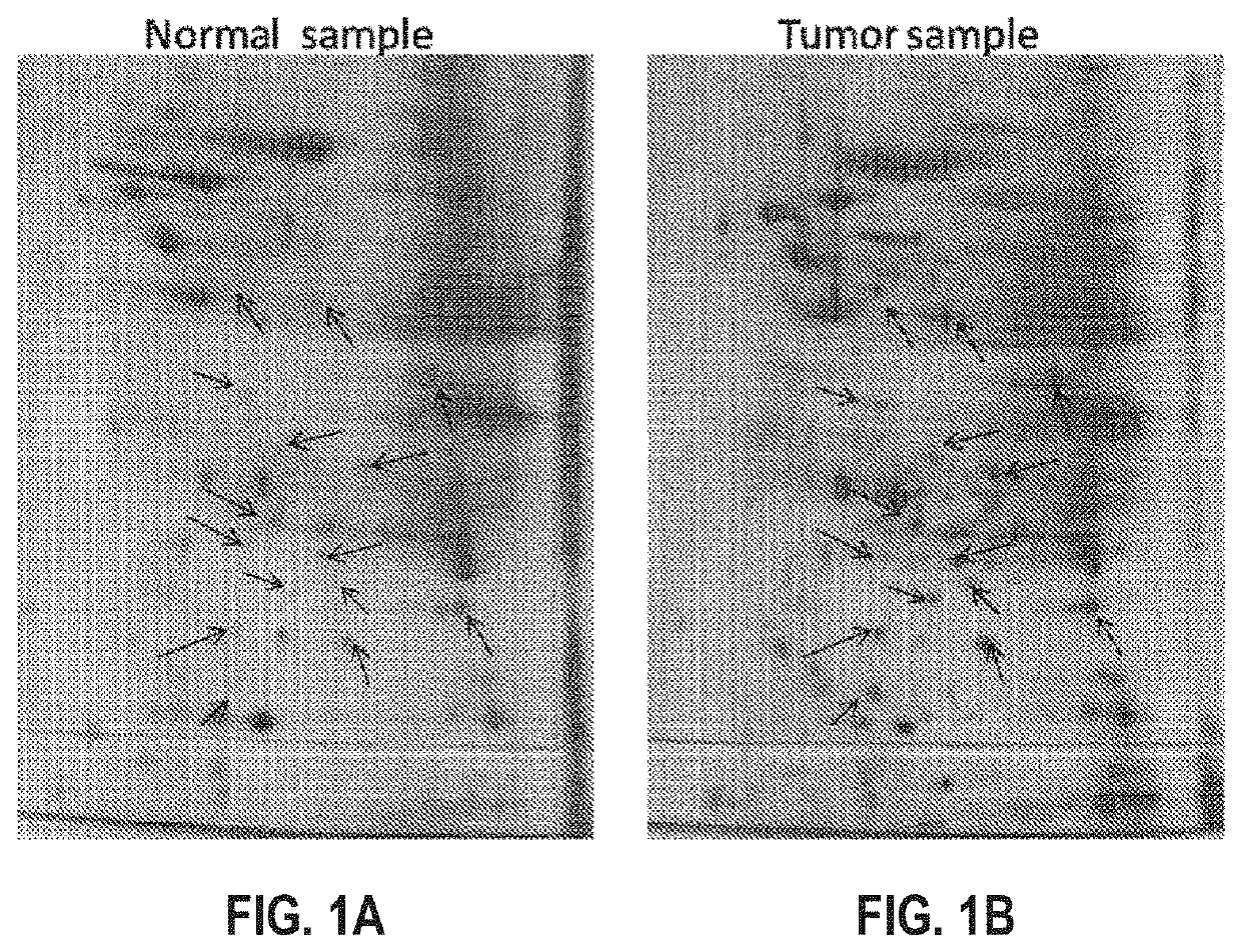
[0121]500 mg of the paired patients' biopsies (tumor biopsy versus juxtaposed normal tissue) are collected and washed with PBS. The tissues were frozen by submerging into liquid nitrogen and immediately homogenized with pestle and mortar. To the homogenized samples, lysis solution (8M Urea, 4% CHAPS, 2% IPG Buffer, 0.2 mg / ml PMSF) was added, then vortex for at least 5 min until the tissues are completely dispersed. The lysates were then clarified by centrifugation at 14,000 rpm for 10 minutes at 4° C. The supernatants were further cleaned up by 2D Clean Up kit (Amersham) to remove the salt and impurities. The pellets were resuspended with minimum volume of Rehydration Solution (No DTT & IPG Buffer added). The protein concentrations were then measured by Bio-Rad™ protein assay and aliquots of 200 g / per tube are stored at −70° C.

example 1b

Proteins by Two-Dimensional Electrophoresis

[0122]To 1 ml rehydration stock solution, 2.8 mg DTT, 5 μl pharmalyte or IPG Buffer, and 2 μl bromophenol blue was added. 50-100 μg of protein sample is added to the 13 cm Immobiline DryStrip™ (IPG strip) containing 250 μl of rehydration solution. After removing the protective cover, the IPG strip was positioned in the strip holder with the gel side facing down, and overlaid with Cover Fluid to prevent dehydration during electrophoresis. The strip was then placed on to Ettan™ IPGphor™ (Amersham) for isoelectric focusing (first dimensional electrophoresis).

[0123]After the first-dimensional electrophoresis, the IPG strip was equilibrated with equilibrate solution (6 M Urea, 2% SDS, 50 mM Tris HCl pH 6.8, 30% Glycerol, 0.002% Bromophenol blue, 100 mg DTT per 10 ml buffer and 250 mg IAA per 10 ml buffer), and then washed with 1×SDS running Buffer for 4-5 times. The IPG strip was placed on top of the second-dimension gel and overlaid with sealin...

example 2a

of HCC Biomarker Set

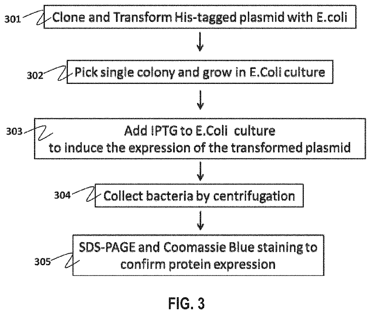
[0125]His tagged plasmids containing cDNA inserts encoding the HCC biomarker set was transformed into DH5 competent cells (301, FIG. 3). A single colony was picked and allowed to grow in bacterial culture (302). The number of plasmid was expanded and extracted from the bacteria by mini-prep. The plasmid was further transformed into BL21DE3 or BL21DE3pLysS competent cells. Transformed bacteria were selected and grew in 2×100 ml LB medium. When the bacterial culture reached the optical density of 0.06, 200 μM of IPTG was added to 100 ml bacterial culture (303). Another 100 ml of bacterial culture without IPTG was used as negative control. The bacterial cultures were incubated at 30° C. with shaking. 500 μl of the bacterial cultures are saved and stored at −20° C. 3 h after the incubation and in the next morning after incubating overnight.

[0126]Bacterial cultures with and without IPTG induction were mixed together in a 500 ml centrifuge bottle. Bacterial cells were ...


PUM


Abstract

Cells within liver tumor mass comprise a unique set of proteins/tumor antigens when compared to the normal liver tissues epithelial cells juxtaposed to the tumor. The presence of tumor antigens couples the production of auto-antibodies against these tumor antigens. The present invention relates to the identification and elucidation of a protein set that can act as a novel marker set for liver cancer diagnosis and prognosis. Specifically, it relates to a kit that enables diagnostic and prognostic measurement of auto-antibodies in serum of liver cancer patients. The present invention provides a non-invasive, specific, sensitive, and cost effective detection and quantification method by evaluating a set of validated liver cancer proteins/tumor antigens, which includes Bmi-1, VCC1, SUMO-4, RhoA, TXN, ET-1, UBE2C, HDGF2, FGF21, LECT2, SOD1, STMN4, Midkine, IL-17A, IL26, or DCP to complement the conventional diagnostic methods.

Description

RELATED APPLICATIONS[0001]This is a continuation-in-part application of U.S. application Ser. No. 15 / 331,472, which was filed on Oct. 21, 2016, which is a continuation of U.S. application Ser. No. 14 / 321,867 filed on Jul. 2, 2014, which issued as U.S. Pat. No. 9,506,925, the contents of each incorporated by reference in their entirety.COPYRIGHT NOTICE / PERMISSION[0002]A portion of the disclosure of this patent document contains material which is subject to copyright protection. The copyright owner has no objection to the facsimile reproduction by anyone of the patent document or the patent disclosure as it appears in the Patent and Trademark Office patent file or records, but otherwise reserves all copyright rights whatsoever. The following notice applies to the processes, experiments, and data as described below and in the drawings attached hereto: Copyright © 2014, Vision Global Holdings Limited, All Rights Reserved.TECHNICAL FIELD[0003]The present invention provides a detection an...

Claims


Application Information

Patent Timeline
IPC IPC(8): G01N33/574G01N33/564G01N21/64
CPCG01N2333/9108G01N21/6428G01N33/57438G01N2021/6441G01N2333/54G01N2333/90283G01N2333/475G01N2458/00G01N2333/4703G01N2333/82G01N2333/50G01N33/564G01N33/54313G01N33/582G01N33/6854
Inventor MAN, CORNELIA WING YINWAI, NORMAN FUNG MANWONG, BING LOUWAI, BENJAMIN CHI YIN
Owner DRAGON VICTORY DEV
Why Eureka
Try Eureka