
Cell-based cancer vaccines and cancer therapies
a cancer vaccine and cell-based technology, applied in the field of cell-based cancer vaccines and immune therapies against cancer, can solve the problems of inability of cisplatin treatment time-consuming and expensive, and the rationale for this strategy did not take into account the lack of ability of ici to enhance tumor immunogenicity, so as to prevent recurrence of cancer, induce tumor regression, and increase the cytotoxic immune response
- Summary
- Abstract
- Description
- Claims
- Application Information
AI Technical Summary
Benefits of technology
Problems solved by technology
Method used
Image

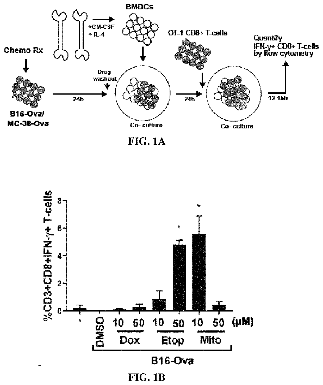
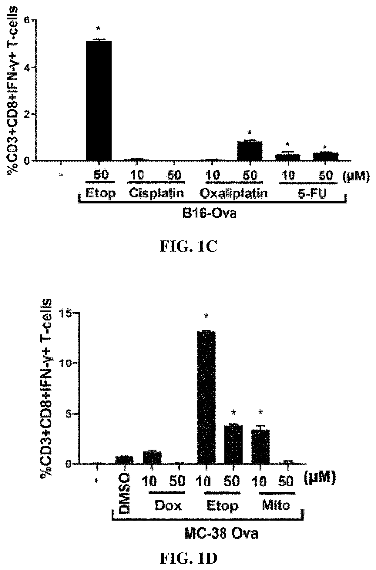
Examples
example 1
and Mitoxantrone-Treated Tumor Cells Induce DC-Mediated OT-I T-Cell Priming In Vitro
[0302]Materials and Methods
[0303]Reagents, Cell Lines and Mouse Strains
[0304]Mouse GM-CSF and AnnV-FITC were purchased from Biolegend. IL-4 was purchased from Thermo Fisher Scientific. Anti-CD3 (FITC) (145-2C11), Anti-CD8 (APC) (53-6.7), Anti-IFNγ (PE) (XMG1.2), Anti-CD45 (BUV395)(30-F11), Anti-CD24 (APC) (M1 / 69), Anti-Ly6C (BV605) (AL-21), Anti-F4 / 80 (BV711) (BM8), Anti-MHCII (PE-Cy7) (M5 / 14.15.2), Anti-CD11b (BV786) (M1 / 70), Anti-CD103 (BV421) (2E7) were purchased from ebioscience or Biolegend. H2-Kb / SIINFEKL (SEQ ID NO:1)-tetramer (PE-conjugated) was purchased from MBL Life Science. Necrostatin-1 and Z-VAD were purchased from Invivogen. Doxorubicin, Etoposide, Mitoxantrone, Cisplatin, Paclitaxel, Camptothecin, Irinotecan, 5-FU and cylcophosphamide were purchased from LC labs or Sigma. Oxaliplatin was purchased from Tocris Biosciences. An antibody against ovalbumin was purchased from Abcam (Cat #ab...
example 2
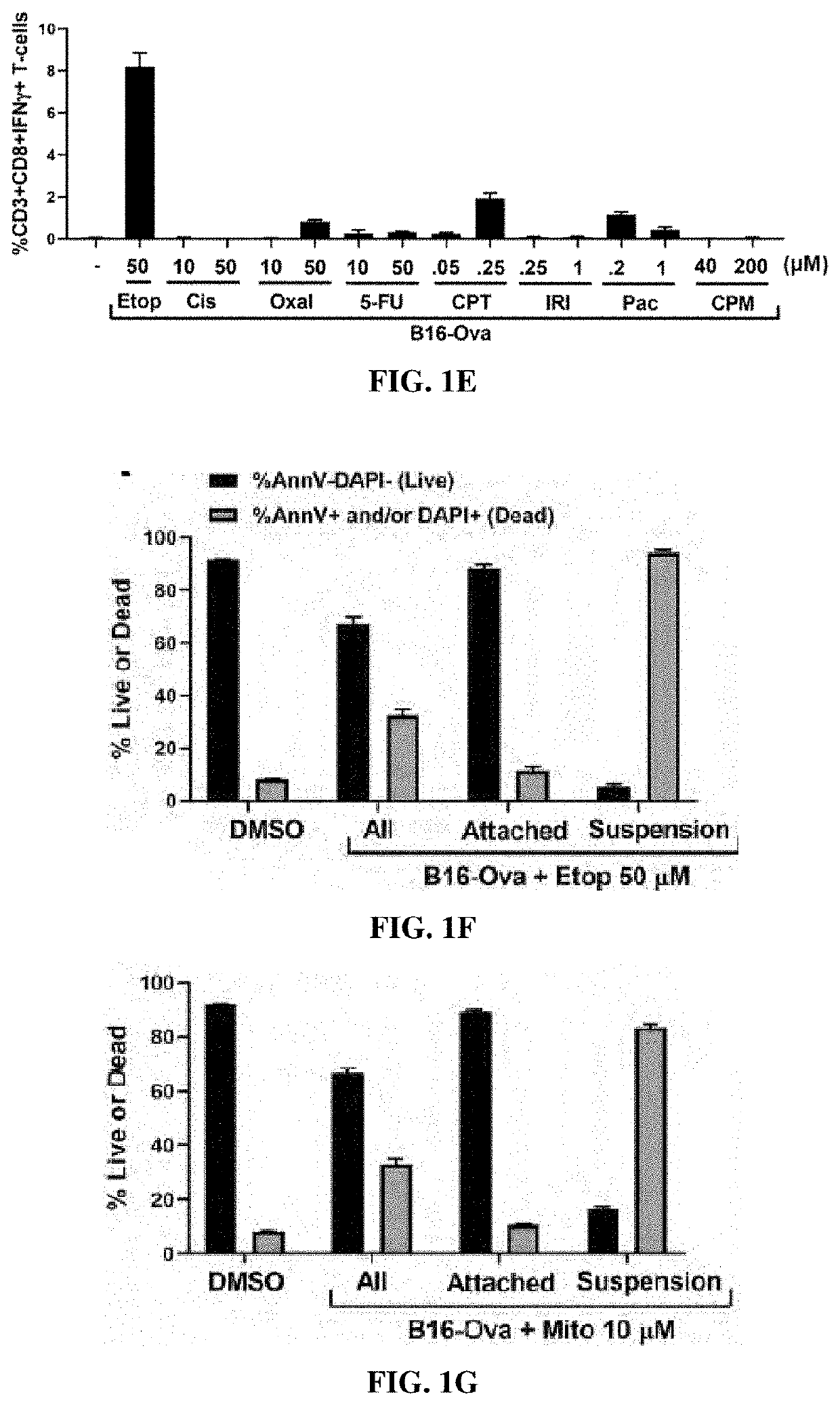
red Cells, Rather than Dead Cells, are Determinants of DC-Mediated IFN-γ Induction in T-Cells in Response to Mitoxantrone and Etoposide Treatment
[0320]Materials and Methods
[0321]Fractionation of Live and Dead Fractions from Chemotherapy-Treated Cells
[0322]B16-Ova cells or MC-38-Ova cells were treated with various doses of chemotherapy as indicated in FIGS. 1F-1I for 24 hours after which the floating fraction of cells was transferred to a separate tube and washed with PBS (for AnnV / DAPI staining) or IMDM (for co-culture with BMDC). The attached fraction was rinsed 1× with PBS, detached using 5 mM EDTA (in PBS), washed with PBS or IMDM and transferred to a separate tube. Separately, cells treated with chemotherapy for 24 h were re-plated at 1 million cells per well of a 24-well plate in 500 ul of IMDM (10% FBS; P / S). Cell-free supernatants were collected after a further 24 h. As shown in FIGS. 1F-1I, staining with AnnV and DAPI of the attached and floating fractions after chemotherapy...
example 3
nal Immunogenic Death Markers do not Predict the Immunogenicity of Etoposide-Treated B16-Ova Cells
[0327]Materials and methods
[0328]Measurement of Immunogenic Cell Death Markers
[0329]For measurement of calreticulin surface exposure, B16-Ova cells were treated for 24 hours with various chemotherapy drugs. All attached and floating cells were harvested and washed in staining buffer (PBS containing 0.5% BSA) and incubated with anti-calreticulin antibodies for 1 hour on ice. Cells were washed once in staining buffer and then incubated with secondary AF488-conjugated secondary antibody for 1 hour at room temperature, washed again, re-suspended in staining buffer and analyzed by flow cytometry.
[0330]For HMGB1 measurement in cell culture media, B16-Ova cells were treated for 24 hours with various chemotherapy drugs, media was collected, and floating cells removed by centrifugation at 250×g for 5 minutes. Cell-free cell culture media was then analyzed by ELISA for HMGB1 according to the manu...
PUM
| Property | Measurement | Unit |
|---|---|---|
| time | aaaaa | aaaaa |
| time | aaaaa | aaaaa |
| time | aaaaa | aaaaa |
Abstract
Description
Claims
Application Information