
Systems and methods for 3D printing of proteins

a technology of proteins and systems, applied in the field of systems and methods for 3d printing of proteins, can solve the problems of limited shape complexity, deterioration of strength, and compromise of biocompatibility, so as to reduce costs, reduce the biocompatibility and degradability of printed structures, and eliminate additives and process steps

Pending Publication Date: 2021-10-14
TRUSTEES OF TUFTS COLLEGE TUFTS UNIV
View PDF4 Cites 1 Cited by
  • Summary
  • Abstract
  • Description
  • Claims
  • Application Information

AI Technical Summary

Benefits of technology

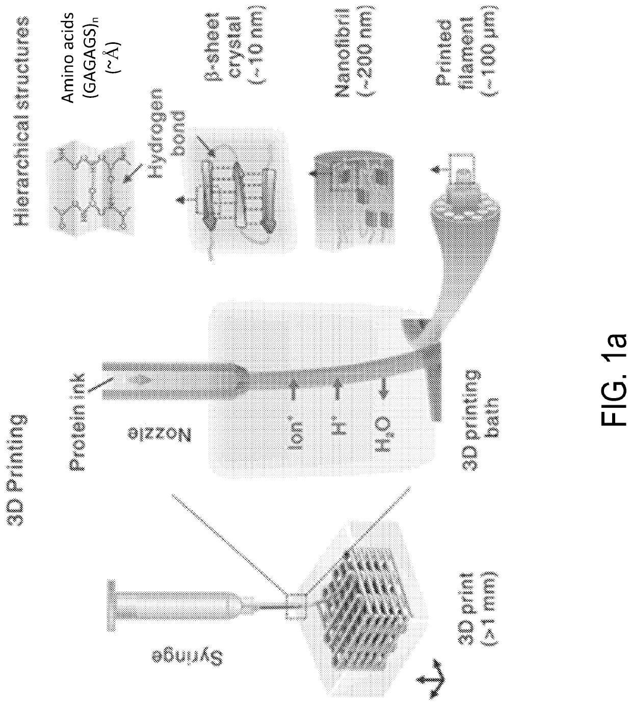
This patent presents a new method for 3D printing structures using biocompatible materials without using chemical solvents or external stimuli. This improves the biocompatibility and degradability of the structures while also increasing their mechanical strength. The method uses induced self-assembly of protein molecules to create structures with molecule and nanoscale precision. This results in the printed structures having less defects and improved mechanical properties. Overall, this patent provides a more energy-efficient and improved manufacturing technique for printing 3D protein structures.

Problems solved by technology

However, it has proven to be challenging to achieve the self-assembly with the similar precision in various manufacturing techniques and three-dimensional (3D) printing, which largely rely on high energy beam / laser, high temperature, organic solvents and chemical crosslinking, thus suffering from deteriorated strength, compromised biocompatibility and limited shape complexity.

Method used


Image

  • Systems and methods for 3D printing of proteins
  • Systems and methods for 3D printing of proteins
  • Systems and methods for 3D printing of proteins

Examples


example 1

[0064]Preparation of Protein Ink and 3D Printing Procedure

[0065]The protein ink was prepared using 5 g of sliced cocoons from Bombyx mori (Tajima Shoji Co., Yokohama, Japan) were degummed in 2 L boiling solution of 0.02 M sodium carbonate for 30 min. The degummed fibers were dried overnight and solubilized in 9.3 M lithium bromide for 4 h in a 60° C. oven, followed by a dialysis (MWCO, 3,500) against DI water with 6 changes over 3 days. The insoluble particulates were removed by centrifugation (two times at 9,000 rpm, 20 min, 4° C.) and syringe filtration (low protein binding PVDF membrane, 5 μm, Merck Millipore, Ireland). The filtered solution was loaded into Slide-a-Lyzer dialysis cassettes (MWCO 3,500, Pierce) and concentrated by drying under 4° C. for 8 days to obtain high concentrations from 14 wt % to 30 wt %. Silk concentration was determined by weighing a dried sample of a known volume. The ink was doped with HRP, ampicillin or nanomaterials (Quantum dots and gold nanorods (...

example 2

[0078]Preparation of Protein Ink and 3D Printing Procedure

[0079]The protein ink was prepared using 5 g of sliced cocoons from Bombyx mori (Tajima Shoji Co., Yokohama, Japan) were degummed in 2 L boiling solution of 0.02 M sodium carbonate for 30 min. The degummed fibers were dried overnight and solubilized in 9.3 M lithium bromide for 4 h in a 60° C. oven, followed by a dialysis (MWCO, 3,500) against DI water with 6 changes over 3 days. The insoluble particulates were removed by centrifugation (two times at 9,000 rpm, 20 min, 4° C.) and syringe filtration (low protein binding PVDF membrane, 5 μm, Merck Millipore, Ireland). The filtered solution was loaded into Slide-a-Lyzer dialysis cassettes (MWCO 3,500, Pierce) and concentrated by drying under 4° C. for around 8 days to obtain high concentrations around 30 wt %. Silk concentration was determined by weighing a dried sample of a known volume. The ink was doped with horseradish peroxidase (HRP)-, labeled antibody (A0293, Sigma-Aldric...


PUM

PropertyMeasurementUnit
soak timeaaaaaaaaaa
soak timeaaaaaaaaaa
inner diameteraaaaaaaaaa

Abstract

Three-dimensional printing methods and systems for forming a three-dimensional protein article are disclosed. The methods and systems involve selecting article formation parameters, such as protein ink parameters, solvent bath parameters, shear force parameters, and mapping parameters. After these parameters are selected, the methods and systems iteratively introduce protein ink into a solvent bath via a three-dimensional printing outlet. The result is a three-dimensional protein article. One exemplary protein is silk fibroin. Further processing can be done, such as drying the article.

Description

CROSS-REFERENCE TO RELATED APPLICATIONS[0001]This application is related to, claims priority to, and incorporated herein by reference for all purposes U.S. Provisional Patent Application No. 62 / 720,016, filed Aug. 20, 2018.STATEMENT REGARDING FEDERALLY SPONSORED RESEARCH[0002]This invention was made with government support under grant EB002520 awarded by the National Institutes of Health. The government has certain rights in the invention.BACKGROUND[0003]Protein is one of the most essential and superior structural materials in nature, from cellular cytoskeleton to spider silk, highly promising in a wide range of applications including regenerative medicine, drug delivery, implantable devices, bioelectronics and biophotonics. The exceptional features of proteins, often unavailable in synthetic materials, closely relate to the manufacturing process through a controlled hierarchical self-assembly with molecule and nanoscale precision. However, it has proven to be challenging to achieve...

Claims


Application Information

Patent Timeline
Patent Type & Authority Applications(United States)
IPC IPC(8): B29C64/106C09D11/04C07K14/435C12N5/073B33Y10/00B33Y70/00B29C64/30B33Y40/20
CPCB29C64/106C09D11/04C07K14/43586C12N5/0605B33Y10/00B29K2105/0032B29C64/30B33Y40/20C12N2533/50B29K2089/00B33Y70/00B33Y80/00C09D11/02B29K2105/0035B29K2105/162B29K2505/14B29K2995/0035
Inventor KAPLAN, DAVID L.MU, XUAN
Owner TRUSTEES OF TUFTS COLLEGE TUFTS UNIV
Learn More