
Methods for optical micropatterning of hydrogels and uses thereof

a hydrogel and micro-patterning technology, applied in the field of optical micro-patterning hydrogels, can solve the problems of less than half the man-hours required by micro-molding methods, and achieve the effects of reducing process time, eliminating the need for a cleanroom, and high throughpu

Pending Publication Date: 2021-12-02
PRESIDENT & FELLOWS OF HARVARD COLLEGE
View PDF1 Cites 2 Cited by
  • Summary
  • Abstract
  • Description
  • Claims
  • Application Information

AI Technical Summary

Benefits of technology

The present invention is about a new method for making small patterns in hydrogels, which can be used for tissue engineering and fluidic device applications. The method is faster and more efficient than previous methods, and it doesn't require the use of toxic chemicals or clean rooms. The method can also guide cell development and allow for single cell micropatterning. Additionally, it can be used for microfluidic technologies and drug screening. Overall, the invention offers a faster, safer, and more precise way to make small patterns in hydrogels.

Problems solved by technology

The optical patterning methods for micropatterning hydrogels described herein, however, surprisingly can be completed within 2 days' time, and require less than half of the man-hours required by the micromolding methods.

Method used


Image

  • Methods for optical micropatterning of hydrogels and uses thereof
  • Methods for optical micropatterning of hydrogels and uses thereof
  • Methods for optical micropatterning of hydrogels and uses thereof

Examples


example 1

or Micropatterning Hydrogel Layers

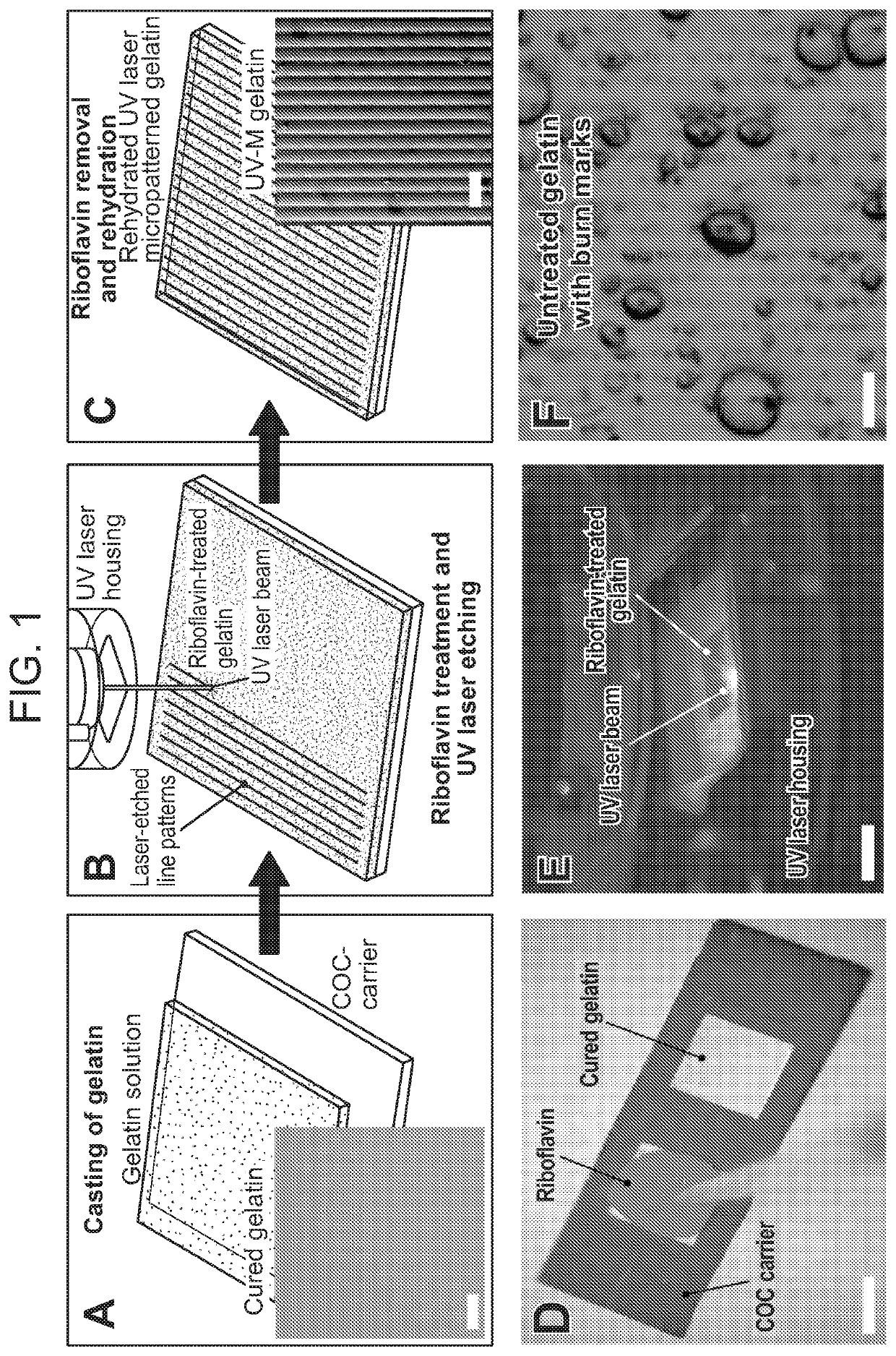
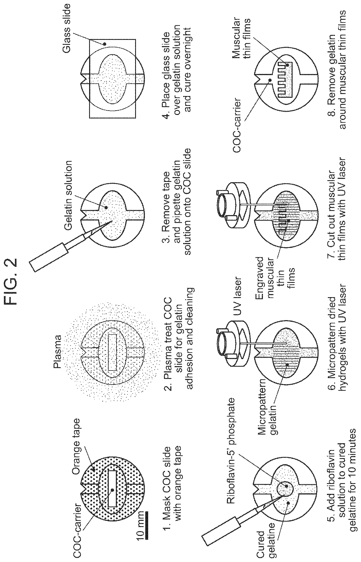
[0220]FIG. 2 is a schematic showing an exemplary method for producing a micropatterned hydrogel by optical patterning in accordance with one embodiment of the invention. The steps shown therein are as follows:[0221]1. A tape masked COC slide was plasma treated to activate and clean the exposed polymer surface.[0222]2. An aqueous solution with 10% w / v gelatin and 4% w / v microbial transglutaminase was deposited onto the plasma treated surface.[0223]3. Gelatin was cast with a glass slide and cured for 12 hours.[0224]4. After 12 hours, the gelatin was hydrated in water to remove the glass slide and tape.[0225]5. The tape was peeled from the COC slide.[0226]6. The gelatin was treated with a riboflavin 5′ phosphate solution for 10 minutes, then rinsed in water.[0227]7. The gelatin was dried.[0228]8. The gelatin was patterned with a 355 wavelength UV laser (LPKF Protolaser U3).[0229]9. The patterned gelatin was rinsed thoroughly with water, e.g., prior to ...

example 2

or Photopatterning Gelatin Hydrogels

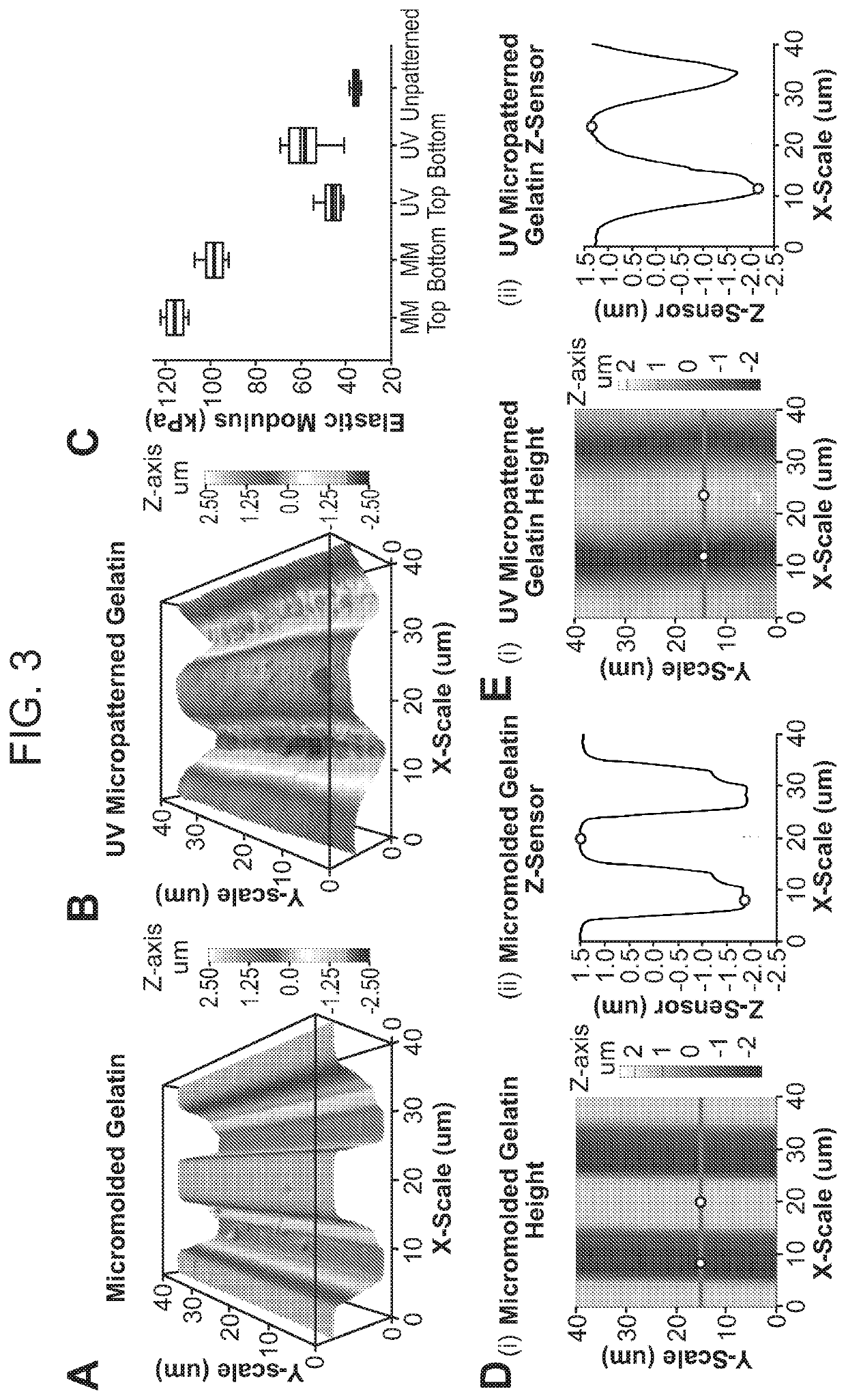
[0236]Organ-on-chip technology combines approaches from cell biology, physiology, and tissue engineering with microsystems engineering and microfluidics to create a microphysiological environment of living cells that recapitulate human tissue and organ-level functions in vitro. The goal of organs-on-chips is to improve preclinical assays for drug safety and development by mimicking the physiology and pathophysiology of healthy and diseased human tissues. However, to become a next-generation tool for drug development and biomedical research in industry, organ-on-chips need to be amenable to large-scale continuous, automated, and quality-controlled fabrication, as opposed to the small-batch manufacture predominant in academic research. In particular, scalable fabrication strategies are needed for producing organ-specific 2D and 3D hydrogel extracellular matrix scaffolds that provide micromechanical cues for cellular adhesion, shape, differentiation,...


PUM


Abstract

The present invention provides methods for optically micropatterning hydrogels, which may be used for, e.g., regenerative medicine, synthetic or cultured foods, and in devices suitable for use in high throughput drug screening assays.

Description

RELATED APPLICATIONS[0001]This application claims the benefit of priority to U.S. Provisional Patent Application No. 62 / 371,385, filed on Aug. 5, 2016, the entire contents of which are incorporated herein by reference.GOVERNMENT SUPPORT[0002]This invention was made with government support under grant number W911NF-12-2-0036, awarded by the Defense Advanced Research Projects Agency (DARPA); and under grant number 4UH3TR000522, awarded by the National Institutes of Health (NIH). The government has certain rights in the invention.FIELD OF THE INVENTION[0003]The present invention provides methods for optically micropatterning hydrogels, which may be used for, e.g., regenerative medicine, synthetic or cultured foods, and in devices suitable for use in high throughput drug screening assays.BACKGROUND OF THE INVENTION[0004]Identification and evaluation of new therapeutic agents and identification of suspect disease associated targets typically employ animal models which are expensive, time...

Claims


Application Information

Patent Timeline
Patent Type & Authority Applications(United States)
IPC IPC(8): C12M3/00B23K26/352B23K26/00C12M1/00C12M3/06C12N5/00
CPCC12M21/08B23K26/355B23K26/0006B23K2103/30C12M23/16C12N5/0075C12M23/20B32B27/08B32B27/24B32B27/32B23K26/364B23K26/402G03F7/038G03F7/16G03F7/168G03F7/2053B23K26/359B23K26/53B23K2103/42G03F7/38C12N2513/00C12N2531/00C12N2533/52C12N2533/54
Inventor PARKER, KEVIN KITNAWROTH, JANNA C.SCUDDER, LISA
Owner PRESIDENT & FELLOWS OF HARVARD COLLEGE
Learn More