Optical clearing and auto-fluorescence quenching solutions and method of use for enhanced microscopy imaging of biological tissues

a technology of autofluorescence and optical clearing, applied in the direction of optical elements, fluorescence/phosphorescence, instruments, etc., can solve the problems of limiting the depth of imaging using light microscopy techniques, unable to resolve the difference between background noise and signal using fluorescent light-based techniques, and unable to image intact organs. , to achieve the effect of reducing autofluorescence, enhancing resolution, and reducing autofluorescen

Pending Publication Date: 2021-12-30
CORNELL UNIVERSITY
View PDF0 Cites 1 Cited by
  • Summary
  • Abstract
  • Description
  • Claims
  • Application Information

AI Technical Summary

Benefits of technology

The present invention provides an improved method for clearing biological tissues, particularly solid tissues, for optical imaging using three-dimensional light microscopy. The invention achieves this improvement by using a specially formulated optical clearing solution that is easy to use, non-toxic, and retains the structural integrity of the biological tissue. The optical clearing solution contains formamide, glycerol, and optionally water, and does not require toxic solvents or dehydration of the tissue. The optical clearing solution also reduces auto-fluorescence in the tissues, which is a limitation in conventional fluorescent imaging techniques. The invention provides a solution for improving the quality of optical imaging of biological tissues.

Problems solved by technology

Imaging intact organs, however, is limited in large part by two challenges: a) biological tissues are not transparent and scatter light, thus limiting the depth of imaging using light microscopy techniques; and b) biological tissues are naturally auto-fluorescent, thus making it hard to resolve the difference between background noise and signal using fluorescent light-based techniques, such as immunofluorescence (IF).
Recent years has seen the development of several optical clearing solutions, but these techniques have a number of limitations (Yu, T. et al., J Biophotonics 11, doi:10.1002 / jbio.201700187, 2018).
Some of these limitations include, for example, technically challenging to use, a resulting increase in auto-fluorescence, high toxicity, denaturing of tissue by presence of alcohols or urea, and damage (loss of integrity) of the tissue.

Method used


Image

  • Optical clearing and auto-fluorescence quenching solutions and method of use for enhanced microscopy imaging of biological tissues
  • Optical clearing and auto-fluorescence quenching solutions and method of use for enhanced microscopy imaging of biological tissues
  • Optical clearing and auto-fluorescence quenching solutions and method of use for enhanced microscopy imaging of biological tissues

Examples


examples

[0036]Synthesis of an Optical Clearing Solution (“Atacama Clear”)

[0037]The following solutions 1-6 were prepared using molecular grade formamide (e.g. Sigma #F7503, ≥99%) and glycerol (Avantor #5092, ≥99.5%), and with ultra-pure distilled water.

1) 30% Formamide in water

2) 30% Formamide, 10% glycerol, in water

3) 30% Formamide, 25% glycerol, in water

4) 30% Formamide, 50% glycerol, in water

5) 30% Formamide, 70% glycerol

6) 20% Formamide, 80% glycerol

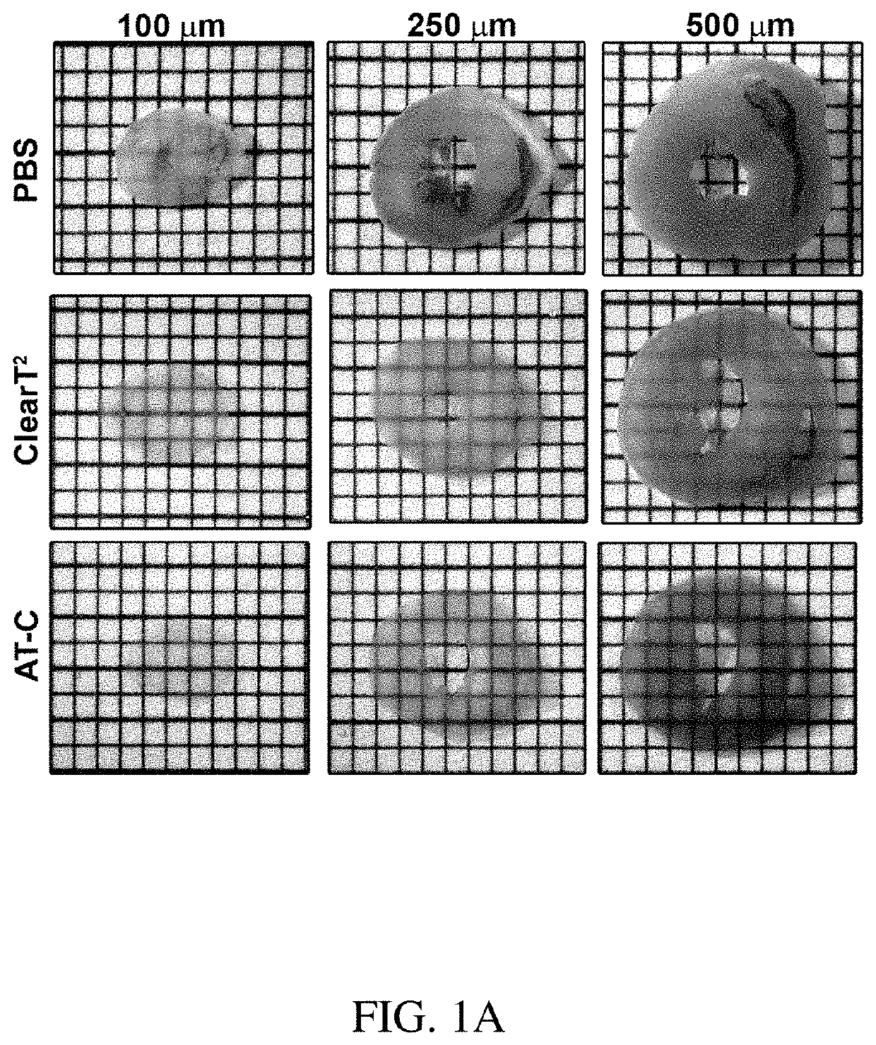
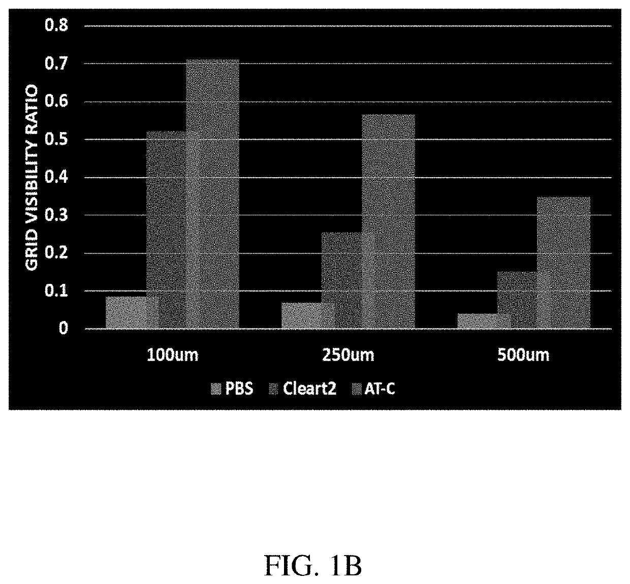
[0038]Using Atacama Clear for Clearing Biological Tissue

[0039]For these studies, heart sections were used because muscle is the most difficult tissue to clear, due to its extensive fibrous cellular structure. Tissues were incubated in increasing grades of glycerol, starting with solution 1 and progressing to solution 6 of the Atacama Clear (AT-C) formulation. Time of incubation can be varied depending on how thick and tough the tissues are, and whether they are embryonic or adult. Smaller and less tough tissues, as well as embryonic tissues,...


PUM

PropertyMeasurementUnit
RIaaaaaaaaaa
RIaaaaaaaaaa
thicknessaaaaaaaaaa

Abstract

A method for rendering biological tissue sufficiently optically transparent for three-dimensional light microscopy imaging, comprising incubating biological tissue with an optical clearing solution, wherein the optical clearing solution comprises: (i) 20-50 wt % formamide, (ii) 10-90 wt % glycerol, and (iii) water as remainder. Also described herein is a method for ridding tissue of blood to make them amenable for optical clearing, comprising incubating biological tissue in a decolorizing solution, wherein the decolorizing solution comprises: (i) 0.5-3 wt % hydrogen peroxide, (ii) 0.05-1 wt % sodium azide, (iii) 5-20 wt % DMSO, and (iv) phosphate buffered saline as a remained. Also described herein is a method for reducing auto-fluorescence in biological tissue to permit imaging of the biological tissue in a fluorescence-based imaging technique with enhanced resolution, wherein the auto-fluorescence quenching solution comprises: 1-100 mM ammonium bicarbonate, (ii) 20-500 M copper sulfate, (iii) 5-20 wt % DMSO, and (iv) water as remainder.

Description

CROSS REFERENCE TO RELATED APPLICATIONS[0001]This application claims the benefit of priority from U.S. Provisional Application No. 62 / 770,208, filed on Nov. 21, 2018, which is herein incorporated by reference in its entirety.GOVERNMENT SUPPORT[0002]This invention was made with government support under grant number NIH R21DK116171 awarded by the National Institutes of Health. The government has certain rights in the invention.FIELD OF THE INVENTION[0003]The present invention generally relates to optical clearing solutions and their use in rendering biologically tissues substantially optically transparent or solutions to reduce tissue auto-fluorescence. In one aspect, the present invention more specifically relates to optical clearing solutions containing formamide and / or glycerol. In another aspect, the present invention relates to solutions to decolorize, or remove blood from tissue to make them more amenable to optical clearing, and which contains hydrogen peroxide, sodium azide, a...

Claims


Application Information

Patent Timeline
Patent Type & Authority Applications(United States)
IPC IPC(8): G01N1/30G01N33/50G01N21/64
CPCG01N1/30G01N33/5061G01N2021/6432G01N21/6458G01N21/6428G01N33/5058G02B21/16G01N21/6486G02B21/0076
Inventor HURTADO, ROMULO
Owner CORNELL UNIVERSITY
Why Eureka
Try Eureka