Gel microdrops in genetic analysis

a microdrop and genetic analysis technology, applied in the field of nucleic acid analysis methods, can solve the problems of limiting the automation and rapid sample processing of samples, and insufficient banding resolution to detect small deletions or additions of chromosomal elements

Inactive Publication Date: 2003-07-01
CELLAY
View PDF7 Cites 198 Cited by
  • Summary
  • Abstract
  • Description
  • Claims
  • Application Information

AI Technical Summary

Benefits of technology

FIG. 2 shows gel electrophoresis of chromosomal DNA. Unencapsulated, freshly isolated chromosomes: b-mouse, e-human K-562, g-human REH; Unencapsulated chromosomes one month after

Problems solved by technology

Current cytogenetic methods are limited to analysis of gene aberrations easily detectable in cells, nuclei, or chromosomes, in part, because slide based methods are highly manual.
However, banding resolution is not sufficient to detect small deletions or additions of chromosomal mass, which occur in a variety of disease conditions, particularly in cancers.
Analysis therefore requires microscopic evaluation of individual slides limiting automation and rapid sample processing.
Fluorescent in situ hybridizations prepared on glass slides rely not only on the assay and reagents but on the instrumentation and the expertise and ingenuity of the scientists using it resulting in poor reproducibility.
An inherent limitation to this technology is that at least 100 kb of DNA sequence in a single cell must be present for detection (68-70).
In addition, harsh conditions fo

Method used


Image

  • Gel microdrops in genetic analysis
  • Gel microdrops in genetic analysis
  • Gel microdrops in genetic analysis

Examples


example 2

Construction of Chromosome-Specific BAC Libraries

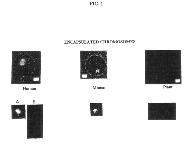
A major unrealized goal of fluorescence in situ hybridization assays has been the use of flow cytometry to isolate specific chromosomes for library construction. Prior to development of the MISH method, after hybridization, only a small fraction of chromosomes remain intact and free in suspension. Without the protection gained using the agarose microspheres, most chromosomes are clumped or fragmented, making them largely unsuitable for flow cytometric analysis (27). We have shown not only that we can hybridize and flow sort encapsulated chromosomes, but also that alkaline-denatured chromosomes can be digested with restriction endonucleases. As a result of this finding, we propose construction of chromosome-specific libraries. Human chromosome 21 was chosen because it is often difficult to identify and sort using conventional dual fluorescent staining since it is small and indistinguishable in the presence of cellular debris (29). The ...

example 3

Use of MISH-sorted Chromosomes for Reverse Chromosome Painting

The development of in situ hybridizations with flow sorted chromosome libraries (25,26), combined with non-isotopic signal detection (30), has become a powerful approach for rapidly analyzing human chromosomal aberrations, such as aneuploidy and translocations. These techniques, termed chromosome painting, are becoming widely used in clinical cytogenetics. However, small rearrangements, additions, or deletions are not detectable using conventional chromosome painting, but these aberrations are detectable by reverse chromosome painting, which is performed using a probe prepared from aberrant chromosomes. A method of using MISH-sorted chromosomes is depicted below.

example 4

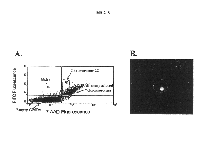
Detection of the Philadelphia Chromosome

The Philadelphia chromosome is a shortened chromosome 22 that results from a balanced translocation between chromosomes 9 and 22 with the translocation breakpoints at 9q34 and 22q11. As a result of this translocation, most of the abl oncogene, located on chromosome 9, is juxtaposed to part of the bcr gene, located on chromosome 22, creating a new bcr-abl gene fusion (40, 51, 52, 53, 54). This gene fusion encodes an abnormal protein with strong tyrosine kinase activity compared with the weak tyrosine kinase activity of the normal abl protein. The abnormal tyrosine kinase produced from bcr-abl causes increased cell proliferation and contributes to leukemogenesis by unknown cellular pathways.

Translocations such as bcr / abl are currently detected by cytogenetic examination of metaphase chromosome preparations prepared from bone marrow cultures. Although this method is adequate to detect most chronic myelogenous leukemia cases (CML) in which Ph.sup....


PUM

PropertyMeasurementUnit
Timeaaaaaaaaaa
Timeaaaaaaaaaa
Fractionaaaaaaaaaa

Abstract

The invention provides methods of nucleic acid analysis. Such methods entail forming a population of gel microdrops encapsulating a population of biological entities, each entity comprising a nucleic acid, whereby at least some microdrops in the population each encapsulate a single entity. The population of gel microdrops is then contacted with a probe under conditions whereby the probe specifically hybridizes to at least one complementary sequence in the nucleic acid in at least one gel microdrop. At least one gel microdrop is then analyzed or detected. The biological entities can be cells, viruses, nuclei and chromosomes.

Description

The present invention resides in the field of genetic analysis.Cytogenetic testing is still in its infancy. Current cytogenetic methods are limited to analysis of gene aberrations easily detectable in cells, nuclei, or chromosomes, in part, because slide based methods are highly manual. Analysis of aberrations present in low frequency is not routinely performed in the clinical setting because many slides of cells, nuclei or chromosomes would have to be evaluated to establish statistical frequency.Banding is the classical approach used for analyzing chromosomes in metaphase spreads. This method is based on staining which results in dark bands in the region of the chromosome where the chromatin occurs at higher density. The banding pattern is specific for each chromosome and allows identification for karyotyping, which is the determination of each chromosome's copy number. However, banding resolution is not sufficient to detect small deletions or additions of chromosomal mass, which o...

Claims


Application Information

Patent Timeline
IPC IPC(8): C12Q1/68C12M1/00C12N1/06C12N5/02C12N15/09G01N33/544C12Q1/6813C12Q1/6816C12Q1/6827C12Q1/6834C12Q1/6841C12Q1/689G01N33/566G01N33/58
CPCC12Q1/68C12Q1/6813C12Q1/6816C12Q1/6827C12Q1/6834C12Q1/6841C12Q1/689C12Q1/70C12Q2523/101C12Q2565/501C12Q2563/161C12Q2600/156C12Q2600/158
Inventor TRNOVSKY, JANMCGRATH, PATRICIA
Owner CELLAY
Why Eureka
Try Eureka