
Apparatuses and methods for performing minimally invasive diagnostic and surgical procedures inside of a beating heart

a technology for diagnosing and surgical procedures inside of beating hearts, applied in applications, manufacturing tools, veterinary instruments, etc., can solve the problems of valvular insufficiency, inability to precisely measure, blind dilation, etc., to minimize the risk of air introduction, reduce the need for cardiopulmonary bypass, and ensure the effect of preventing dilated blood vessels

Inactive Publication Date: 2005-01-11
UNIV OF MARYLAND
View PDF10 Cites 341 Cited by
  • Summary
  • Abstract
  • Description
  • Claims
  • Application Information

AI Technical Summary

Benefits of technology

The present invention is drawn to apparatuses and methods for performing minimally invasive diagnostic and surgical procedures inside of a beating heart that alleviate the need for cardiopulmonary bypass (CPB), minimizes the risk of air introduction into the heart during surgery and emboli, and maintains sufficient intracardial pressure during the diagnostic and surgical procedures. In addition, the apparatuses and methods are compatible with several types of diagnostic and surgical techniques, including mitral valve repair, repair of atrial or ventricular septal defects, endovascular aortic surgery, and electrophysiologic studies.
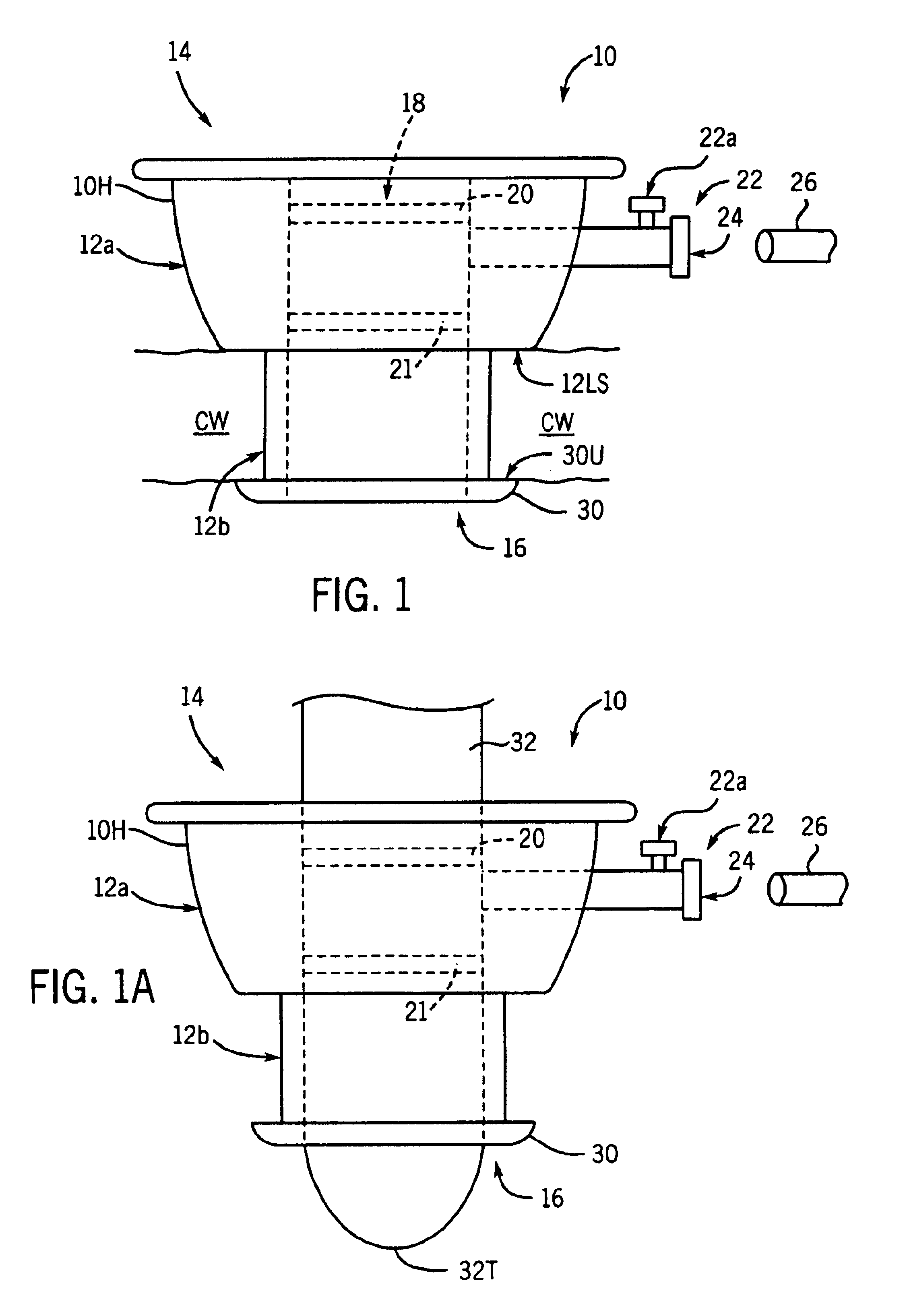
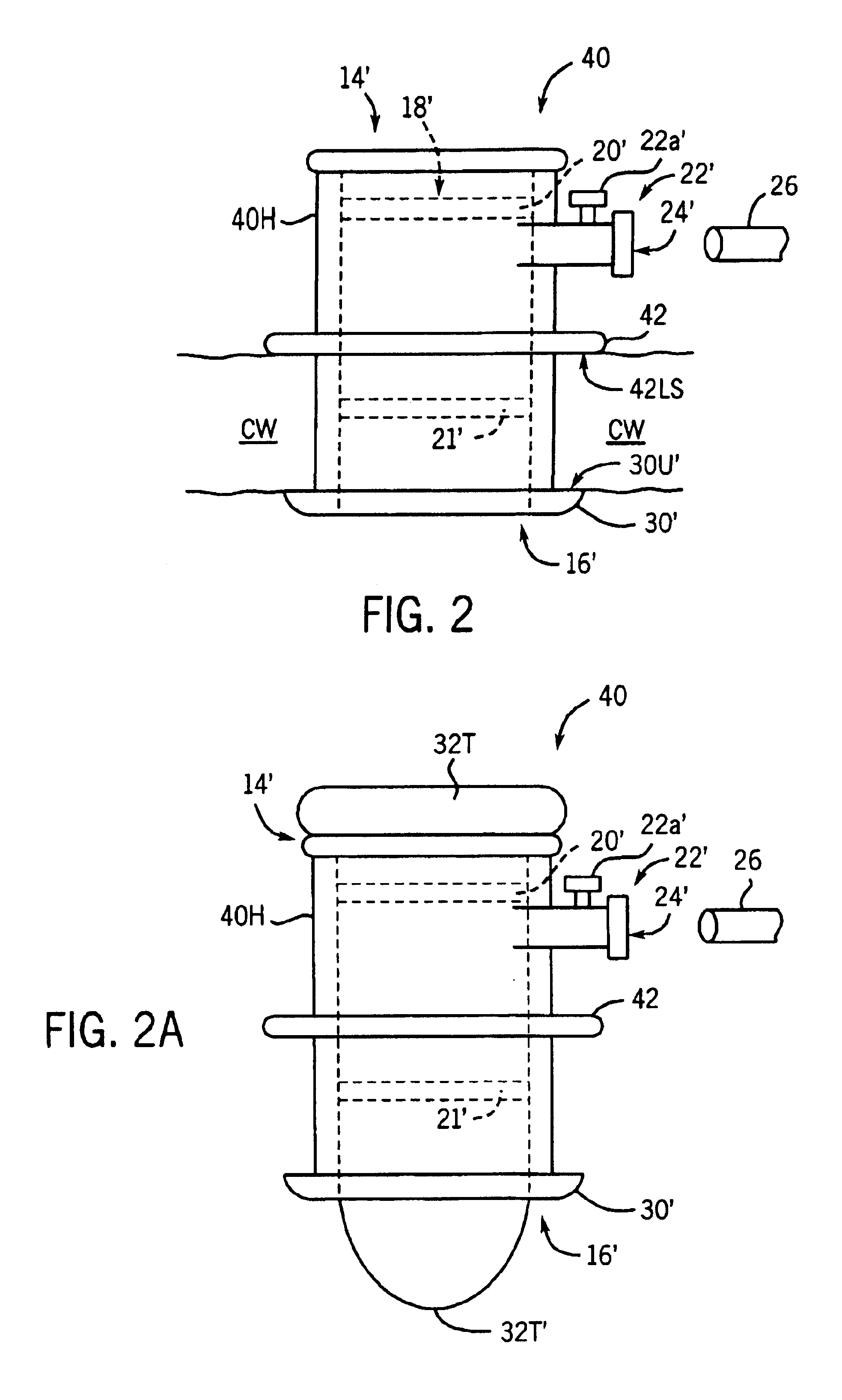
The apparatuses and methods of the present invention provide several advantages over known cardiac surgical instrumentation and procedures. For example, the apparatuses and methods of the present invention alleviate the need for CPB or cardioplegic arrest by combining a cardiac port with advances in cardiac imaging and endoscopic suture placement devices and stapling devices and simplified valve and septal defect repair techniques. The cardiac port of the present invention is configured to be inserted through a chamber wall of a beating heart and to enable passage of medical instruments into a heart chamber, while minimizing injection of air into the heart chambers and consequent emboli. The method of the present invention provides access to a heart chamber via the cardiac port so that the surgical instruments can be inserted through the heart chamber to gain access to the operative site.
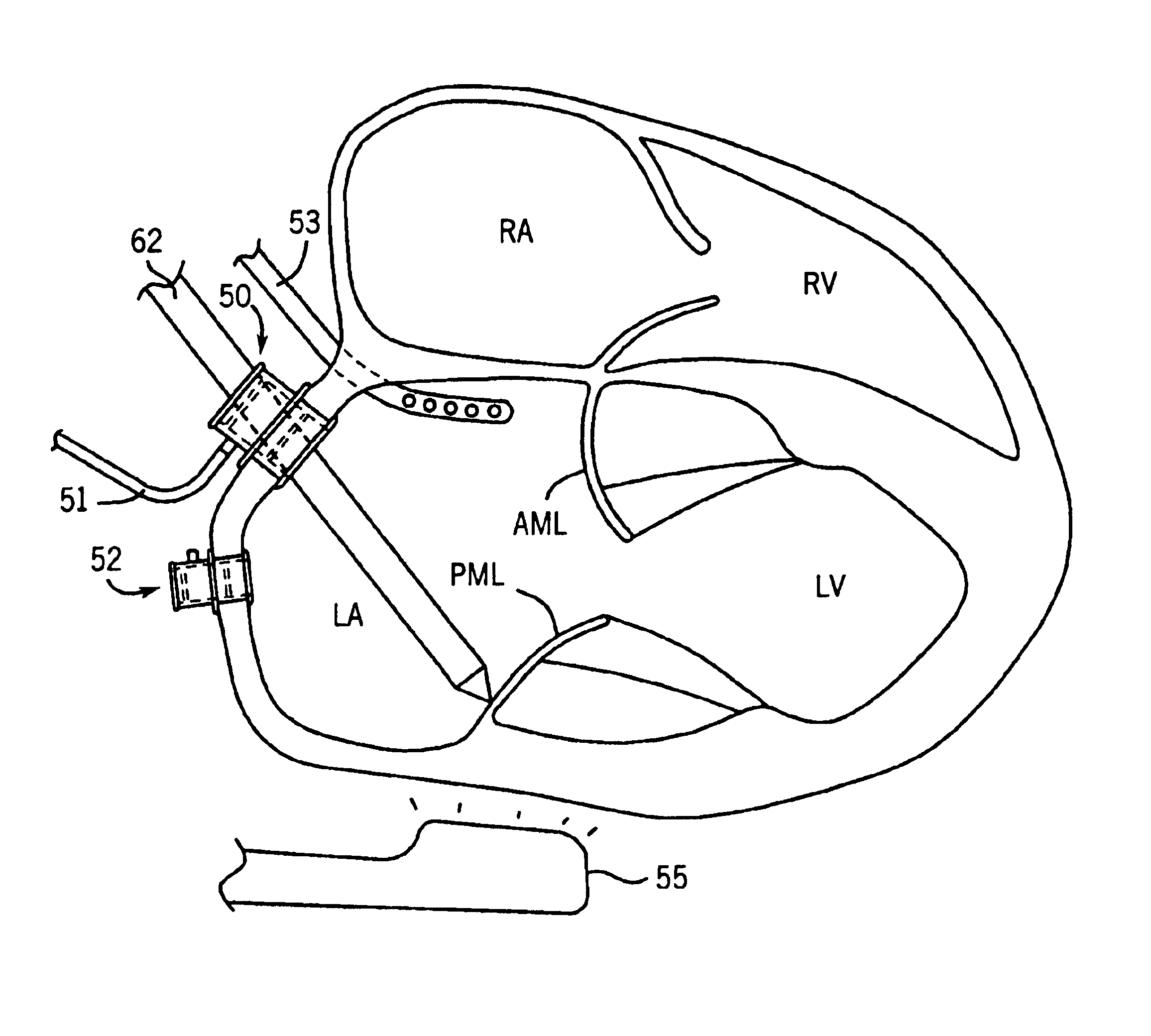
The advances in cardiac imaging include multiplane trans-esophageal echocardiography (TEE). For example, in mitral valve repair, TEE images the mitral valve well enough to allow surgical manipulation of the valve via instruments, such as endoscopic suture placement devices, inserted into the heart chamber through the cardiac port. In one surgical technique, the endoscopic suture placement device can grasp the edge of a moving mitral valve leaflet and pass a suture through the valve leaflet. The endoscopic suture placement device, in essence, sews the valve. This surgical technique involves suturing the free edges of the anterior and posterior mitral valves together to replicate an Alfiori (“bow tie”) mitral valve repair. The method of the present invention also enables investigation of the hemodynamic consequences of temporary acute mitral valve incompetence that will occur with valve manipulation.
In addition, the apparatuses and methods of the present invention enable the surgeon to enter the heart through the atrium. The atrial approach allows the surgeon to place additional sutures and annuloplasty devices, including staples, sutures, and rings, in the appropriate locations in the heart. Further, an atrial approach, as compared to a ventricular approach, minimizes blood loss and maximizes the types of surgical techniques that can be performed.

Problems solved by technology

These dilations were blind in that the patient's blood blocked the surgeon's sight to the valve.
These various techniques were somewhat effective to repair heart valves, but operating “blind” made precision impossible, and dilation often tore the valve, leading to valvular insufficiency.
In addition, embolic brain and other organ injuries occurred due to air entering the heart chambers and to loosening calcific debris.
Like the valve repairs, this procedure was a blind one, due to the surgeon's inability to see through blood that collected in the heart chamber.
In addition, blood loss was unpredictable.
Further, it was easy to entrain air into the cardiac chamber during this procedure, which could cause heart failure, strokes, and death.
Several creative methods were devised to temporarily stop blood flow or briefly turn off the circulation or to support the circulation using another person's circulatory system, but these methods were cumbersome and dangerous, and only the simplest repairs could be performed.
Despite advances, many open-heart surgical procedures still have a 5-9% mortality rate and a 20% morbidity rate.
The advances have not reduced the major complication rates, however, and they are associated with an increased risk of aortic dissection and stroke.
Further, they increase the cost per case by approximately $5000.
The major complication rate has not been reduced because the procedures have not eliminated the need for cardioplegia and CPB, which together account for the majority of the complications.
However, cardiac function is always depressed after cardioplegic arrest, and the longer the duration of arrest, the greater the injury.
In addition to the risks associated with cardioplegia, CPB, in its own right, can be damaging in multiple ways.
This leads to pulmonary dysfunction, third space loss, and neurohumoral imbalances.
Additionally, CPB has negative effects on the immune system.
Surgery of the interior of the heart is not possible with these methods, however, because the methods do not allow the cardiac chambers to be opened.
Opening the cardiac chambers of a beating heart would lead to massive blood loss and air being drawn into the circulation in large amounts.
The clip technique has some potential drawbacks, however, including possible device embolization.
In addition, entering the heart through the apex of the left ventricle increases the risk of blood loss due to the high intracavitary pressures of the ventricle and does not allow for any other work to be performed on the mitral valve.

Method used


Image

  • Apparatuses and methods for performing minimally invasive diagnostic and surgical procedures inside of a beating heart
  • Apparatuses and methods for performing minimally invasive diagnostic and surgical procedures inside of a beating heart
  • Apparatuses and methods for performing minimally invasive diagnostic and surgical procedures inside of a beating heart

Examples


Embodiment Construction

The present invention provides apparatuses and methods for performing minimally invasive diagnostic and surgical procedures inside of a beating heart, thereby obviating the need for administration of cardioplegia and cardiopulmonary bypass (CPB). Procedures contemplated by the present invention can include mitral and tricuspid valve repair, aortic and pulmonary valve repair, and repair of intracardiac defects, such as atrial and ventricular septal defects. Repair of the mitral valve in turn can include correcting mitral regurgitation (i.e., annular dilation and heart failure, myxomatous degeneration, and chordal rupture) and mitral stenosis. Other procedures include adjuncts to endovascular surgery and access for electrophysiologic studies. One of ordinary skill in the art will recognize other applications for the present apparatuses and methods.

In this connection, the present invention provides a valved cardiac port that can be temporarily inserted in a chamber wall of a heart cham...


PUM

PropertyMeasurementUnit
pressureaaaaaaaaaa
atmospheric pressureaaaaaaaaaa

Abstract

Diagnostic and surgical procedures may be performed on a beating heart using an assembly which includes a port and a fluid transport device. The port has a housing for insertion through a wall of the heart chamber and may include one valve disposed in the housing and an inlet connected to the housing. Methods for repair and diagnosis of the heart are also described. A specific method for repairing a mitral valve uses staples which may be banded together with a strip of material.

Description

FIELD OF THE INVENTIONThe present invention relates generally to an apparatuses and methods for performing minimally invasive diagnostic and surgical procedures inside of a beating heart. More particularly, the present invention relates to an apparatuses and methods for accessing the heart chamber and performing minimally invasive diagnostic and surgical procedures, including diagnosing and repairing pathology of the heart valves (aortic, mitral, pulmonary and tricuspid) and the cardiac chambers (atrial and ventricular septums), inside of a heart while the heart is still beating.BACKGROUND OF THE INVENTIONDiagnostic and reparative surgical procedures have been performed on the heart since at least the 1920s. The first attempts at repairing heart valves began in 1923 with Cutler and Levine performing blind dilations of the mitral valve with cutting instruments. These dilations were blind in that the patient's blood blocked the surgeon's sight to the valve. Thereafter, Sutar reported ...

Claims


Application Information

Patent Timeline
Patent Type & Authority Patents(United States)
IPC IPC(8): A61F2/24
CPCA61F2/2466A61B2017/3425A61B2017/3441A61F2/2445
Inventor DOWNING, STEPHEN W.
Owner UNIV OF MARYLAND
Learn More