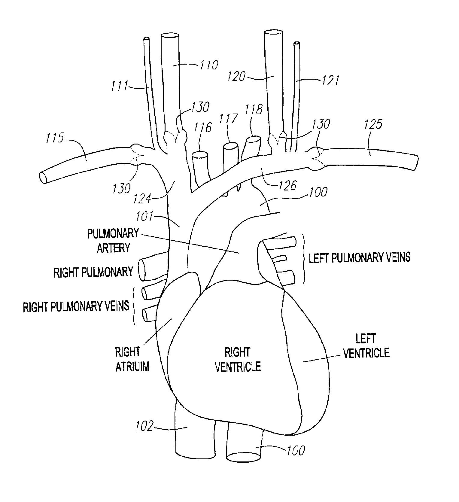
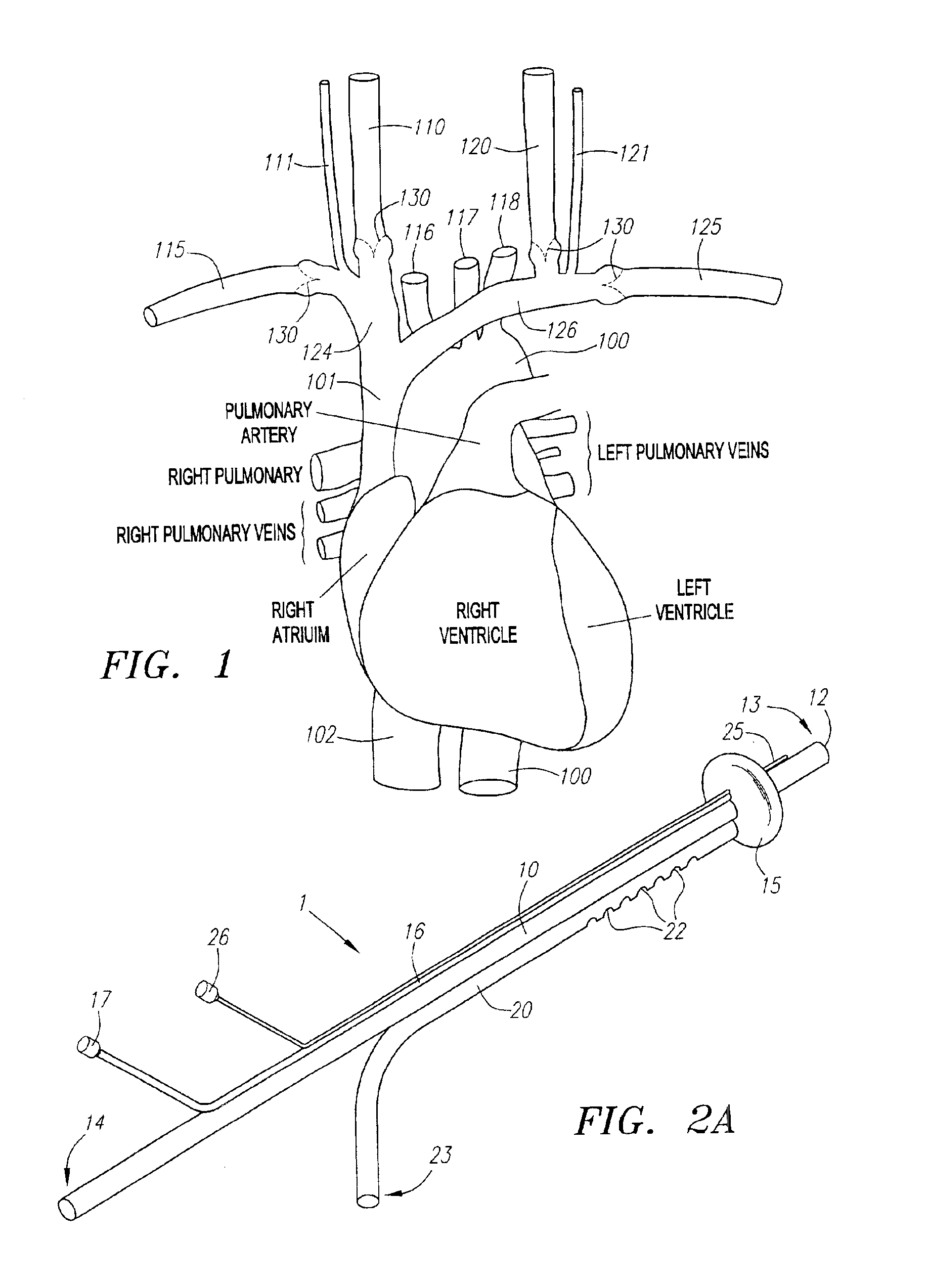
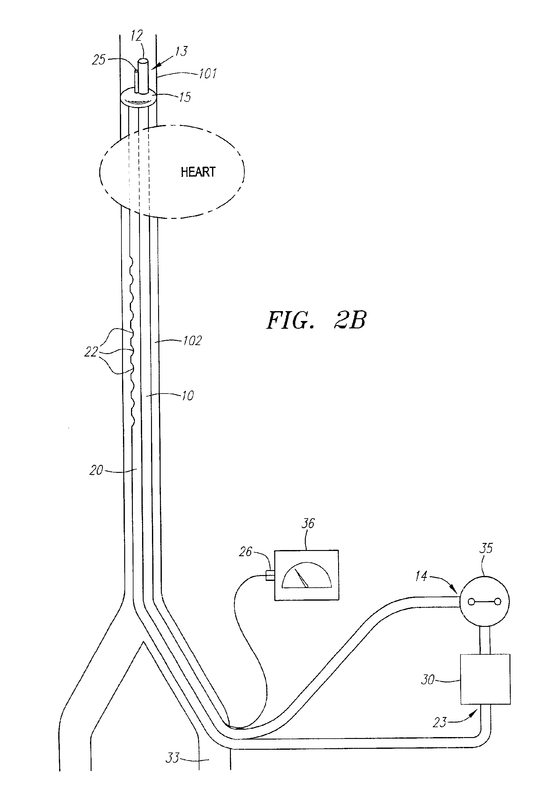
[0012]In a first method of using the devices, the distal end of the catheter is inserted through a
peripheral vein, e.g., the
femoral vein, into the
vena cava. The distal region of the catheter and the occluder are positioned in the
superior vena cava (SVC), and the occluder is expanded to isolate
blood flow in the SVC. Deoxygenated blood is withdrawn from the drainage port(s) and passed through the second lumen and the proximal end of the catheter, which is connected to an
oxygenator. The oxygenated blood or medium is then infused into the first lumen and through the infusion port of the catheter to perfuse the cerebral tissues through the SVC in a retrograde fashion, thereby obviating the need for IVC clamping and
backflow of oxygenated blood into the
right atrium and the lungs which occurs with existing methods for retrograde cerebral venous perfusion (RCP). Infusion of oxygenated blood may be facilitated by a pump connected to the
oxygenator and the proximal end of the first lumen. The infusion rate of oxygenated blood can be varied according to the
venous pressure recorded by the manometer, which is mounted distal to the occluder.
[0013]In still another embodiment, retrograde venous perfusion can be achieved by inserting the distal end of the catheter in the right or the left
internal jugular vein. The occluder is expanded to occlude the
internal jugular vein. Deoxygenated blood is withdrawn from the
vena cava, oxygenated by an oxygenator, and returned to the internal
jugular vein to perfuse the cerebral tissues in a retrograde direction through the lumen and infusion port of the catheter. By positioning the catheter in the internal
jugular vein to minimize
backflow of oxygenated blood to the arm instead of the SVC, and by positioning the occluder distal to the jugular
venous valves to minimize obstruction of blood flow to the intracranial venous
system, the infusion rate of oxygenated blood can be reduced. The jugular
venous pressure (JVP) is often obtained indirectly by measurement of the
pulmonary artery pressure from a Swan-Ganz catheter, which is commonly inserted in patients having aortic
surgery or hemodynamic
instability. The
pulmonary artery pressure, however, often fails to reflect the JVP due to the presence of
jugular vein valves. The manometer, which is mounted distal to the occluder in the internal jugular
vein, will provide a more accurate measurement of the JVP.
[0014]In another method, deoxygenated blood withdrawn from the drainage port is passed through an oxygenator and a cooling system. The cooled oxygenated blood or medium is then infused through the first lumen and the distal port of the catheter to provide isolated hypothermic and retrograde venous perfusion to the brain. In this way, complication associated with systemic hypothermic perfusion, e.g., disseminated intravascular
coagulopathy, is avoided.
[0015]In another method, after
venous blood withdrawn from the SVC is passed through an oxygenator, the oxygenated blood is passed through a pump which connects with the proximal end of the first catheter and a proximal end of a second catheter. The second catheter has a lumen communicating with a distal end inserted through the
femoral artery and positioned within the
femoral artery, the
iliac artery, or the
descending aorta. In this way, oxygenated blood is delivered to the brain and the lower extremities and / or other vital organs, such as the kidneys. During aortic surgeries, the space available for
instrumentation on the aorta, e.g.,
insertion of cannula for
cardiopulmonary bypass, is often limited. The methods described above provide perfusion to the
peripheral organs during cardiac arrest without the need for
instrumentation on the aorta, and therefore provide a superior alternative to conventional
cardiopulmonary bypass.
[0017]It will be understood that there are several advantages in using the devices and methods disclosed herein for protecting the brain and cerebral vasculature of patients suffering from global or focal ischemia. For example, compared with known techniques for retrograde cerebral venous perfusion (RCP), the retrograde venous perfusion of the invention (1) minimizes
backflow of blood to the heart and / or the arms, thereby allowing lower infusion rates to be used, (2) eliminates the need for IVC clamping, thereby minimizing damage to the IVC, (3) positions the catheters distal to the jugular
venous valves, thereby minimizing obstruction of blood flow by the valves, (4) provides isolated cerebral hypothermic perfusion, (5) provides an accurate measurement of the jugular
venous pressure, and (6) provides perfusion to the brain, the lower extremities, and other vital organs during cardiac arrest. In addition, significant cerebral
embolization associated with retrograde aortic perfusion (RAP) and selective antegrade cerebral perfusion (SCP) is avoided using the venous retrograde perfusion disclosed herein.

