Amphiphilic polymeric vesicles

Inactive Publication Date: 2005-07-12
BIOCURE
View PDF12 Cites 136 Cited by
  • Summary
  • Abstract
  • Description
  • Claims
  • Application Information

AI Technical Summary

Benefits of technology

[0010]Vesicles are made from segmented amphiphilic A+B copolymers, where A is hydrophilic and B is hydrophobic, which self-assemble when dispersed in oil or water. In one embodiment, the vesicles are made from an ABA triblock copolymer, and where the inner core is hydrophilic, the middle layer is hydrop

Problems solved by technology

Self-assembled structures known in the prior art have often exhibited limited stability that induces, for example, a rapid clearance of drug-loaded vesicles from the blood after their intravascular adm

Method used


Image

  • Amphiphilic polymeric vesicles
  • Amphiphilic polymeric vesicles
  • Amphiphilic polymeric vesicles

Examples


example 1

Synthesis of PMOXA-PDMS-PMOXA Diblock and Triblock Copolymers

[0102]Bifunctional poly(dimethylsiloxane)

[0103]In a 250 mL round bottom two-necked flask with a Soxhlet extractor (filled with molecular sieve (4 A)), a condenser and a septum on the second ground joint, 34.2 g (6.34 mmol) α-ω-bis(3-hydroxypropyl)-polydimethylsiloxane (I−1) were dissolved in 90 mL hexane and distilled under reflux for 17 h in a nitrogen atmosphere. After this drying procedure, the solution still contained 21 ppm water. Subsequently, the solution was concentrated to 60 mL hexane, cooled to 0° C. and 3.6 g (45.5 mmol) of dry pyridine were added. Then, 12.4 g (43.9 mmol) trifluoromethane sulfonic acid anhydride were added over 15 minutes and the mixture was stirred for another 30 min at a temperature of 0° C. After the addition of 20 mL chloroform (water content <10 ppm), the resulting suspension was filtered under vacuum using a G4 glass filter funnel. The solvent was evaporated under high vacuum.

[0104]The y...

example 2

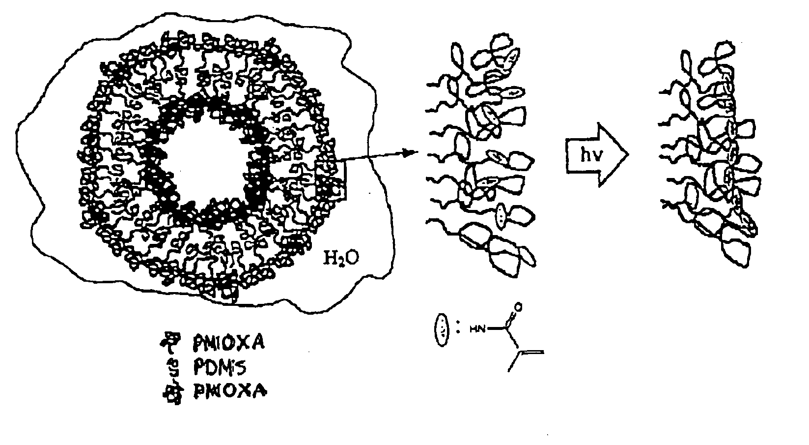
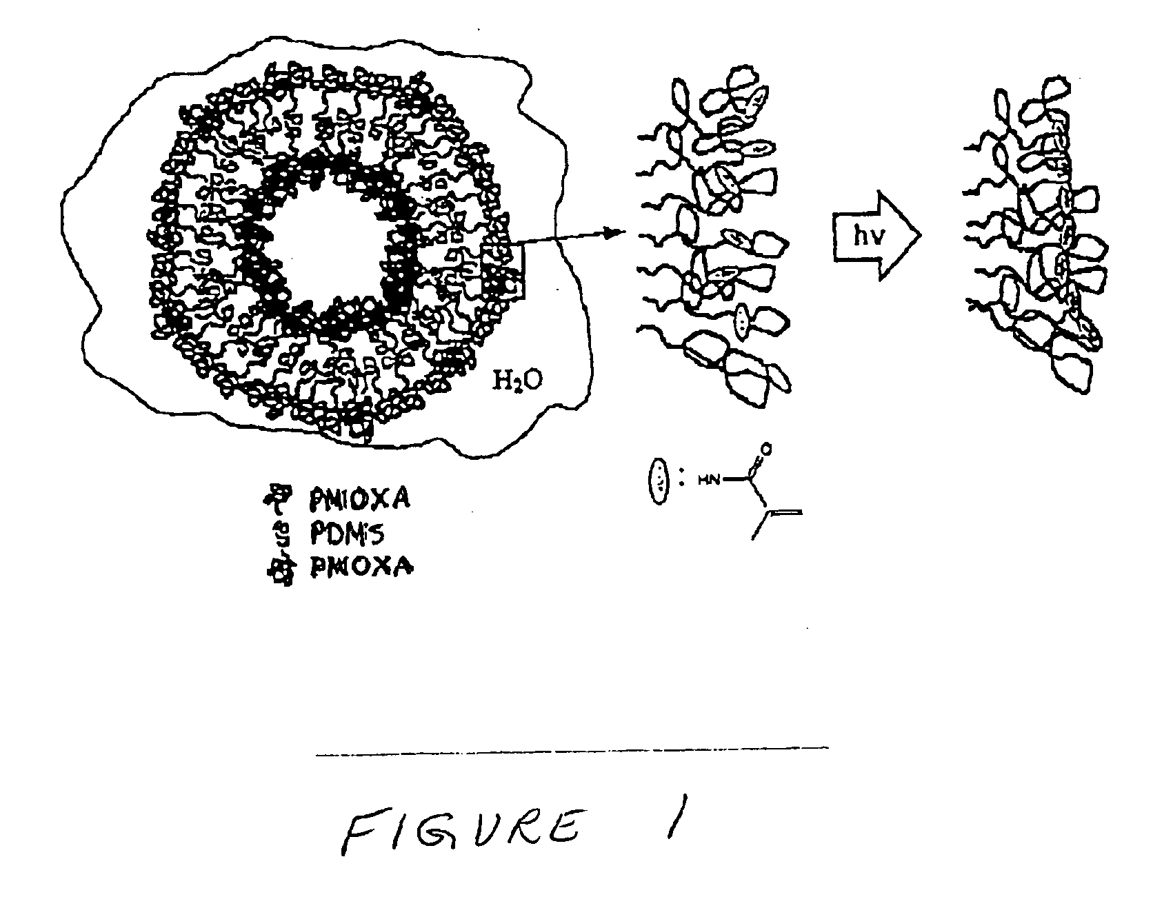
Preparation of Vesicles from PMOXA-PDMS-PMOXA

[0114]The formation of small unilamellar vesicles from the triblock copolymer was achieved according to the following procedure.

[0115]The end-group functionalised PMOXA-PDMS-PMOXA triblock copolymer was dissolved in ethanol to yield a clear, homogeneous solution containing 17 wt % polymer. This solution was added dropwise under vigorous stirring to the respective volume of doubly distilled water. The procedure led to a dispersion of triblock copolymer vesicles of a broad size distribution.

[0116]The polydispersity was reduced by extrusion of the vesicular dispersion through a Nucleopore filters (Millipore) having a pore size of 200 nm.

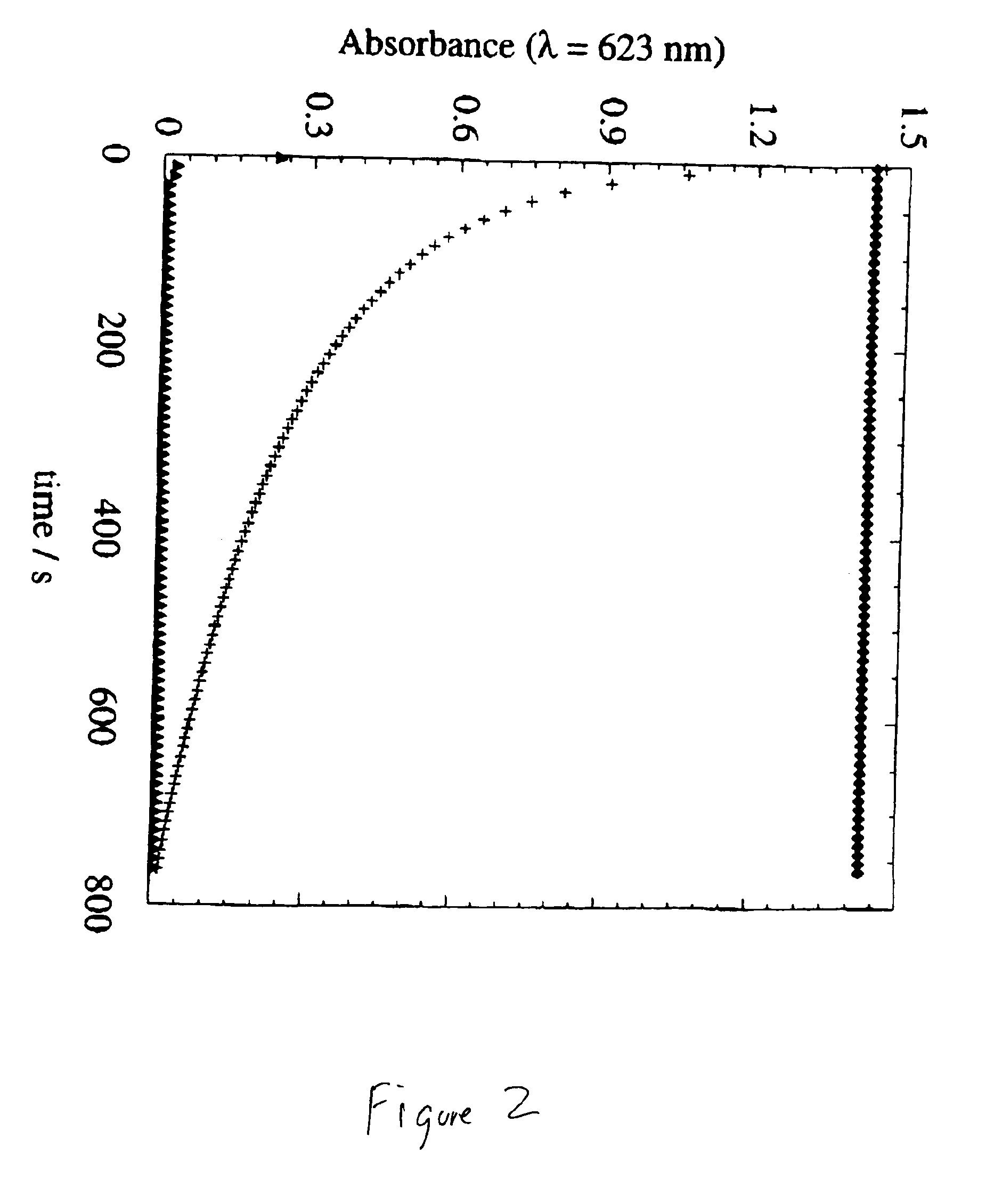
[0117]Polymerization of the vesicles was achieved by irradiating the dispersion for 15 min with an UV lamp (Ultratech 400 W, wavelength=254 nm, Osram AG). If stored in the dark to prevent their polymerization, the PMOXA-PDMS-PMOXA triblock copolymer vesicles were stable over several weeks and displayed no cha...

example 3

Characterization of Vesicles from PMOXA-PDMS-PMOXA

Freeze-fracture Replication Transmission Electron Microscopy

[0118]A sample of approximately 10 microliters of the vesicle dispersion was brought onto a gold platelet at room temperature and was quenched by hand plunging into a mixture of 15% 2-methyl butane and 85% propane at 83 K. After quenching, the sample was transferred into liquid nitrogen and clamped on a brass block (Balzer). It was mounted on a Balzer freeze etch device (BAF 300), and subsequently the pressure was reduced to 5 10−9 mbar. After evacuation, the sample was fractured with a liquid nitrogen cooled microtome. To enhance the contrast of the surface structure, the sample was warmed to 153 K and etched for 10 min. Thereafter, the sample was cooled again with liquid nitrogen and shadowed with W / Ta under an angle of 30°. After the samples were warmed up to room temperature and brought to atmospheric pressure, the replica was washed with chloroform, put on a 400 mesh co...


PUM

PropertyMeasurementUnit
Hydrophilicityaaaaaaaaaa
Hydrophobicityaaaaaaaaaa
Biodegradabilityaaaaaaaaaa

Abstract

Vesicles made from amphiphilic copolymers are disclosed. The amphiphilic copolymers can be ABA copolymers, where one of A and B is hydrophilic and the other is hydrophobic. AB copolymers can also be used. The copolymers may be crosslinked to form nanocapsules. Crosslinking can be accomplished using a variety of methods, including end to end polymerization of copolymers having terminal unsaturated groups. Molecules, such as membrane proteins, can be incorporated into the wall of the vesicles or nanocapsules.

Description

RELATED APPLICATION[0001]This application claims priority to U.S. provisional application Ser. No. 60 / 163,678, filed on Nov. 5, 1999.FIELD OF THE INVENTION[0002]The present invention is generally in the field of vesicles. More specifically, the present invention is in the field of self-assembling polymeric vesicles with enhanced stability.BACKGROUND OF THE INVENTION[0003]Self-organizing, or self-assembling, structures are known. A common example is liposomes. Liposomes are made by emulsifying amphiphilic (and optionally hydrophobic or lipophilic) molecules in water, preferably in the presence of surfactant. Liposomes are either unilamellar or multilamellar spheres that are manufactured from a variety of lipids. Drugs, for example, can be encapsulated within liposomes or captured within the liposome membrane.[0004]Self-assembled structures known in the prior art have often exhibited limited stability that induces, for example, a rapid clearance of drug-loaded vesicles from the blood ...

Claims


Application Information

Patent Timeline
IPC IPC(8): A61K9/51A61K9/127A61K47/30A61K47/34C08G81/00
CPCA61K9/5146A61K9/5192
Inventor MEIER, WOLFGANGHIRT, THOMASNARDIN, CORINNE
Owner BIOCURE
Why Eureka
Try Eureka