Method of imaging lymphatic system using nanocapsule compositions

a composition and nanocapsule technology, applied in the field of nanocapsule compositions for imaging the lymphatic system, can solve the problems of nerve damage risk, reduced immune system function, and selective lymph node dissection

Inactive Publication Date: 2005-05-26
HORIZON TECH FUNDING CO LLC
View PDF43 Cites 36 Cited by
  • Summary
  • Abstract
  • Description
  • Claims
  • Application Information

AI Technical Summary

Problems solved by technology

Elective lymph node dissection, however, benefits only those patients with micrometastases and is unnecessary in many cases.
This is a matter of concern because dissection of the entire lymphatic drainage basin is a major surgical procedure associated with a number of potential short- and long-term complications including increased surgical trauma and scarring, risk of nerve damage, possible reduced immune system function, and lymphedema.
These technologies can only detect the presence of a sizeable, well-defined tumor mass and have not been routinely applied to the identification of the sentinel nodes.
However, application of this imaging modality for sentinel node identification has not heretofore been considered because an ultrasonic contrast agent with the necessary size spectrum and acoustic properties has yet to be described.
Larger bubbles, though more easily detected, fail to pass through the capillary network and smaller bubbles, though unrestrained by the capillary network, are detected poorly.
Microbubbles in the 1 to 10 micron size range, however, are not suitable for use in the lymphatic system.
There is as yet no consensus on the optimal radiopharmaceutical agent for sentinel node identification.
Solid and liquid particles, and particularly those less than a micron in diameter, have been shown to be poor backscatterers of ultrasound and hence have not traditionally been useful as echographic contrast agents.
Air trapping particles of a size range of less than about 900 nanometers, while potentially superior to solid or liquid nanoparticles, presents special challenges not only in their manufacture but also for optimizing a lymphatic ultrasound contrast agent due to the influence of size on its acoustic properties.
However, due to a of this rapid drop-off of signal with diameter, signal levels can quickly fall below the measurement threshold.
Thus backscatter from intact nanobubbles normally cannot be detected.

Method used


Image

  • Method of imaging lymphatic system using nanocapsule compositions
  • Method of imaging lymphatic system using nanocapsule compositions
  • Method of imaging lymphatic system using nanocapsule compositions

Examples


example 1

Preparation of Albumin Coated Polycaprolactone Nanobubbles

[0053] A 6% aqueous solution was prepared from a 25% solution of USP grade human serum albumin (HSA) by dilution with deionized water. Separately, 1 part by weight polycaprolactone and 5 parts cyclooctane were dissolved in 55 parts isopropyl acetate at approximately 70° C. Once dissolution was complete, the organic solution was then thoroughly emulsified into an equal volume of the prepared HSA solution using a rotor / stator homogenizer. The emulsion was then diluted into 17 volumes of deionized water maintained at 30° C. and containing glutaraldehyde to crosslink the HSA. During the addition, the pH of the bath was monitored to insure that it remained between 7 and 8. Low shear mixing was continued for approximately 2½ hours until the isopropyl acetate had completely volatilized. Poloxamer 188 was then dissolved into the bath. Final concentration of the poloxamer was 0.25%. The suspension was allowed to come to room temperat...

example 2

Preparation of Albumin Coated Polylactide Nanobubbles

[0056] A 50.0 gm 6% aqueous solution was prepared from a 25% solution of USP grade human serum albumin by dilution with deionized water. Separately, a 25 gm organic solution containing 0.98% poly-d,l-lactide, 6.91% cyclooctane, and 92.1% isopropyl acetate. The organic solution was then thoroughly emulsified into the prepared aqueous solution using a Virsonic sonicator homogenizer. The emulsion was then diluted into 350 ml deionized water maintained at 30° C. and containing 1.25 ml of 1N NaOH. After the emulsion was fully dispersed, 1.0 ml 25% gluteraldehyde was added to crosslink the HSA. During the addition, the pH of the bath was monitored to insure that it remained between 7 and 8. Low shear mixing was continued for approximately 2½ hours until the isopropyl acetate had completely volatilized. Poloxamer 188 in the amount of 0.75 gm was then dissolved into the bath. The suspension was allowed to come to room temperature and the...

example 3

In-Vitro Acoustic Study

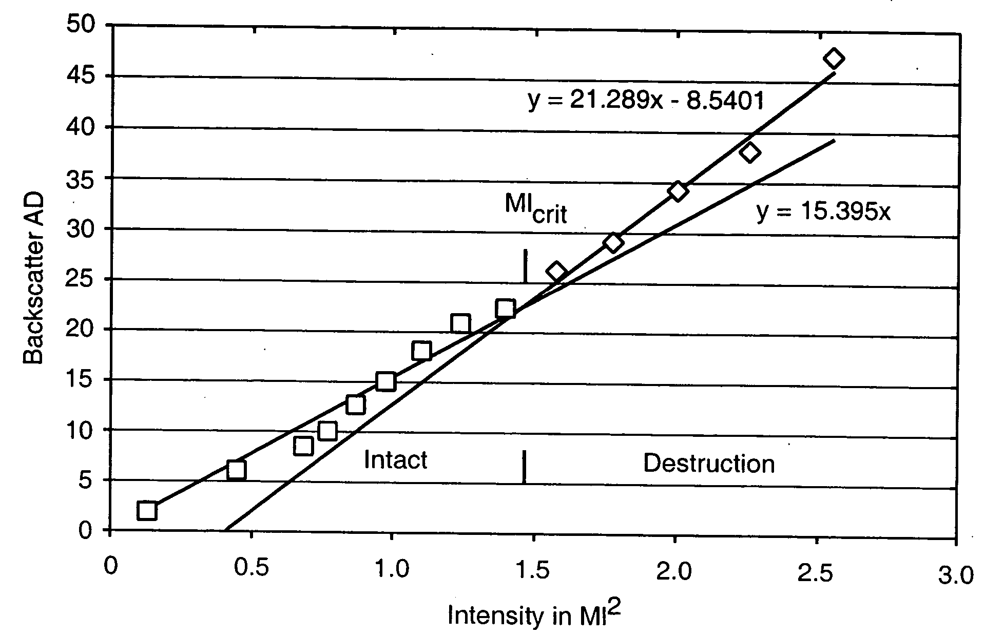
[0059] Nanobubbles fashioned in accordance with the procedures of Example 2 were tested for acoustic backscatter. For the study, an open loop flow circuit was assembled to include an ATS Laboratories Doppler flow phantom having a 6.0 mm diameter flow channel, a VWR variable flow mini-pump, and a 500 ml beaker positioned on a magnetic stir plate to serve as the reservoir for the nanobubble suspension. Flow through the phantom was adjusted to a rate of approximately 95 ml / min.

[0060] The backscatter measurements were made using an ATL HDI 5000 ultrasound system equipped with an L7-4 linear array probe. The probe was positioned onto the flow phantom so that a longitudinal image of the flow channel could be obtained. All measurements were made in harmonic B-mode at a Mechanical Index of 1.0 and focused to a depth of 2.9 cm. For each test run, a total of 30 images were taken at a triggering rate of 30 pulses per minute and then digitally stored.

[0061] A vial of t...


PUM


Abstract

A composition is provided accompanying nonoparticles having diameters in the range of about 100 to 800 nanometers with hollow cores and outer shells with mechanical properties such that they rupture on exposure to predetermined ultrasound energy. The composition is useful for identifying sentinel lymph nodes.

Description

[0001] This invention relates to hollow gas-filled nanoparticles in a size range optimized for uptake by the lymphatic system and to their method of use in identifying sentinel lymph nodes around tumors by means of echographic imaging techniques. BACKGROUND OF THE INVENTION [0002] Solid tumor cancers often spread through lymph channels to regional lymph nodes. Because of this tendency, elective lymph node dissection has become a recognized treatment for patients with early stage melanoma, breast, and other cancers. Many surgeons believe that elective lymph node dissection in a patient with micrometastases can prolong the patient's life and, if conducted early enough, cure the disease. Elective lymph node dissection, however, benefits only those patients with micrometastases and is unnecessary in many cases. This is a matter of concern because dissection of the entire lymphatic drainage basin is a major surgical procedure associated with a number of potential short- and long-term com...

Claims


Application Information

Patent Timeline
Patent Type & Authority Applications(United States)
IPC IPC(8): A61B8/00A61K41/00A61K49/22
CPCA61B8/481A61K49/223A61K41/0028
Inventor OTTOBONI, THOMAS B.SHORT, ROBERT E.GABE, JEFFREY D.
Owner HORIZON TECH FUNDING CO LLC
Why Eureka
Try Eureka